Prognostic Factors of Idiopathic Facial Palsy:
A Retrospective Study
Gi Nam Park, Jeong Kyo Jeong, Eun Seok Kim, Jung Ho Kim and Young Il Kim
*Dept. of Acupuncture & Moxibustion, College of Korean Medicine, Daejeon University
[Abstract]
Objectives : The purpose of this study was to evaluate the clinical prognostic factors affecting facial palsy in 98 idiopathic facial palsy patients who were hospitalized and treated in 2015, using retrospective statistical analysis.
Methods : We investigated patients with idiopathic facial nerve palsy, admitted to a Korean medical hospital in 2015, and examined patients’ variables and therapeutic variables. For analysis of clinical data, an independent sample t-test, analysis of variance (ANOVA), and simple regression analysis were performed using IBM SPSS version 24.0.
Results : 1. The initial degree of facial palsy showed statistical significance with age. The older the age, the more severe the initial palsy.
2. Following treatment degree of facial palsy was statistically significant with age, hypertension, and fasting blood sugar (FBS). The higher the value, the slower the recovery from facial palsy.
There was a statistical significance with the number of treatments in a Korean medical hospital.
The more frequent the treatment, the faster the facial palsy recovery.
3. Degree of facial palsy after 12 months was statistically significant with age, hypertension, diabetes, FBS, and the initial severity of facial palsy. The higher the value, the slower the facial palsy recovery.
4. Sex, left or right sided palsy, alcohol consumption, smoking, history of facial palsy, season of onset, total number of treatments and bio chemistry (BC), complete blood cell count (CBC), urinalysis (UA) factors had no statistical significance with prognosis of facial palsy.
Conclusion : Age, season of onset, hypertension, diabetes, FBS, initial severity of facial palsy, and the number of treatments at a Korean medical hospital showed statistical significance.
The number of treatments at the Korean medical hospital positively correlated with facial palsy prognosis, and the others variables showed a negative correlation with facial palsy prognosis.
✱ Corresponding author : 156, Daedeok-daero, Seo-Gu, Daejeon, Korea Tel : +82-42-470-9137 E-mail : [email protected]
Key words : Facial palsy;
Prognostic factors;
Retrospective analysis
Received : 2017. 07. 04.
Revised : 2017. 07. 22.
Accepted : 2017. 07. 27.
On-line : 2017. 08. 20.
This is an Open-Access article distributed under the terms of the Creative Commons Attribution Non-Commercial License (http://creativecommons.org/licenses/by- nc/3.0) which permits unrestricted non-commercial use, distribution, and reproduction in any medium, provided the original work is properly cited.
The Acupuncture is the Journal of Korean Acupuncture & Moxibustion Medicine Society. (http://www.TheAcupuncture.org)
Copyright 2017 KAMMS. Korean Acupuncture & Moxibustion Medicine Society. All rights reserved.
Ⅰ. Introduction
Bell’s palsy is a paralysis or weakness of the muscle on one side of face due to facial nerve dam- age. The most common symptoms in patients with Bell’s palsy are facial palsy or asymmetry. And it sometimes accompanied by other symptoms, such as otalgia, loss of taste, dry eye discomfort, tinnitus, hearing loss, and excessive tears
1).
The unilateral facial weakness associated with Bell’s palsy is thought to result from facial nerve inflammation and edema induced by reactivation of the Herpes simplex or Varicella zoster virus. In the temporal bone, the facial nerve travels in a narrow canal; and swelling of the nerve may result in compression and subsequent damage
2,3).
Diagnosis of Bell’s palsy is based on excluding other causes of unilateral facial paralysis, and 30%
to 60% of cases of facial palsy result from an un- derlying disorder that mimics Bell palsy, including a central nervous system lesion (stroke, demyeli- nating disease), Ramsay Hunt syndrome, trauma, granulomatousdisease, otitismedia, cholesteatoma, and Guillain-Barre syndrome. Many of these con- ditions have associated features that help distin- guish them from Bell’s palsy
4).
The treatment of Bell’s palsy focuses on maxi- mizing recovery and minimizing associated com- plications. In Korean medicine, the treatment of Bell’s palsy is based on acupuncture, moxibustion, and combined with herbal medicine
5). There are some clinical reports have indicated that elec- troacupuncture and moxibustion are effective for Bell’s palsy
6,7). The conventional medical treatments of Bell’s palsy are corticosteroids, antiviral therapy, and surgical decompression, etc. The main treatment of Bell’s palsy is steroid prescrip- tion. The anti-inflammatory effect of steroids is assumed to minimize facial nerve swelling, com- pression and damage, so reducing the length of re- covery time
8,9). Recently clinical research has reported on attempts to implement a combination of more traditional approaches to medicine
derived from Korea and conventional medical ap- proaches
5,10).
The annual incidence of Bell’s palsy is 15-30 per 100,000 person, with no predilection for sex or eth- nicity. It can affect people at any age, but the inci- dence is slightly higher after age 40
11).
Most Bell’s palsy patients recover completely. In adults, upto30% ofuntreatedpatientsfailtomake a complete recovery and have residual symptoms after 6 months. Furthermore, 7 to 12% of patients have a recurrence
4,12).
Facial dysfunction has a dramatic effect on a pa- tient’s appearance, psychological wellbeing and quality of life. Therefore, being able to predict of a Bell’s palsy patient’s prognosis and recovery is very valuable. There have been many studies of Bell’s palsy treatments in relation to acupuncture, moxibustion, herbalmedicine
5-7)andBell’spalsydi- agnosis of ENG
13), EMG
13,14), and HRV
14). However retrospective studies of factors related to Bell’s palsy prognosis are uncommon. This retrospective study statistically analyzed factors related to Bell’s palsy prognosis and prepare basis of Bell’s palsy progress.
Ⅱ. Subjects and Methods
1. Participants
The study included 98 patients diagnosed with
Bell’s palsy admitted to a Korean medical hospital
in 2015. All patients were diagnosed as having
acute facial palsy (only of the idiopathic type, ex-
cluding central, infectious, and traumatic palsy)
and were admitted within 1 week of onset and had
taken corticosteroid medication. Those who could
be contacted by telephone after one year were in-
cluded in the study.
2. Methods 1) Data source
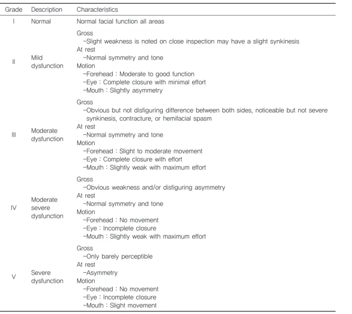
Data were collected from medical records and a telephone survey. The House-Brackmann (HB) scale was used to determine the degree of Bell’s palsy severity (Table 1). Collected data included pa- tients’ personal information, blood test and urinal- ysis results, and the number of treatments. (Table 2)
2) Statistical analysis
To analyze the statistical relationship between the clinical data and the degree of Bell’s palsy
severity(HB scale), IBM SPSS version 24.0 was used. An independent sample t-test was used to verify the correlation between sex, the affected side of face, alcohol consumtion, smoking, diabetes mellitus, hypertension, history of Bell’s Palsy, blood test and urinalysis, using the HB scale for initial, following treatment, and after 12 months.
Analysis of variance (ANOVA) was used to con- firm a relationship between the season of onset, ageandtheHBscaleatinitial, followingtreatment, and after 12 months.
Simple regression analysis was used to confirm a correlation between the total number of treatments at a Korean medical hospital and the total number
Table 1. House-Brackmann (HB) Facial Nerve Grading System Grade Description Characteristics
I Normal Normal facial function all areas
II Mild dysfunction
Gross
-Slight weakness is noted on close inspection may have a slight synkinesis At rest
-Normal symmetry and tone Motion
-Forehead : Moderate to good function -Eye : Complete closure with minimal effort -Mouth : Slightly asymmetry
III Moderate dysfunction
Gross
-Obvious but not disfiguring difference between both sides, noticeable but not severe synkinesis, contracture, or hemifacial spasm
At rest
-Normal symmetry and tone Motion
-Forehead : Slight to moderate movement -Eye : Complete closure with effort -Mouth : Slightly weak with maximum effort
IV Moderate severe dysfunction
Gross
-Obvious weakness and/or disfiguring asymmetry At rest
-Normal symmetry and tone Motion
-Forehead : No movement -Eye : Incomplete closure
-Mouth : Slightly weak with maximum effort
V Severe
dysfunction
Gross
-Only barely perceptible At rest
-Asymmetry Motion
-Forehead : No movement
-Eye : Incomplete closure
-Mouth : Slight movement
of treatments within 1 year, using the HB scale at initial, following treatment, and after 12 months.
The statistical significance level of this study was set at a p-value under 0.05(p<0.05).
Ⅲ. Results
1. Clinical analysis of Patients 1) Sex
The 98 Bell’s palsy patients comprised 39 males and 59 females. Statistical analysis of sex and the
HB scale showed no significance between sex and the HB scale at initial, following treatment, and after 12 months (p>0.05)(Table 3).
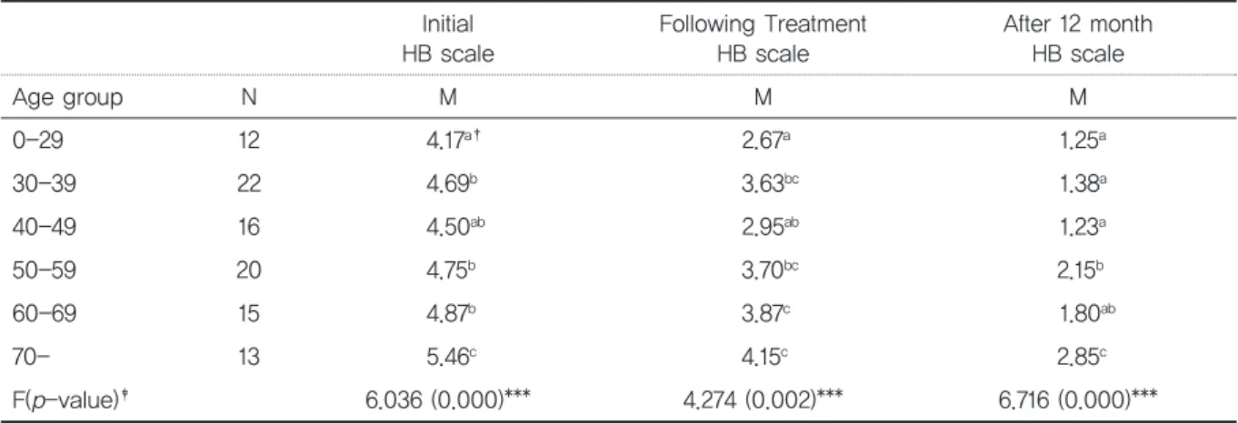
2) Age
Bell’s palsy patients were divided into six groups.
The groups comprised a 0-29 year old group (N=12), 30-39 year old group (N=16), 40-49 year old group (N=22), 50-59 year old group (N=20), 60- 69 year old group (N=15), and an older than or equal to 70 years of age group (N=13). Statistical analysis of the age groups (ANOVA) and the initial HB scale grading showed statistical significance (p
<0.05). There was also statistical significance for HB scale following treatment and after 12 months Table 2. Research data
Data Composition
Related with
patient Sex, Age, Palsy affected side of the face, alcohol consumption, Smoking, Season of onset, Hypertension, Diabetes, A past history of Bell’s palsy
Treatment number
Number of treatments in a Korean medical hospital (Total number of inpatient and outpatient treatments),
Total number of treatments for Bell’s palsy (Number of treatments in a Korean medical hospital and the number of treatments in other hospitals)
Blood test BC Total protein, Albumin, T.bilirubin, AST, ALT, ALP, Cholesterol, LDH, FBS
CBC WBC, RBC, Hemoglobin, Hematocrit, ESR, Platelet, MCV, MCH, MCHC, RDW, PDW, PCT, MPV Urinalysis Protein, S.G, P.H, Leukocyte, Nitrite, Glucose, Urobilinogen, Ketone, Bilirubin, Blood, RBC, WBC,
Epithelial
Table 3. The sex effects on Bell’ s palsy patients*
Sex Bell’s palsy
(N=98) Mean (M) Standard deviation
(SD)
Initial Male 39 4.77 0.742
HB scale Female 59 4.69 0.701
t(p-value)
†0.502 (0.617)
Following Treatment Male 39 3.26 1.163
HB scale Female 59 3.63 1.097
t(p-value) -1.598 (0.113)
After 12 months Male 39 1.64 1.063
HB scale Female 59 1.81 1.090
t(p-value) -0.774 (0.441)
*Statistical significance was evaluated using an independent sample t-test.
† **p<0.05, ***p<0.01.
(p<0.05) (Table 4).
According to Duncan’s new multiple range test (MRT) for post-hoc analysis, the initial HB scale grade in the groups, other than the 0-29 year old group, showed increased average grade of HB scale, which suggests that the older the patient, the more severe the initial palsy. Using Duncan’s post-hoc analysis on the HB scale grade following treatment showed an increased average grade of HB scale in groups other than 0-29 years old, meaning that older the patient, the slower the re- covery rate of the palsy. Duncan’s post-hoc analy- sis on the HB scale after 12 months showed similar recovery rates in age groups with similar averages of HB scale: 0-29 year old, 30-39 year old, and 40- 49 year old. These age groups showed a better re- covery rate than the older age groups. Patients greater than or equal to 50 years of age showed slower rates of recovery (the 60-69 year old group was both in the a and b sections within Duncan’s post-hoc analysis, which lowers statistical signifi- cance) (Table 4).
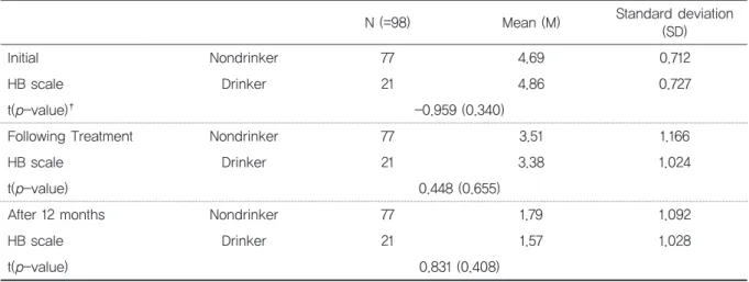
3) Alcohol consumption and smoking
There were 21 drinkers and 77 nondrinkers among the 98 Bell’s palsy patients, and 17 smokers and 81 non-smokers. Drinking and smoking had no statistical significance with the initial HB scale
grade after statistical analysis( p>0.05). Further- more there was no statistical significance with the HB scale following treatment and after 12 months after ( p>0.05) (Table 5,6).
4) Facial palsy affected side of the face
Bell’s palsy had occurred on the right side of the face in 51 patients, and on the left side in 47 pa- tients. The 40 male patients with Bell’s palsy com- prised 20 patients with right palsy and 20 patients with left palsy. The 58 female patients with Bell’s palsy comprised 31 right palsy and 27 left palsy pa- tients (Table 7).
Statistical analysis of the affected side of the face in the Bell’s palsy patients showed no statisti- calsignificancewithHBscalegrade(p>0.05). Also, there was no statistical significance shown with the HB scale following treatment and after 12 months ( p>0.05) (Table 8).
5) Season of onset
Seasons were classified into spring (March to May, N=22), summer (June to August, N=23), fall (September to November, N=25) and winter (De- cember to February, N=28). The season of onset showed no statistical significance with the initial HB scale grade, following treatment, and after 12 month (p>0.05) (Table 9).
Table 4. The age group effects on Bell’s palsy patients*
Initial
HB scale Following Treatment
HB scale After 12 month
HB scale
Age group N M M M
0-29 12 4.17
a†2.67
a1.25
a30-39 22 4.69
b3.63
bc1.38
a40-49 16 4.50
ab2.95
ab1.23
a50-59 20 4.75
b3.70
bc2.15
b60-69 15 4.87
b3.87
c1.80
ab70- 13 5.46
c4.15
c2.85
cF(p-value)
‡6.036 (0.000)*** 4.274 (0.002)*** 6.716 (0.000)***
*Statistical significance was evaluated using ANOVA and Duncan’ s post-hoc analysis.
†a, b, c are Duncan’ s post-hoc analysis results, meaning a>b>c.
‡ **p<0.05, ***p<0.01.
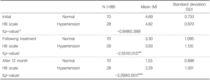
6) Hypertension
28 patients had hypertension and 70 patients did not. Statistical analysis of the hypertension history of Bell’s palsy patients showed no statistical significance with the initial HB scale(p>
0.05). However, there was statistical significance
shown for a history of hypertension in relation to the HB scale grade following treatment and after 12 months (p<0.05) (Table 10). This result suggests that a history of hypertension slows the recovery of facial palsy.
Table 5. The effects of alcohol consumption on Bell’ s palsy patient*
*Statistical significance was evaluated using an independent sample t-test.
† **p<0.05, ***p<0.01.
N (=98) Mean (M) Standard deviation
(SD)
Initial Nondrinker 77 4.69 0.712
HB scale Drinker 21 4.86 0.727
t(p-value)
†-0.959 (0.340)
Following Treatment Nondrinker 77 3.51 1.166
HB scale Drinker 21 3.38 1.024
t(p-value) 0.448 (0.655)
After 12 months Nondrinker 77 1.79 1.092
HB scale Drinker 21 1.57 1.028
t(p-value) 0.831 (0.408)
Table 6. The effects of smoking on Bell’ s palsy patients*
*Statistical significance was evaluated using an independent sample t-test.
† **p<0.05, ***p<0.01.
N (=98) Mean (M) Standard deviation
(SD)
Initial Nonsmoker 81 4.70 0.679
HB scale Smoker 17 4.82 0.883
t(p-value)
†-0.626 (0.533)
Following Treatment Nonsmoker 81 3.51 1.108
HB scale Smoker 17 3.35 1.272
t(p-value) 0.505 (0.615)
After 12 months Nonsmoker 81 1.79 1.104
HB scale Smoker 17 1.53 0.943
t(p-value) 0.906 (0.367)
Table 7. The characteristics of the palsy affected side of face in Bell’ s palsy patients
Sex N %
Right Men 20 20.4
Women 31 31.6
Left Men 20 20.4
Women 27 27.6
Table 8. The palsy direction effects on Bell’ s palsy*
*Statistical significance was evaluated using an independent sample t-test.
† **p<0.05, ***p<0.01.
N (=98) Mean (M) Standard deviation
(SD)
Initial Left 47 4.72 0.743
HB scale Right 51 4.73 0.695
t(p-value)
†-0.014 (0.989)
Following Treatment Left 47 3.47 1.231
HB scale Right 51 3.49 1.046
t(p-value) -0.096 (0.924)
After 12 month Left 47 1.70 1.102
HB scale Right 51 1.78 1.064
t(p-value) -0.376 (0.708)
Table 9. The season of onset effects on Bell’ s palsy*
Initial
HB scale Following Treatment
HB scale After 12 months
HB scale
Season N M M M
Spring 22 4.68
a†3.55
ab1.68
aSummer 23 4.91
a3.39
ab1.91
aFall 25 4.60
a3.08
a1.52
aWinter 28 4.71
a3.86
b1.86
aF(p-value)
‡0.809(0.492) 2.227(0.090) 0.669(0.573)
*Statistical significance was evaluated using ANOVA and Duncan’ s post-hoc analysis.
†a, b are Duncan post-hoc analysis results, meaning a>b.
‡ **p<0.05, ***p<0.01.
Table 10. The hypertension effects on Bell’ s palsy patients*
*Statistical significance was evaluated using an independent sample t-test.
† **p<0.05, ***p<0.01.
N (=98) Mean (M) Standard deviation
(SD)
Initial Normal 70 4.69 0.733
HB scale Hypertension 28 4.82 0.670
t(p-value)
†-0.848(0.399)
Following treatment Normal 70 3.30 1.095
HB scale Hypertension 28 3.93 1.120
t(p-value) -2.551(0.012)**
After 12 month Normal 70 1.53 0.896
HB scale Hypertension 28 2.29 1.301
t(p-value) -3.299(0.001)***
7) Diabetes mellitus
From the 98 patients, 17 patients had diabetes mellitus and 81 patients did not. A statistical analysis of diabetes history of Bell’s palsy patients showed no statistical significance with the initial HB scale and following treatment (p>0.05). How- ever, there was statistical significance between a history of diabetes and the HB scale after 12 months (p<0.05) (Table 11). This result indicates that a history of diabetes slows the recovery of fa- cial palsy.
8) Past history of Facial palsy
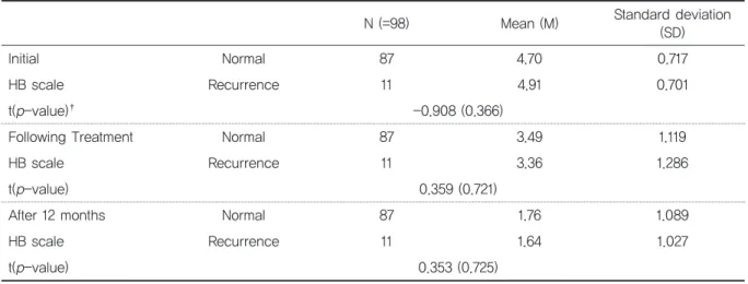
Of the 98 Bell’s palsy patients, 11 patients had a history of Bell’s palsy. A history of Bell’s palsy showed no statistical significance with the initial HB scale grade, following treatment, and after 12 months (p>0.05) (Table 12). This result shows that a past history of facial palsy does not affect the re- covery of facial palsy.
9) Degree of initial Bell’s palsy severity
Among the 98 Bell’s palsy patients, 3 patients
Table 11. The diabetes effects on Bell’ s palsy patients*
*Statistical significance was evaluated using an independent sample t-test.
† **p<0.05, ***p<0.01.
N (=98) Mean (M) Standard deviation
(SD)
Initial Normal 81 4.69 0.701
HB scale Diabetes 17 4.88 0.781
t(p-value)
†-1.002 (0.319)
Following Treatment Normal 81 3.40 1.069
HB scale Diabetes 17 3.88 1.364
t(p-value) -1.626 (0.107)
After 12 months Normal 81 1.62 0.969
HB scale Diabetes 17 2.35 1.367
t(p-value) -2.637 (0.010)**
Table 12. A history of Bell’s palsy effects on Bell’ s palsy patients*
*Statistical significance was evaluated using an independent sample t-test.
† **p<0.05, ***p<0.01.
N (=98) Mean (M) Standard deviation
(SD)
Initial Normal 87 4.70 0.717
HB scale Recurrence 11 4.91 0.701
t(p-value)
†-0.908 (0.366)
Following Treatment Normal 87 3.49 1.119
HB scale Recurrence 11 3.36 1.286
t(p-value) 0.359 (0.721)
After 12 months Normal 87 1.76 1.089
HB scale Recurrence 11 1.64 1.027
t(p-value) 0.353 (0.725)
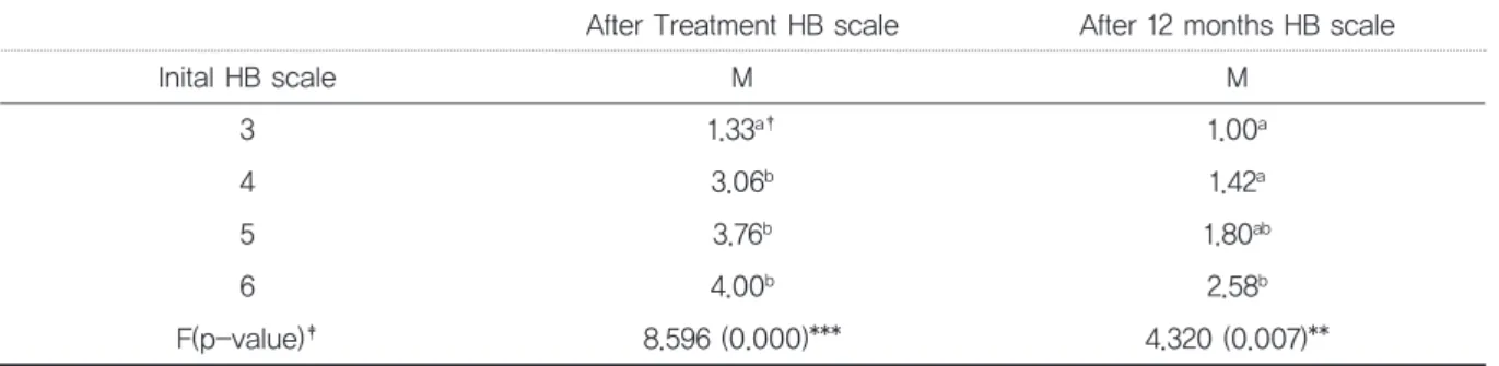
were evaluated as HB scale step 3, 33 patients as 4, 50 patients as 5, and 12 patients as 6 (Table 13).
The degree of initial Bell’s palsy severity had sta- tisticalsignificancewiththeHBscalegradefollow- ing treatment and after 12 months (p<0.05).
AccordingtoDuncan’spost-hocanalysis, themore severe the degree of initial Bell’s palsy, the lower the recovery rate (Table 14).
2. Clinical analysis of blood and urine tests
Thepurposeofanalyzingofbloodandurinetests is to identify the statistical significance of the initial condition of Bell’s palsy patients in relation to their prognosis.
Table 13. The HB scales of Bell’ s palsy patients Initial
HB scale Following Treatment
HB scale After 12 months
HB scale
Scales N Scales N % Scales N %
3 3 1 2 66.7 1 3 100
2 1 33.3 2 - -
4 33
1 1 3.0 1 23 69.7
2 10 30.3 2 6 18.2
3 10 30.3 3 4 12.1
4 12 36.4 4 - -
5 50
1 1 2.0 1 30 60.0
2 8 16.0 2 3 6.0
3 7 14.0 3 14 28.0
4 20 40.0 4 3 6
5 14 28.0 5 - -
6 12
1 - - 1 5 41.2
2 - - 2 1 8.3
3 3 25.0 3 2 16.7
4 6 50.0 4 2 16.7
5 3 25.0 5 2 16.7
Table 14. The initial HB scales effects on Bell’ s palsy patients*
After Treatment HB scale After 12 months HB scale
Inital HB scale M M
3 1.33
a†1.00
a4 3.06
b1.42
a5 3.76
b1.80
ab6 4.00
b2.58
bF(p-value)
‡8.596 (0.000)*** 4.320 (0.007)**
*Statistical significance was evaluated using ANOVA and Duncan’ s post-hoc analysis.
†a, b are Duncan post-hoc analysis results, meaning a>b.
‡ **p<0.05, ***p<0.01.
1) Blood chemistry (BC)
BC (blood chemistry) consists of total protein, al- bumin, total bilirubin, aspartate aminotransferase (AST), alanine transferase (ALT), alkaline phos- phatase (ALP), Cholesterol, low density lipoprotein (LDL), and FBS. Only FBS showed a statistical sig- nificance with HB scale grade following treatment and after 12 months (p<0.05). No other BC factors showedastatisticalsignificancewiththeinitialHB scale grade, following treatment, and after 12 months (p>0.05) (Table 15). ALP and LDL could not be statistically analyzed due to small abnormal number of patients with abnormal values (N<5).
2) Complete blood cell count (CBC)
A CBC consists of white blood cell (WBC), red blood cell (RBC), hemoglobin, hematocrit, erythro- cyte sedimentation rate (ESR), platelet, mean cor- puscular volume (MCV), mean corpuscular hemoglobin (MCH), mean corpuscular hemoglobin concentration (MCHC), red cell distribution width
(RDW), platelet distribution width (PDW), platelet- crit (PCT), and mean platelet volume (MPV). All the factors of CBC had no statistical significance with HB scale at initial, following treatment, and after 12 months (p>0.05) (Table 16). Platelet, RDW, and PDW could not be statistically analyzed due to small number of patients with abnormal values (N
<5).
3) Urinalysis
UA (Urinalysis) involves analyzing urine, which comprises protein, specific guavity (S.G), power of hydrogen (P.H), leukocyte, nitrite, glucose, uro- bilinogen, ketone, bilirubin, blood, RBC, WBC, and epithelial. No UA factors showed any statistical significance with the initial HB scale grade, follow- ing treatment, and after 12 months (p>0.05) (Table 17). Levels of P.H, nitrite, urobilinogen, and bilirubin could not be statistically analyzed due to the small number of patients with abnormal values (N<5).
Table 15. The blood chemisty (BC) factor effects on Bell’s palsy patients*
Initial HB scale
Following Treatment
HB scale
After 12 months HB scale
Total protein Normal (N=76)
Abnormal (N=22) t(p-value)
†0.316 (0.752)
-0.107 (0.915)
-0.585 (0.560) Albumin Normal (N=97)
Abnormal (N=1) t(p-value) -0.386
(0.701) -1.355
(0.179) -1.173
(0.244) T.bilirubin Normal (N=93)
Abnormal (N=5) t(p-value) 1.042
(0.300) 0.565
(0.574) -0.541
(0.589)
AST Normal (N=94)
Abnormal (N=4) t(p-value) 0.639 (0.524)
0.863 (0.390)
0.462 (0.645)
ALT Normal (N=87)
Abnormal (N=11) t(p-value) 0.881
(0.381) -0.204
(0.839) -0.238
(0.812)
ALP Normal (N=97)
Abnormal (N=1) t(p-value) - - -
Cholesterol Normal (N=49)
Abnormal (N=49) t(p-value) 0.141 (0.888)
-1.526 (0.130)
-0.093 (0.926)
LDL Normal (N=98)
Abnormal (N=0) t(p-value) - - -
Glucose(FBS) Normal (N=42)
Abnormal (N=55) t(p-value) -1.233
(0.221) -2.517
(0.014)** -2.286 (0.024)**
*Statistical significance was evaluated using an independent sample t-test.
† **p<0.05, ***p<0.01.
3. Clinical analysis of therapeutic variation
1) The number of treatments in Korean Medical Hospital
The number of treatments in a Korean medical hospital (sum of inpatient and outpatient) in rela- tiontotheHBscalegradefollowingtreatment, and after 12 months, was analyzed through simple re- gression analysis. According to the analysis, the number of treatments at a Korean medical hospital showed no statistical significance with the HB scale after 12 months (p>0.05), but showed statis-
tical significance with the HB scale following treat- ment ( p<0.05) (Table 18). Patients who were treated in a Korean medical hospital demonstrated early recovery.
2) Total number of treatments for Facial palsy
Simple regression analysis was used to identify the relationship between the total number of treat- ments (sum of the number of treatment in a Korean medical hospital and the number of treat- ments in other hospitals within 12 months) and the HB scale that following treatment and after 12 Table 16. The complete blood cell count (CBC) factors effects on Bell’s palsy patients*
Initial HB scale
Following treatment HB scale
After 12 months HB scale
WBC Normal (N=75)
Abnormal (N=23) t(p-value)
†-0.112
(0.911) -0.834
(0.406) -0.411
(0.682)
RBC Normal (N=78)
Abnormal (N=20) t(p-value) 0.171 (0.865)
0.793 (0.430)
0.672 (0.503) Hemoglobin Normal (N=80)
Abnormal (N=18) t(p-value) 0.743
(0.459) -0.774
(0.441) -0.384
(0.702) Hematocrit Normal (N=84)
Abnormal (N=14) t(p-value) 1.689
(0.094) -0.072
(0.942) -0.152
(0.279)
ESR Normal (N=74)
Abnormal (N=24) t(p-value) -1.526 (0.130)
-2.219 (0.029)
-0.898 (0.372) Platelets Normal (N=98)
Abnormal (N=0) t(p-value) - - -
MCV Normal (N=86)
Abnormal (N=12) t(p-value) 0.298
(0.767) 1.578
(0.118) 1.420
(0.159)
MCH Normal (N=84)
Abnormal (N=14) t(p-value) 0.864 (0.390)
0.181 (0.857)
0.114 (0.909)
MCHC Normal (N=57)
Abnormal (N=41) t(p-value) -1.234
(0.220) -0.601
(0.549) -0.087
(0.931)
RDW Normal (N=97)
Abnormal (N=1) t(p-value) - - -
PDW Normal (N=98)
Abnormal (N=0) t(p-value) - - -
PCT Normal (N=77)
Abnormal (N=21) t(p-value) 0.073
(0.942) 0.448
(0.655) -0.537
(0.593)
MPV Normal (N=61)
Abnormal (N=37) t(p-value) -0.638 (0.525)
-0.966 (0.336)
-0.147 (0.640)
*Statistical significance was evaluated using an independent sample t-test.
† *p<0.1, **p<0.05, ***p<0.01.
months. The total number of treatments within one year had no statistical significance with the
initial HB scale and after 12 months (p>0.05) (Table 19).
Table 17. The UA factors effect on facial palsy*
Initial HB scale
Following Treatment HB scale
After 12 months HB scale
Protein Normal (N=92)
Abnormal (N=6) t(p-value)
†-0.383
(0.702) -1.164
(0.247) -0.597
(0.552)
S.G Normal (N=91)
Abnormal (N=7) t(p-value) 0.039 (0.969)
-0.222 (0.825)
-1.014 (0.313)
P.H Normal (N=98)
Abnormal (N=0) t(p-value) - - -
Leukocyte Normal (N=86)
Abnormal (N=12) t(p-value) 0.728
(0.468) -0.882
(0.380) 0.552
(0.582) Nitrite Normal (N=97)
Abnormal (N=1) t(p-value) - - -
Glucose Normal (N=78)
Abnormal (N=20) t(p-value) 0.171
(0.865) 0.130
(0.897) -0.954
(0.343) Urobilinogen Normal (N=96)
Abnormal (N=2) t(p-value) - - -
Ketone Normal (N=84)
Abnormal (N=14) t(p-value) 0.460 (0.647)
0.946 (0.346)
0.918 (0.361) Bilirubin Normal (N=96)
Abnormal (N=2) t(p-value) - - -
Blood Normal (N=86)
Abnormal (N=12) t(p-value) -0.131
(0.896) -2.001
(0.048) -1.752
(0.083)
RBC Normal (N=84)
Abnormal (N=14) t(p-value) 0.057 (0.954)
0.181 (0.857)
1.189 (0.237)
WBC Normal (N=85)
Abnormal (N=13) t(p-value) 0.589
(0.557) 0.061
(0.951) 0.188
(0.851) Epithelial Normal (N=76)
Abnormal (N=22) t(p-value) 0.316 (0.752)
0.117 (0.907)
-0.776 (0.440)
*Statistical significance was evaluated using an independent sample t-test.
† **p<0.05, ***p<0.01.
Table 18. The number of treatments in a Korean medical hospital effects on Bell’ s palsy patients*
Non-standardized Standardized
F( p-value)
†Constant SD beta
Following Treatment HB scale
Model 3.920(0.000)*** 0.208
6.277 (0.014)**
Treatment
number -0.022(0.014)** 0.009 -0.248
After 12 months HB scale
Model 1.937(0.000)*** 0.023
1.253 (0.266) Treatment
number -0.010(0.266) 0.009 -0.114
*Statistical significance was evaluated using simple regression.
† **p<0.05, ***p<0.01.
Ⅳ. Discussion
Idiopathic facial palsy, also known as Bell’s palsy, is the most common type of facial nerve paralysis Bell’s palsy can occur at any age
8), with a prevalence in pregnant women
9), and in those with diabetes mellitus, hypertension, and a history of Bell’s palsy
10,11). However, according to a study of Ahn et al
12), age, sex, post-auricular pain, and the initial HB grade were statistically significant, while the palsy affected side of the face, a history of facial palsy, the presence of diabetes mellitus and hypertension were shown to have no statistical significance. In a study of Min et al
13), sex, age, post-auricular pain, and the time from onset, the presence of diabetes mellitus and hyper- tension, and a history of facial palsy did not show statistical significance, while the palsy-affected side of the face, the initial degree of palsy severity, the time of initial recovery, and recovery in three weeks from onset, showed statistical significance.
There have been few studies on the prognosis and related factors in respect of facial palsy, with dif- fering results.
This study identified the statistical significance of factors related to the prognosis of facial palsy, andaimedtoprovidefurtherinformationforongo- ing research on the prognosis and treatment of fa- cial palsy patients. This study was conducted on 98 facialpalsypatientswhowerehospitalizedinaKo-
rean medical hospital from January 1, 2015 to De- cember 31, 2015. Patients with facial palsy were followed up retrospectively at the initial, following treatment, and after 12 months. A statistical analysis was performed to confirm the association of facial palsy recovery with various factors related to facial palsy. To minimize statistical error, this study only included facial palsy patients who were admitted to the hospital within one week from the onset of the illness and who were pre- scribed steroids. The medical data were divided into three categories: data related to patient char- acteristics, data related to medical examinations, and data related to the number of treatments
Concerning patient characteristics, sex, age, the palsy-affected side of the face, alcohol consump- tion, smoking, a history of Bell’s palsy and the sea- son of onset were not statistically significant with the initial HB grade, following treatment, and after 12 months. Sex, the palsy-affected side of the face, alcohol consumption, smoking, a history of Bell’s palsy, and the season of onset were not significant as factors for the occurrence and prog- nosis of Bell’s palsy.
Age had a statistical significance with the initial HB grade, following treatment, and after 12 months. According Duncan’s post-hoc analysis, initial HB grade and following treatment showed a statistical difference in the 0-29 year old group and in the 70-79 year old group. It appears that Table 19. The total number of treatment effects on Bell’s palsy patients*
Non-standardized Standardized
F( p-value)
†Constant SD beta
Initial HB scale
Model 4.539(0.000)*** 0.126
3.150 (0.079) Treatment
number 0.007(0.079) 0.004 0.178
After 12 months HB scale
Model 1.662(0.000)*** 0.193
0.268 (0.606) Treatment
number 0.003(0.606) 0.006 0.053
*Statistical significance was evaluated using simple regression.
† **p<0.05, ***p<0.01.
the older the patient, the more severe the initial degree of facial palsy. Furthermore, the older the patient, the more difficult it is for recovery. Ac- cording to Duncan’s post-hoc analysis, the HB grade after 12 months, the 0-29 year old, 30-39 year old, and 40-49 year old showed similar recov- ery rates, while the groups older than 50 years showing slowed recovery rates.
Patients with a history of hypertension had a high initial average HB grade leading to severe ini- tial degree, but it did not have statistical signifi- cance. However, there was a statistically significant difference with the HB grade following treatment and after 12 months. The mean value of the HB grade was higher than those without a his- tory of hypertension. Also, patients with a history of diabetes mellitus had a high initial HB grade tending toward severity, with no statistical signif- icance. The average of the HB grades following treatment was also high, which was not statistically significant. However, the HB grade after 12 months was statistically significant, and the average of HB scale grades was higher than those without a history of diabetes mellitus, which showsthatapasthistoryofdiabetesmellitusslows the recovery of facial palsy. Therefore, patients with hypertension and diabetes mellitus have slower rates of recovery from facial palsy and a high possibility of having sequela.
HB grade at initial and after 12 months showed statistical significance with initial HB grade, and the higher the initial HB grade, the slower the re- covery from facial palsy.
Except for FBS, none of blood and urine test fac- tors was statistically significant with the prognosis of facial palsy. As with a history of diabetes mellitus, a FBS also had a negative effect on the prognosis of facial palsy. There were 55 patients who had FBS abnormality. In contrast, 17 patients were diagnosed with diabetes mellitus. The FBS can be tested easily, so it is possible to identify those who are unaware of possible diabetes mellitus, and it is more useful as a prognostic factor than a history of diabetes mellitus. However,
there was no statistical significance between uri- narysugarandfacialpalsyprognosis. Thismayre- sult from the fact that glucose can be detected in urine for reasons other than renal dysfunction due to diabetes mellitus. Therefore, urinary sugar has a limited significance in relation to the prognosis of facial palsy, which calls for further studies.
In relation to the number of treatments and the prognosisoffacialpalsy, thegreaterthenumberof treatments at a Korean medical hospital, the more improvement in HB grade following treatment, which was statistically significant. This result shows that treatment at a Korean medical hospital leads to faster recovery. This is consistent with a study of Won et al
20)that more treatment sessions positively affect recovery.
The total number of treatments for one year was not statistically significant with the initial HB grade and following treatment. In conclusion, re- covery from facial palsy is dependent on personal characteristics such as age, the initial degree of palsy severity, hypertension, and diabetes mellitus.
As noted, age, hypertension, diabetes mellitus, FBS, and initial degree of palsy were statistically significant with prognosis of facial palsy. The study of Ahn et al
18)and Min et al
19), also found that, the initial degree of palsy severity was significant with the prognosis of facial palsy, and this finding was consistent with the conclusion in the study of Won et al
20). However, the results of this study differ from previous studies undertaken in Korea
18-21)