Introduction
Oxidative stress is generated from the toxic levels of oxygen-derived reactive oxygen species (ROSs) such as sin- glet oxygen, superoxide anion (O
2-), hydrogen peroxide (H
2O
2), and the highly reactive hydroxyl radical (OH
.) via the metal-catalyzed Haber-W eiss and Fenton reaction [5,12]. These ROSs are the result of normal metabolism, in- cluding the energy generation process of aerobic respira- tion, and the β-oxidation of fatty acids. The toxic materials are also generated through the metabolism of foreign stimulants such as high temperature, osmotic pressure, ir- radiation and chemicals including ethanol, H
2O
2, mena- dione (MD) and xenobiotics or the host immune system [21]. The produced ROSs can lead to damage of a wide range of cellular biological molecules: DNA fragmentation and mutations, lipid peroxidation, the disassembly of iron-sulfur clusters, disulfide bond formation, and other types of protein oxidation [6].
Under normal physiological conditions, antioxidant de-
fense systems are well organized to adjust ROSs at a basal level and to repair cellular damage or degrade oxidized molecules. However, an imbalance between oxidants and the ability of detoxification is introduced under oxidative stress [12]. In order to overcome transient or continuous ROS chal- lenges, cells have evolved a variety of enzymatic and non-enzymatic antioxidant defense systems, which are capa- ble of removing free radicals and their by-products leading to the repair of stress-induced cellular damage, and therefore can protect the cellular constituents [7,12,19].
As the industrial yeast, Saccharomyces cerevisiae has been used in fermentation and brewing industries for more than 8,000 years for favorite characteristics including high yields of ethanol, production of good flavor, and resistance to etha- nol and toxic materials such as sulfite [11,22]. Today, the impact of yeasts on industry extends beyond the original food and beverage production as to production of in- gredients for food processing such as B vitamins, proteins, peptides, amino acids and trace minerals, biocontrol of spoil- age microorganisms, and biotherapeutic and probiotic agents [26]. For example, S. cerevisiae var. boullardii has been successfully used over the last 20 years as an oral bio- therapeutic agent to treat patients with severe cases of diar- rhea and other gastrointestinal disorders [23,26]. S. cerevisiae
Increased Antioxidative Activities against Oxidative Stress in Saccharomyces cerevisiae KNU5377
Il-Sup Kim, Hae-Sun Yun
1, Ji Young Yang, Oh-Seok Lee
2†, Heui-Dong Park
2, Ingnyol Jin and Ho-Sung Yoon*
School of Life Science, Kyungpook National University, Daegu 702-701, Korea
1Division of Enteric and Hepatitis Viruses, Center for Infectious Diseases, National Institute of Health, Seoul 122-701, Korea
2Department of Food Science and Biotechnology, Institute of Fermentation Biotechnology, Kyungpook National University, Daegu 702-701, Korea Received January 14, 2009 /Accepted March 11, 2009
Oxidative stress is a consequence of an imbalance of the defense system against cellular damage gen- erated by reactive oxygen species (ROSs) such as superoxide anions (menadione; MD). Most organ- isms have evolved a variety of defense systems to protect cells from adverse conditions. In order to evaluate stress tolerance against oxidative stress generating MD, comparative analyses of antioxidant capacity, or free radical scavenger ability, were performed between S. cerevisiae KNU5377 (KNU5377) and three wild-type S. cerevisiae strains. In a medium containing 0.4 mM MD, the KNU5377 strain showed higher cell viability and antioxidant ability, and contained higher levels of trehalose, super- oxide dismutase, thioredoxin system, glucose-6-phosphate dehydrogenase, and some heat shock proteins. The KNU5377 strain also produced a lower level of oxidative stress biomarker than the other three yeast strains. These results indicate that S. cerevisiae KNU5377 has a higher level of tolerance to oxidative stress due to the increased expression of cell rescue proteins and molecules, thus alleviat- ing cellular damage more efficiently than other S. cerevisiae strains.
Key w ords : Saccharomyces cerevisiae K N U5377, oxidative stress, antioxidant enzym es, heat shock protein, stress tolerance
*Corresponding author
*Tel +82-53-950-5348, Fax +82-53-953-3066: :
*E-mail : hyoon@knu.ac.kr
KNU5377 (KNU5377) was isolated from sewage [15] for in- dustrial applications such as alcohol fermentation under high temperature and stressful conditions in order to reduce the cost of ethanol production. The KNU5377 was charac- terized as having thermotolerant and stress tolerances against various types of environmental stressors including hydrogen peroxide, ethanol, high osmotic pressure, and sul- furic acid than that of reference strains such as S. cerevisiae S288C (S288C), S. cerevisiae W 303-1A (W 303-1A), and an ethanol-tolerant yeast S. cerevisiae ATCC 24858 (ATCC24858) [13,14].
As a model eukaryote, laboratory strain S. cerevisiae has been extensively studied to oxidative stress response.
However, at present the stress responses to peroxide or su- peroxide stresses in wild industrial S. cerevisiae are not de- scribed, especially to superoxide stress. Therefore, under- standing the oxidative stress response could give insights into how microorganisms survive in detrimental environ- ments such as extreme temperature, oxidative stress, and os- motic shock. As a first step towards comprehending the de- fensive mechanisms, the stress response against superoxide anion by menadione was investigated in S. cerevisiae KNU5377, and the levels of induction of protective cell res- cue proteins containing antioxidant enzymes and anti- oxidant molecules in stress response.
Materials and Methods Strains and growth conditions
The KNU5377 was isolated from sewage in Korea [15].
ATCC 24858 as an ethanol tolerant strain, W 303-1A and S288C was purchased from the American Type Culture Collection (ATCC), Euroscarf (http://web.uni-frankfurt.de/
fb15/mikro/euroscarf/) and Korean Culture Center of Microorganisms (KCCM), respectively. Yeast cells were aer- obically grown in a nutrient-rich YPD media (1% yeast ex- tract, 2% peptone, 2% dextrose) for 20 h at 30
oC, with shak- ing at 160 rpm. To monitor cell viability, the mid-log cul- tured cells (OD
600=1.0) following pre-culture at 30
oC over- night were exposed to 0.4 mM of menadione (MD) for 1 h at 30
oC in YPD liquid media. Cultures were properly di- luted, spread on YPD agar plates and then incubated for 24 hr at 30
oC. Cell viability was calculated by colony numbers. For growth kinetics, cells were inoculated in YPD media supplemented 40 µM MD, and then cell growth was monitored at 600 nm using a spectrophotometer. For analy-
sis of stress sensitivity, the mid-log cultured cells were ex- posed to a variety of concentration of MD for 1 hr at 30
oC.
The cells were properly diluted, spotted onto YPD agar plates, and incubated at 30
oC.
Measurement of hydroperoxide, carbonyl content and malondialdehyde
The intracellular hydroperoxide levels were determined by ferrous ion oxidation in the presence of a ferric ion in- dicator, xylenol orange [13]. Carbonyl contents were meas- ured via the spectrometric method [13]. Malondialdehyde (MDA) levels were examined via thiobarbituric acid (TBA) assay [13].
Trehalose and glycogen assay
Cell pellets (4-10 mg) were put on ice, washed twice with 10-20 volumes of cold-distilled water, resuspended in 0.25 ml of 0.25 M sodium carbonate, and incubated at 95
oC for 4 hr. The pH of the mixture was brought to 5.2 by adding 0.15 ml of 1.0 M acetic acid and 0.6 ml of 0.2 M Na-acetate (pH 5.2). One half of the suspension was incubated over- night with trehalase (0.05 U/ml) at 37
oC for measurement of trehalose content and the second half with Aspergillus ni- ger amyloglucosidase preparation (1.2 U/ml) at 57
oC under constant agitation for measurement of glycogen content.
After incubation, these suspensions were centrifuged for 3 min at 5,000× g [20]. The resulting glucose was measured by the Somogy-Nelson method. The trehalose and glycogen contents were defined as nmol/mg protein.
Protein extracts
Crude cellular protein extracts were prepared by the glass bead method. Briefly, cells were washed with saline (0.85%
of NaCl) three times, and resuspended in a lysis buffer (20 mM HEPES, 10% glycerol, 1 mM PMSF, 2 µM pepstatin A and protease inhibitor cocktails) with an equal volume of glass beads (425-600 microns; Sigma, USA). After vigorously vortex-mixing for 1 min 5 times with 2 min interval on ice, the cell extract was collected by centrifuging at 12,000 rpm for 10 min at 4
oC. The protein concentration was determined by the Bradford method (Bio-Rad, USA).
Immunoblot assay
Electrophoresis via protein extract for SDS-PAGE and
two-dimensional gel electrophoresis was performed by a
previously employed protocol [13,14]. Briefly, 40 µg of dena-
tured proteins were analyzed in a 10% or 15% poly- acrylamide gel and electrophoretically transferred to a PVDF membrane (Bio-Rad, USA) in a transfer buffer (25 mM of Tris-base, 192 mM of glycine and 20% methanol). The PVDF membranes were blocked for 60 min at room temperature in a TTBS buffer (0.05% Tween-20, 10 mM of Tris-HCl, pH 7.6, 150 mM of NaCl) containing 5% non-fat skim milk and 0.02% sodium azide. The blotted membranes were incubated overnight at 4
oC with the primary antibodies: rabbit anti-glu- cose-6-phosphate dehydrogenase (Sigma, St. Louis, USA), rabbit anti-Hsp104, mouse anti-Hsp60 and rabbit anti-alco- hol dehydrogenase (Stressgen, Canada), rabbit anti-hex- okinase (Rockland, USA), rabbit anti-Hsp90, rabbit anti-Ssa and rabbit anti-Ssb (kindly provided by Dr. Elizabeth A.
Craig, USA), rabbit anti-Tsa1p (kindly provided by Dr. Park Jeen W oo, Korea), rabbit anti-Trx2p, Trx3p and anti-Trr1p (kindly provided by Dr. Kim Kang-Hwa, Korea), and rabbit anti-Cpr1p (kindly provided by Dr Joseph Heitman, USA).
After washing four times with a TTBS, the membranes were incubated for 90 min with a secondary antibody such as an- ti-rabbit IgG (H+L) HRP Conjugate (Promega, USA) or an- ti-mouse IgG (Amersham Biosciences, Sweden), then wash- ed four times with a TTBS, developed by enhanced chem- illuminescence (ECL kit; Amersham Biosciences), and processed.
Results
Viability and growth assay against menadione
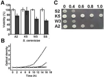
To determine the sensitivity of yeast strains against me- nadione (MD), ATCC24858, KNU5377, W303-1A and S288C cells were challenged with 0.4 mM of MD for 1 hr, and subsequently spread onto YPD agar plates. After MD treat- ment all of the percent viability was decreased in all four strains. From 30 to 40 % decrements were observed in three strains, ATCC24858, W 303-1A and S288C however, only about a 10 percent decrement in KNU5377 could be observed (Fig. 1A). Although three strains could not grow under 40 µM of MD, KNU5377 could do so after a delayed lag phase, for 8 hr (Fig. 1B). These results were strongly supported by a stress sensitivity assay. As shown in Fig.
1C, the KNU5377 strain exposed to a variety of concen- trations of MD did grow at 1.0 mM of MD, whereas three reference strains did not grow at this concentration.
Therefore, ATCC24858 of a high ethanol tolerance strain, W 303-1A of a wild type strain and S288C of a general S.
Fig. 1. Cell viability and growth rate of S. cerevisiae KNU 5377 and three reference strains. For cell viability, aerobically grown cells (A600=1.0) were exposed to 0.4 mM m ena- dione (M D) for 1 h at 30oC, properly diluted, spread on YPD agar plates, and incubated for 24-36 h at 30oC. Cell viability was calculated by colony num bers. A2: S. cer- evisiaeATCC24858; K5: S. cerevisiae KNU5377; W 3: S. cer- evisiaeW 303-1A; S2: S. cerevisiae S288C; Open bar: with- out M D; Closed bar: with M D (A). For growth kinetics, cells were cultured in liquid YPD media with 40 µΜMD and then, optical density was measured at 600 nm . Values are shown for one representative of at least three independent experiments. ATCC24858 ( ); KNU5377□ ( ); KNU5377; W 303 ( ); S288C ( ) (B). Aerobically○ △ ▽ grown cells (A600=1.0) were exposed to a variety of con- centration of menadione (M D) for 1 h at 30oC, properly diluted, spotted on YPD agar plates, and then incubated for 24 h at 30oC. S2: S288C; K5: KNU5377; W 3: W303-1A;
S2: S288C.
cerevisiae strain displayed greater sensitivity than that of KNU5377 against MD.
Hydroperoxide, carbonyl and malondialdehyde assay
To examine intracellular oxidative biomarkers by Ros,
hydroperoxide, carbonyl and malondialdehyde concen-
trations were measured after 0.4 mM MD treatment for 1
hr. Hydroperoxide was measured during MD treatment
with and without adaptation by ferrous ion oxidation in the
presence of a ferric ion indicator, xylenol orange. As shown
in Fig. 2A, the levels were increased after treatment in four
strains, however the lowest increment of KNU5377 could be
observed in the strains. Patterns of oxidatively damaged
proteins were measured by carbonyl content. Although the
contents were also increased after treatment, it could be ob-
served that the intrinsic cellular carbonyl content of
Fig. 2. Cellular levels of hydroperoxide, carbonyl content and malondialdehyde upon exposure to menadione (M D) stress. Cells grown to the mid-log phase were exposed to 0.4 mM M D for 1 hr at 30oC with shaking. Then the cells were collected to measure cellular hydroperoxide (A), carbonyl content (B), and malondialdehyde (C).
Values are shown from at least three independent experim ents. A2: ATCC24858; K5: KNU5377; W 3:
W 303-1A; S2: S288C; Open bar: without MD; Closed bar:
with M D.
KNU5377 was the lowest of four strains (Fig. 2B). Over 2-fold induction of malondialdehyde as a product of lipid peroxidation were observed in W 303-1A and S288C how- ever, the increment of the reactive intermediate was not sig- nificant in KNU5377 (Fig. 2C).
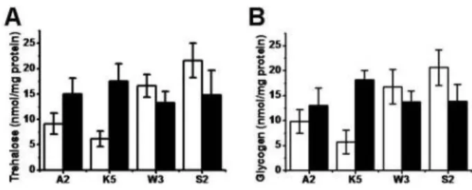
Trehalose and glycogen accumulation after menadione stress
Intracellular storage dicarbohydrates, trehalose and glyco- gen concentrations were various depending on S. cerevisiae strains. Different accumulation patterns were observed be- tween ATCC24858 and KNU5377, and W303-1A and S288C.
The former strains showed lower intrinsic dicarbohydrate levels than those of the latter strains however, cellular accu- mulation of these two components were well induced in the latter strains after MD treatment (Fig. 3). Moreover, over 2-fold inductions of the two components were observed in KNU5377.
Expression changes of antioxidant enzymes during menadione stress
Immunoblot analysis was performed in order to detect general antioxidant enzymes and molecules including Sod1 (cytosolic Cu/Zn Sod), Sod2 (mitochondrial Mn Sod), Trx3 (thioredoxin 3), Tsa1 (cytosolic thioredoxin peroxidase), Ssa
Fig. 3. Cellular level of trehalose and glycogen to m enadione.
Cells were aerobically grown in nutrient rich YPD media at 30oC with shaking. Once mid-log phase to OD600 of 1.0 is reached, the cells were challenged with 0.4 mM menadione for 1 hr at 30oC. Cell cultures were collected to analyze trehalose content (A) and glycogen content (B). The data of the trehalose and the glycogen concen- trations are mean values±SD from three independent experiments. A2: A TCC24858; K5: KNU5377; W 3:
W 303-1A; S2: S288C; Open bar: without MD; Closed bar:
with M D.
Fig. 4. Im munoblot analysis of cell rescue proteins and anti- oxidant molecules. Cells were aerobically grown in nu- trient rich YPD media at 30oC with shaking. Once mid-log phase to OD600of 1.0 is reached (without M D), the cells were challenged with 0.4 mM menadione (M D) for 1 hr at 30oC (with MD). Then proteins from different strains were extracted and loaded (40 µg) on SDS-PAGE gels (10, 12, and 17% ). The gels were transferred to a PVDF membrane and developed. Sod1: Cu/Zn super- oxide dismutase; Sod2: M n superoxide dismutase; Trx3:
thioredoxin 3; Tsa1: cytosolic thioredoxin peroxidase I;
Zwf1: glucose-6-phosphate dehydrogenase; Ald: alde- hyde dehydrogenase; Cpr1: cyclophilin A; Ssa: heat shock protein family 70; Hsp104: heat shock protein 104.
Actin was detected for housekeeping control. A2:
ATCC24858; K5: KNU5377; W 3: W 303-1A; S2: S288C (A).
Relative intensity of immunoblot analyses was expressed relative to the expression levels of KNU5377 without MD (100%). Values are means with standard deviations from at least three independent experim ents (B).
(heat shock protein 70 family), Hsp104, Cpr1 (cyclophilin),
Ald (aldehyde dehydrogenase) and Zwf1 (glucose-6-phos-
phate dehydrogenase) (Fig. 4). All expression levels were
compared with that of KNU5377 without or with MD. The Sod1 expressions of W 303-1A and S288C were higher than that of KNU5377 without MD however, down-regulated af- ter MD treatment (Fig. 4A). As antioxidant molecules, Trx3 expression was significantly up-regulated over 3-fold result- ing from MD treatment in KNU5377 and S288C. Moreover, the expressions of Tsa1, Cpr1 and Zwf1 were extremely up-regulated in KNU5377 after MD treatment. The differ- ences of Ssa, Hsp104 and Cpr1 could not be observed be- tween four strains without MD.
Discussion
S. cerevisiae has long been studied as a model for the re- search of the stress response because yeast takes the energy by aerobic respiratory process. A major stress faced to yeast cells during aerobic growth is oxidative stress that cause re- active oxygen species (ROS) such as superoxide anion (O
2-), hydrogen peroxide (H
2O
2) and hydroxyl radical (OH
.) [4].
These ROS in yeast may be generated by various ex- tra-environmental conditions or during the fermentation process. The produced ROS causes cellular damage to vari- ous cellular components such as DNA fragmentation, pro- tein or enzyme inactivation via protein carbonylation, and change of membrane fluidity by lipid peroxidation [8].
Menadione (MD) is a quinone extensively used in studies of cellular oxidative stress as well as a therapeutic agent dis- playing anticancer activity. The main postulated mechanism of its toxicity is oxidative stress caused by the process of redox cycling and yielding peroxide radicals [18].
The S. cerevisiae KNU5377 strain is capable of production of a high concentration of (bio)ethanol via fermentation at high temperature [15]. During this fermentation process, a variety of stressors could be provoked, which are mainly oxidative stresses. The stress tolerance of S. cerevisiae KNU5377 was evaluated by comparing with three other S.
cerevisiae strains under MD stress. Among the S. cerevisiae strains analyzed, it was found that S. cerevisiae KNU5377 has the most stress tolerance against MD (Fig. 1). Some bio- chemical and physiological properties of the KNU5377 strain were therefore analyzed. As seen in Fig. 3 and 4, the KNU5377 strain represented a significant accumulation of trehalose and glycogen (Fig. 3), massive induction of anti- oxidant enzymes such as Trx3p (thioredoxin 3), Tsa1p (cytosolic thioredoxin peroxidase I), molecular chaperones and cyclophilin (Hsp104p, Ssap and Cpr1p) and G6PDH
(glucose-6-phosphate dehydrogenase) (Fig. 4). These pro- teins and molecules are very important in the process of in- tracellular detoxification of the by-products produced by MD-induced oxidative stress. Sod protein has an antioxidant function by catalyzing the disproportionation of superoxide anion to hydrogen peroxide, whose activity requires redox active metal ions. Mutants or down-regulation of Sod1p ex- hibit stress-sensitive phenotypes [12,19]. Thioredoxins are small thiol oxidoreductases, which contain two conserved cysteine residues at the active site participating in protein thiol reduction. The thioredoxin system is completed by thoredoxin reductase, which reduces oxidized thioredoxin to the active thiol form by using NADPH and thioredoxin peroxidase. Thioredoxin peroxidase reduces peroxides and peroxinitrites, with thioredoxins acting usually as electron donors [4-8,12]. The cytosolic thioredoxin system of S. cer- evisiae is required for the defense against exogenous hydro- peroxides [12,19,26]. In addition, the protective role of the cytosolic thioredoxin system against reductive conditions operates through the chaperone activity of Tsa1, which would prevent the aggregation of mis-folded ribosomal pro- teins occurring under reductive stress [26]. NADPH is an important cofactor in many biosynthesis pathways and the regeneration of reduced thioredoxin, critically important in cellular defense against oxidative damage. It is mainly pro- duced by glucose 6-phosphate dehydrogenase (G6PDH). The role of G6PDH in the cell response to oxidative stress is well established in yeast, human erythrocytes and the mouse em- bryonic stem cells [12,17]. Aldehyde dehydrogenase (ALD) catalyzes the oxidation of aldehydes to their corresponding carboxylic acids by using NAD or NADP as a cofactor [31]
and is known to involve in stress tolerance [19].
Secondly, the nonreducing disaccharide, trehalose (α-D-
glucopyranonyl α-D-glucopyranoside) is widespread in
nature. For a long time it had been assigned a role of only
as a storage compound, but more recently its stress-pro-
tection properties have been elucidated [26]. In industrial
yeasts, for example, improved stress tolerance is often corre-
lated with cellular trehalose levels as a function of protective
roles. Further evidence for stress-protective roles of trehalose
has been provided by yeast strains genetically engineered
in trehalose metabolism, revealing a clear link between tre-
halose levels and tolerance against different stress types such
as freeze, heat shock, dehydration, ethanol, osmotic and oxi-
dative stress [26,27]. This observation has also been extended
to other yeast species such as Schizosaccharomyces pombe [25],
Candida albicans [1] Zygosaccharomyces rouxii [16] and Hansenula polymorpha [24]. How trehalose provides pro- tective roles to cells is not entirely clear.
Finally, denaturation of proteins is a major cellular dam- age following stress and, not surprisingly, the action of mo- lecular chaperones conserved microbes and man is a major stress tolerance mechanism in yeast cells. Molecular chaper- one proteins such as heat shock proteins (Hsps) grouped by their molecular weight and high degree of amino acid homology can stabilize macromolecules to prevent them from aggregating. They recognize, selectively bind and re- assemble proteins with an aberrant structure [2,26]. Among Hsps, Hsp104, Hsp70 (Ssa) and cyclophilin A (Cpr1) are important in maintaining tolerance, whose expression is in- duced by various stresses and is involved in protein fold- ing [3]. Hsp104 facilitates disaggregation and reactivates aggregated proteins with the assistance of Hsp70 (Ssa1) and Hsp40 (Ydj1) in S. cerevisiae [10]. Although the direct interactions between Hsp104 and Ssa1 were not elucidated, the up-regulated of the two Hsps were observed in KNU5377 (Fig. 4) that might help to maintain a low degree of protein denaturation and reassemble damaged proteins during and after the imposition of the stress leading to the tolerance against MD. In addition, there is increasing evi- dence that trehalose and molecular chaperones act synergi- cally as stress protectants [9]. During heat shock, trehalose has been shown to suppress the aggregation of denaturated proteins in yeast and can be activated by molecular chaper- ones [26,28-30].
In this paper, it has been shown that adaptive response of yeast S. cerevisiae KNU5377 induces multiple components of cellular processes to oxidative stress. A higher activation of ROS-scavenging systems in KNU5377 as compared with control strains probably resulted in a decreased level of oxi- dative stress biomarkers such as hydroperoxide level, car- bonyl content and MDA level. Although the molecular basis of the greater stress resistance of KNU5377 could not be clearly understood, it may be, at least partially, explained in this paper.
Acknowledgements
This work was supported by Korea Research Foundation Grant (KRF-2004-005-F00063), and a grant (20070401-034- 001) from BioGreen 21 Program, Rural Development Administration, Republic of Korea.
References
1. Arguelles, J. C. 1997. Thermotolerance and trehalose accu- m ulation induced by heat shock in yeast cells of Candida albicans. FEMS Microbiol. Lett. 146, 65-71.
2. Buchner, J. 1996. Supervising the fold: functional principles of molecular chaperones. FASEB J. 10, 10-19.
3. Burine, J. P., T. L. Carter, S. J. Hodgetts, and R. C. Matthews.
2006. Fungal heat shock proteins in human disease. FEMS Microbiol. Rev. 30, 53-88.
4. Christian, G., L. Gilles, L. Jaekwon, J. M . Buhler, K. Sylvie, P. M ichel, B. Helian, B. T. M ichael, and L. Jean. 1998. The H2O2 stimulon in Saccharomyces cerevisiae. J. Biol. Chem. 273, 22480-22489.
5. Costa, V. and P. M oradas-Ferreira. 2001. Oxidative stress and signal transduction in Saccharomyces cerevisiae: insights into ageing, apoptosis and diseases. Mol. Aspects Med. 22, 217-246.
6. Crmel-Harel, O. and G. Storz. 2000. Roles of the glutathione- and thioredoxin-dependent reduction systems in the Escherichia coliand Saccharomyces cerevisiae responses to oxi- dative stress. Annu. Rev. Microbiol. 54, 439-461.
7. Dawes, I. W . 2000. Response of eukaryotic cells to oxidative stress. Agric. Chem. Biotechnol. 43, 211-217.
8. Elisa, C., P. Eva, E. Pedro, H. Enrique, and R. Joaquim. 2000.
Oxidative damage stress promotes specific protein damage in Saccharomyces cerevisiae. J. Biol. Chem. 275, 27393-27398.
9. Francois, J. and J. L. Parrou. 2001. Reserve carbohydrates m etabolism in the yeast Saccharomyces cerevisiae. FEMS Microbiol. Rev. 25, 125-145.
10. Glover, J. R. and S. Linquist. 1998. Hsp104, Hsp70, and Hsp40: A novel chaperone system that rescues previously aggregated proteins. Cell 94, 73-82.
11. Hauser, N. C., K. Fellenberg, R. Gil, S. Bastuck, J. D.
Hoheisel, and J. E. Perez-Ortin. 2001. W hole genome analy- sis of a wine yeast strain. Comp. Funct. Genom. 2, 69-79.
12. Jamieson, D. J. 1998. Oxidative stress response of the yeast Saccharomyces cerevisiae. Yeast 14, 1511-1527.
13. Kim, I. S., H. S. Yun, H. Iwahashi, and I. N. Jin. 2006.
Genome-wide expression analyses of adaptive response against menadione-induced oxidative stress in Saccharomyces cerevisiae KNU5377. Process Biochem. 41, 2305-2313.
14. Kim, I. S., H. S. Yun, and I. N. Jin. 2007. Comparative pro- teomic analyses of the yeast Saccharomyces cerevisiae KNU 5377 strain against menadione-induced oxidative stress. J. Microbiol. Biotechnol. 17, 207-217.
15. Kim , J. W ., I. N. Jin, and J. H. Seu. 1995. Isolation of Saccharomyces cerevisiaeF38-1, a thermotolerant yeast for fuel alcohol production a high tem perature. Kor. J. Appl.
Microbiol .Biotechnol. 23, 617-623.
16. Kwon, H. B., E. T. Yeo, S. E. Hahn, S. C. Bae, D. Y. Kim, and M. O. Byun. 2003. Cloning and characterization of genes encoding trehalose-6-phosphate synthase (TPS1) and treha- lose-6-phosphate phosphatase (TPS2) in Zygosaccharomyces rouxii. FEMS Yeast Res. 3, 433-440.
17. Lee, S. M ., H. J. Koh, D. C. Park, B. J. Song, T. L. Huh, and J. W . Park. 2002. Cytosolic NA DP(+)-dependent iso- citrate dehydrogenase status m odulates oxidative dam age to cells. Free Radic. Biol. Med. 32, 1185-1196.
18. M auzeroll, J., A . J. Bard, O. Owhadian, and T. J. M onks.
2004. M enadione metabolism to thiodione in hepato- blastom a by scanning electrochemical microscopy. Proc.
Natl. Acad. Sci .USA 101, 17582-17587.
19. Moradas-Ferreira, P. and V. Costa. 2000. Adaptive response of the yeast Saccharomyces cerevisiae to reactive oxygen spe- cies: defenses, damage and death. Redox Rep. 5, 277-285.
20. Parrou, J. L. and J. Francois. 1997. A simplified procedure for a rapid and reliable assay of both glycogen and trehalose in whole yeast cells. Anal. Chem. 248, 186-188.
21. Perreira, M . D., E. C. Eleutherio, and A . D. Panek. 2001.
Acquisition of tolerance against oxidative damage in Saccharomyces cerevisiae. BMC Microbiol. 1, 11.
22. Pretorious, I. S. 2000. Tailoring wine yeast for the new mil- lennium : novel approaches to the ancient art of wine making. Yeast 16, 675-729.
23. Querol, A., M . T. Fernández-Espinar, M . I. del Olmo, and E. Barrio. 2003. Adaptive evolution of wine yeast. Int. J. Food Microbiol. 86, 3-10.
24. Reinders, A ., I. Romano, A. W iem ken, and C. de Virgilio.
1999. The therom ophilic yeast Hansenula polymorpha does not require trehalose synthesis for growth at high temper-
atures but does for normal acquisition of thermotolerances.
J. Bacteriol. 181, 4665-4668.
25. Ribeiro, M . J., A . Reinders, T. Boller, A. W iemken, and C.
de Virgilio. 1997. Trehalose synthesis is important for the acquisition of thermotolerance in Schizosaccharomyces pombe.
Mol. Microbiol. 25, 571-581.
26. Querol, A. and G. H. Fleet. 2006. Yeasts in food and beverage. New York, Springer Verlag.
27. Sano, F., N. A sakawa, Y. Inoue, and M . Sakurai. 1999. A dual role for intracellular trehalose in the resistance of yeast cells to water stress. Cryobiology 39, 80-87.
28. Singer, M . A. and S. Lindquist. 1998a. M ultiple effects of trehalose on protein folding in vitro and in vivo. Mol. Cell 1, 639-648.
29. Singer, M . A . and S. Lindquist. 1998b. Thermotolerance in Saccharomyces cerevisiae: the Yin and Yang of trehalose.
Trends Biotechnol. 16, 460-468.
30. Van Dijck, P., P. Ma, M. Versele, M. F. Gorwa, S. Colombo, K. Lem aire, D. Bossi, A . Loiez, and J. M . Thevelein. 2000.
A baker’s yeast mutant (fil1) with a specific, partially in- activating m utation in adenylate cyclase maintains a high stress resistance during active ferm entation and growth. J.
Mol. Microbiol. Biotechnol. 2, 521-530.
31. Yoshida, A ., A. Rzhetsky, L. C. Hsu, and C. Chang. 1998.
Human aldehyde dehydrogenase family. Eur. J. Biochem.
251, 549-557.