Journal of Life Science 2009 Vol. 19. No. 6. 705~710 ⓒJLS / ISSN 1225-9918
Sodium Butyrate Alters Cell-Cell Interactions through Up-Regulation of E-Cadherin in Human Hepatocellular Carcinoma Cells
Hyun Jin Kwun and Kyung Lib Jang*
Department of Microbiology, College of Natural Sciences, Pusan National University, Busan 609-735, South Korea
Received March 10, 2009 /Accepted March 17, 2009Sodium butyrate (NaBt), a naturally occurring short chain fatty acid derived from carbohydrate me- tabolism in the gut, is known to exhibit strong anti-cancer potentials in various human cancer cells;
however, its action mechanism is poorly understood. In the present study, we demonstrated that NaBt up-regulates levels of E-cadherin, a key cell adhesion molecule implicated as a tumor suppressor, in a cell type-specific manner. Although levels of p21, a potential activator for E-cadherin expression, were also up-regulated by treatment with NaBt in several types of cells, it does not seem to be asso- ciated with the activation of E-cadherin in the NaBt-treated cells. Instead, the data from promoter analysis suggest that NaBt up-regulates expression of E-cadherin at the transcription level by enhanc- ing its promoter strength via a CCAAT-box. The elevated E-cadherin in the presence of NaBt was pri- marily localized at the cell-cell contacts, converting Hep3B cells into a more differentiated form.
Key words : CCAAT-box, CTF, E-cadherin, hepatocellular carcinoma, sodium butyrate
*Corresponding author
*Tel:+82-51-510-2178, Fax:+82-51-514-1778
*E-mail : [email protected]
Introduction
E-cadherin has been shown to execute important func- tions in embryogenesis and tissue architecture by forming intercellular junction complexes and establishing cell polar- ization [28]. In addition, E-cadherin is a subject of intense interest in cancer research. Especially, loss of E-cadherin ex- pression has been closely correlated with tumor invasiveness [15]. It is frequently suppressed or reduced in carcinoma tis- sues of the breast and liver, and many carcinoma cell lines derived from colon, stomach, and prostate [20]. Introduction of E-cadherin into tumor cells can reduce invasiveness, an effect that can be blocked by anti-E-cadherin antibodies [23].
Consequently, knowledge of the molecular mechanism that controls its expression or function is of primary importance in understanding the process of tumor invasion.
Sodium butyrate (NaBt), a naturally occurring short chain fatty acid resulting from carbohydrate metabolism in the gut, is known to arrest cell growth and to induce differ- entiation in various cell types [9,26]. NaBt is known to in- duce cell cycle arrest by activating the expression of p21, a potent inhibitor of cyclin-dependent kinases (CKI) [22].
However, the mechanism by which NaBt induces differ- entiation is poorly understood. In the present study, we
show that NaBt up-regulates levels of E-cadherin, a key cell adhesion molecule implicated as a tumor suppressor, in a cell type-specific manner. In addition, we investigate the mechanism by which NaBt up-regulates E-cadherin ex- pression in human hepatocellular carcinoma cells (HCC).
Considering the importance of E-cadherin in the main- tenance of cell shape and cell-cell adhesion, the effect must be important for the morphological change during differ- entiation induced by NaBt.
Materials and Methods Plasmids
The E-cadherin promoter (GenBank accession No.
AY341818) was obtained from genomic DNA of HepG2 cells
by PCR amplification using a primer pair, Ecad forward
(5'-ACC GCT CGA GCC CAG GAG TT-3', -453 to -433) and
Ecad reverse (5'-CCG CAA GCT TAC AGG TGG T-3', +34
to +15). The E-cadherin promoter from -448 to +29 was
subcloned into the Xho I and Hind III sites of the luciferase
reporter vector, pGL2-basic (Promega) to create Ecad
-448. The
fragment from -70 to +29 was amplified using a primer set,
E-box 2 (5’-GCA GAG GTA CCC TCA GCC AAT CA-3’,
-76 to -53) and Ecad reverse and was cloned into the Kpn
I and Hind III sites of the luciferase vector to generate Ecad
-70.
For Ecad
-60, the fragment from -60 to +29 was amplified us-
ing a primer set, CAAT (5’-AGC CAG CTA GCG GTA CGG
GGG GC-3’, -66 to -43) and Ecad reverse, and was cloned
into the Nhe I and Hind III sites of the luciferase vector. For Ecad
-21, the fragment from -448 to -22 in Ecad
-448was deleted using the Pst I site located at -21. Ecad
ΔCAATthat has nucleo- tide substitutions to destruct the CCAAT box (5’-GCC AAT-3’ to 5’-GCT AGC-3’) was constructed by PCR-directed mutagenesis. HW
1-347and HW
mC/EBP-2, containing human en- dogenous virus type W (HERV-W) long terminal repeat (LTR) in an intact and a CCAAT box-defective forms, re- spectively, were described previously [18]. Both TK and TK
mC/EBP,containing the herpes simplex virus type 1 (HSV-1) thymidine kinase (TK) promoter were also described pre- viously [17].
Transfection and luciferase assay
Human epithelial cell lines, HCC HepG2 (KCLB 58065) and Hep3B (KCLB 58064), breast carcinoma MCF-7 (ATCC HTB-22) were obtained from the American Type Culture Collection (ATCC) whereas MDA-MB-231 (KCLB 30026), cervical carcinoma HeLa (KCLB 30022), embryonal kidney HEK 293 (ATCC CRL-1573), and colon carcinoma HCT 116 (KCLB 10002) were from Korean Cell Line Bank (KCLB).
Cells were seeded at 2×10
5cells per 60-mm diameter plate and transfected the next day with the use of the WelFect-EX
TMPLUS (WelGENE) following the manu- facturer’s instruction. To control for variation in transfection efficiency, 1 μg of plasmid pCH110 (Pharmacia) containing the E. coli lac Z gene under control of the SV40 promoter was cotransfected. When appropriate, cells were treated with either NaBt (Sigma) or cisplatin (Sigma) at the indicated con- centration for 24 hr. At 48 hr after transfection, the level of expression from the target gene (luciferase activity) was analyzed and values obtained were normalized to the β -galactosidase activity measured in the corresponding cell extracts. Each experiment was repeated at least three times.
Western blot analysis
Cells were lysed in buffer (50 mM Tris-HCl, pH 8, 150 mM NaCl, 0.1% SDS, 1% NP-40) supplemented with pro- tease inhibitors. 10 μg of cell extracts was separated by SDS-PAGE and transferred onto a nitrocellulose membrane (Hybond PVDF; Amersham). Western blotting was per- formed with either anti-p53 monoclonal antibody (Santa Cruz), anti-p21 rabbit polyclonal IgG (Santa Cruz), an- ti-E-cadherin monoclonal antibody (Calbiochem), anti-β -catenin monoclonal antibody (BD Transduction Laboratories) or anti-actin monoclonal IgG (Santa Cruz), and
subsequently detected by chemiluminescent ECL kit (Amesham) as recommended by the manufacturer.
Immunofluorescence
For indirect immunofluorescence, the cells were fixed and permeabilized with 95% methanol in phosphate-buffered saline at -20
oC for 10 min. Cells were then reacted with 1:100 dilution of a mouse anti-E-cadherin monoclonal antibody (Calbiochem), followed by 1:80 dilution of anti-mouse im- munoglobulin-fluorescein (Chemicon International). The cells were photographed on a Carl Zeiss microscope equip- ped for fluorescent illumination at a magnification of ×400 with Kodak Gold 400 film.
Results and Discussion
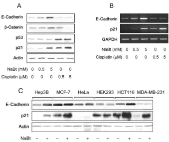
First, we investigated the effect of NaBt on the expression of E-cadherin in a human hepatoma cell line, HepG2. As a result, both RNA and protein levels of E-cadherin were up-regulated by NaBt in a dose-dependent manner (Fig. 1A and 1B). To assess whether the effect is common in human epithelial cells, we employed six additional cell lines of
Fig. 1. p21-independent activation of E-cadherin expression by NaBt. A: HepG2 cells were treated with either NaBt (lanes 2 and 3) or cisplatin (lanes 4 and 5) for 24 hr at the indicated concentration. Levels of E-cadherin, β -catenin, p53, p21, and Actin were measured by Western blots. B: Levels of E-cadherin, p21, and GAPDH RNA transcaripts in HepG2 cells prepared as in Fig. 1A were determined by RT-PCR. C: Protein levels of E-cadherin, p21, and Actin in Hep3B, MCF-7, HeLa, HEK298, HCT116, MDA-MB-231 cells with or without NaBt treat- ment (5 mM) were determined by Western blots.
708 생명과학회지 2009, Vol. 19. No. 6
Hep3B, HCT116, and MCF7 cells while it was not responsive to cisplatin treatment (Fig. 2A and 2B). Taken together, we conclude that p21 is not involved in the NaBt-mediated acti- vation of E-cadherin expression.
To provide a mechanism by which NaBt activates E-cad- herin transcription, we tried to define a NaBt-response ele- ment(s) in the E-cadherin promoter. For this purpose, we cloned E-cadherin promoter composed of approximately 400 bp upstream of the human E-cadherin transcription site [4,11] from human genomic DNA isolated from HepG2 cells.
The E-cadherin promoter is known to contain several pos- itive regulatory elements including a CCAAT box and a Sp1 binding site (GC box) as well as two E-boxes (CANNTG) to which a repressor role is ascribed (Fig. 2C) [11,19]. To get insight into what might be involved in the NaBt-medi- ated activation of the E-cadherin promoter, we analyzed the activity of serial 5’ deletion constructs of the promoter (Fig.
2C). Deletion of the fragment spanning -448 to -71 reduced the basal promoter activity up to 50% of the full-length pro- moter, suggesting the presence of a positive regulatory ele- ment(s) in addition to the negative regulatory E-box 2 in the deleted region [11,19]. However, the activation fold by NaBt was little affected by the deletion. Further deletion up to -61 as in Ecad
-60resulted in not only additional reduction in basal activity but also almost complete abolishment of the promoter activation by NaBt, both of which possibly re- sulted from removal of the positive regulatory CCAAT box located at -65 to -61. Consistently, Ecad
ΔCAATthat contains a mutated CCAAT box by nucleotide substitutions was not responsive to NaBt treatment (Fig. 2C), indicating that the CCAAT box plays a role in the activation of E-cadherin by NaBt.
To provide additional evidence that NaBt activates tran- scription of E-cadherin via the CCAAT box, we employed other CCAAT box-containing promoters and tested their re- sponse to NaBt. According to our previous reports, a func- tional CCAAT box is critical for the promoter activity in both HERV-W LTR and HSV-1 TK promoter [17,18]. Both pro- moters were activated in HepG2 cells by treatment with NaBt, approximately 4 and 6 folds, respectively (Fig. 2D).
However, neither of the two promoters containing a de- fective CCAAT box was responsive to NaBt. Therefore, we concluded that NaBt activates expression of the E-cadherin gene via the CCAAT box probably by stimulating the CCAAT-box binding transcription factor (CTF).
To explain the cell type-specific activation of E-cadherin
Fig. 3. Morphological alteration of hepatocellular carcinoma cells by treatment with NaBt. Hep3B cells were either mock- treated (A) or treated (B) with 5 mM NaBt for 24 hr.
Indirect immunoflorescence assay was performed to lo- cate E-cadherin in cells before and after NaBt treatment.
by NaBt, we tested the promoter activity of E-cadherin in various cell lines in the absence or presence of NaBt.
Consistently to the results shown in Fig. 2A, the E-cadherin promoter in Ecad
-448was successfully activated by NaBt in Hep3B, HCT 116 and breast carcinoma MCF-7 cells whereas the promoter in E-cad
-80was not (Fig. 2B). A similar level of E-cadherin promoter activation by NaBt was also ob- served in other cell lines such as human embryonal kidney HEK 293 and breast carcinoma MDA-MB-231 cells (data not shown). Therefore, the cell type-specific up-regulation of E-cadherin by NaBt might not result from the differential activation of CCAAT box by NaBt. Instead, it might reflect the difference in the mechanism of down-regulation of E-cadherin in various carcinoma cells. The inactivation of E-cadherin promoter in carcinoma cells seems to occur through different mechanisms such as hypermethylation of CpG sites [12,29], the presence of two E-boxes [11], loss of activating protein-2 (AP-2) expression [2,14], up-regulation of transcription repressor Snail [1,24], and a single nucleotide polymorphism in the E-cadherin promoter [16]. Therefore, the elevated CTF activity by NaBt might be not sufficient to activate the E-cadherin promoter in some cell lines that contain dominating mechanism for its expression.
Finally, we investigated the morphological changes of hepatoma cells following induction of E-cadherin upon NaBt treatment. Hep3B cells tend to proliferate individually, ex- hibiting extremely rare cell-cell interactions (Fig. 3A).
Following NaBt treatment, Hep3B cells contact each other,
resembling HepG2 in growth pattern (Fig. 3B). The indirect
immunofluorescence data show that the elevated E-cadherin
in the presence of NaBt primarily localizes at the cell-cell
contact faces, inducing the carcinoma cells into more differ-
entiated form.
More extensive studies are required to understand the ex- act mechanism of the NaBt-mediated E-cadherin activation.
In particular, it is necessary to reveal the pathway through which NaBt stimulates CTF activity, leading to the sub- sequent induction of E-cadherin transcription. The CTF tran- scription factors are known to regulate gene expression ini- tiated through a number of signal transduction pathways, including those controlled by insulin [7], TGF-β [27], c-AMP [8], steroid hormones [5] and others. In most cases, only a single pathway-specific gene has been studied, making gen- eralization impossible. Since the expression of CTF proteins can be affected by the growth and differentiation state of cells [16], it is difficult whether the effects of agents on CTF proteins are direct or indirect. In addition, although post- translational modifications such as phosphorylation [6] and O-glycosylation [13] of CTF-proteins were demonstrated, yet it is still unclear whether these modifications affect CTF function in vivo. To resolve these issues, it is necessary to determine the specific member of CTF family present in the cells and demonstrate the specific biochemical pathways by which NaBt affects its expression or modification.
Nonetheless, the present study shows that NaBt induces up-regulation of E-cadherin and consequent morphological changes, which might contribute to the anti-cancer activity of NaBt.
Acknowledgement
This work was supported for two years by Pusan National University Research Grant.
References
1. Batlle, E., E. Sancho, C. Franci, D. Dominguez, M. Monfar, J. Baulida, and A. Garcia De Herreros. 2000. The tran- scription factor snail is a repressor of E-cadherin gene ex- pression in epithelial tumour cells.
Nat. Cell Biol.
2, 84-89.2. Batsche, E., C. Muchardt, J. Behrens, H. C. Hurst, and C.
Cremisi. 1998. RB and c-Myc activate expression of the E-cadherin gene in epithelial cells through interaction with transcription factor AP-2.
Mol. Cell Biol.
18, 3647-3658.3. Bukholm, I. K., J. M. Nesland, R. Kåresen, U. Jacobsen, and A. L. Børresen-Dale. 1997. Expression of E-cadherin and its relation to the p53 protein status in human breast carcinomas.
Virchows Arch.
431, 317-321.4. Bussemakers, M. J., L. A. Giroldi, A. van Bokhoven, and J. A. Schalken. 1994. Transcriptional regulation of the hu- man E-cadherin gene in human prostate cancer cell lines:
characterization of the human E-cadherin gene promoter.
Biochem. Biophys. Res. Commun.
203, 1284-1290.5. Chaudhry, A. Z., A. D. Vitullo, and R. M. Gronostajski.
1999. Nuclear factor I-mediated repression of the mouse mammary tumor virus promoter is abrogated by the co- activators p300/CBP and SRC-1.
J. Biol. Chem.
274, 7072-7081.6. Coke, D. W. and M. D. Lane. 1988. A sequence element in the GLUT4 gene that mediates repression by insulin.
J.
Biol. Chem.
273, 6210-6217.7. Cooke, D. W. and M. D. Lane. 1999. The transcription factor nuclear factor I mediates repression of the GLUT4 promoter by insulin.
J. Biol. Chem.
274, 12917-12924.8. Cooke, D. W. and M. D. Lane. 1999. Transcription factor NF1 mediates repression of the GLUT4 promoter by cy- clic-AMP.
Biochem. Biophys. Res. Commun.
260, 600-604.9. Couchie, D., N. Holic, M. N. Chobert, A. Corlu, and Y.
Laperche. 2002.
In vitro
differentiation of WB-F344 rat liver epithelial cells into the biliary lineage.Differentiation
69, 209-215.10. Eastman, A. 1990. Activation of programmed cell death by anticancer agents: cisplatin as a model system.
Cancer Cells
2, 275-280.11. Giroldi, L. A., P. P. Bringuier, M. de Weijert, C. Jansen, A.
van Bokhoven, and J. A. Schalken. 1997. Role of E boxes in the repression of E-cadherin expression.
Biochem Biophys.
Res. Commun.
241, 453-458.12. Graff, J. R., J. G. Herman, R. G. Lapidus, H. Chopra, R.
Xu, D. F. Jarrard, W. B. Isaacs, P. M. Pitha, N. E. Davidson, and S. B. Baylin. 1995. E-cadherin expression is silenced by DNA hypermethylation in human breast and prostate carcinomas.
Cancer Res.
55, 5195-5199.13. Jackson, S. P. and R. Tjian. 1988. O-glycosylation of eukary- otic transcription factors: implications for mechanisms of transcriptional regulation.
Cell
55, 125-133.14. Jean, D., J. E. Gershenwald, S. Huang, M. Luca, M. J.
Hudson, M. A. Tainsky, and M. Bar-Eli. 1998. Loss of AP-2 results in up-regulation of MCAM/MUC18 and an increase in tumor growth and metastasis of human melanoma cells.
J. Biol. Chem.
273, 16501-16508.15. Jeanes, A. and C. J. Gottardi, and A. S. Yap. 2008. Cadherins and cancer: how does cadherin dysfunction promote tumor progression?
Oncogene
27, 6920-6929.16. Kulkarni, S. and R. M. Gronostajski. 1996. Altered ex- pression of the developmentally regulated NFI gene family during phorbol ester-induced differentiation of human leu- kemic cells.
Cell Growth Differ.
7, 501-510.17. Kwun, H. J., S. W. Yim, D. H. Lee, and K. L. Jang. 1997.
Activation of the thymidine kinase promoter by herpes sim- plex virus type 1 immediate early proteins.
Mol. Cells
9, 277-280.18. Lee, W. J., H. J. Kwun, H. S. Kim, and K. L. Jang. 2003.
Activation of the human endogenous retrovirus W long ter- minal repeat by herpes simplex virus type 1 immediate ear- ly protein 1.
Mol. Cells
15, 75-80.19. Li, L. C., R. M. Chui, M. Sasaki, K. Nakajima, G. Perinchery, H. C. Au, D. Nojima, P. Carroll, and R. Dahiya. 2000. A
710 생명과학회지 2009, Vol. 19. No. 6
single nucleotide polymorphism in the E-cadherin gene pro- moter alters transcriptional activities.
Cancer Res.
60, 873-876.20. Momparler, R. L. and V. Bovenzi. 2000. DNA methylation and cancer.
J. Cell Physiol.
183, 145-154.21. Mueller, S., E. Cadenas, and A. H. Schönthal. 2000.
p21WAF1 regulates anchorage-independent growth of HCT116 colon carcinoma cells via E-cadherin expression.
Cancer Res.
60, 156-163.22. Nakano, K., T. Mizuno, Y. Sowa, T. Orita, T. Yoshino, Y.
Okuyama, T. Fujita, N. Ohtani-Fujita, Y. Matsukawa, T.
Tokino, H. Yamagishi, T. Oka, H. Nomura, and T. Sakai.
1997. Butyrate activates the WAF1/Cip1 gene promoter through Sp1 sites in a p53-negative human colon cancer cell line.
J. Biol. Chem.
272, 22199-22206.23. Perl, A. K., P. Wilgenbus, U. Dahl, H. Semb, and G.
Christofori. 1998. A causal role for E-cadherin in the tran- sition from adenoma to carcinoma.
Nature
392, 190-193.24. Poser, I., D. Dominguez, A. G. de Herreros, A. Varnai, R.
Buettner, and A. K. Bosserhoff. 2001. Loss of E-cadherin ex-
pression in melanoma cells involves up-regulation of the transcriptional repressor Snail.
J. Biol. Chem.
276, 24661- 24666.25. Sadot, E., B. Geiger, M. Oren, and A. Ben-Ze'ev. Down-reg- ulation of beta-catenin by activated p53.
Mol. Cell Biol.
21, 6768-6781.26. Saito, H., H. Ebinuma, M. Takahashi, F. Kaneko, K.
Wakabayashi, M. Nakamura, and H. Ishii. 1998. Loss of bu- tyrate-induced apoptosis in human hepatoma cell lines HCC-M and HCC-T having substantial Bcl-2 expression.
Hepatology
27, 1233-1240.27. Sun, P., P. Dong, K. Dai, G. J. Hannon, and D. Beach. 1998.
p53-independent role of MDM2 in TGF-beta1 resistance.
Science
282, 2270-2272.28. van Roy, F. and G. Berx. 2008. The cell-cell adhesion mole- cule E-cadherin.
Cell Mol. Life Sci.
65, 3756-3788.29. Yoshiura, K., Y. Kanai, A. Ochiai, Y. Shimoyama, T.
Sugimura, and S. Hirohashi. 1995. Silencing of the E-cadher- in invasion-suppressor gene by CpG methylation in human carcinomas.