Protective Effects of Quercetin-3-O-glucuronide against 1-methyl-4- phenylpyridinium-induced Neurotoxicity
Ramesh Pariyar, Tonking Bastola and Jungwon Seo*
Institute of Pharmaceutical Research and Development,College of Pharmacy, Wonkwang University, Iksan 570-749, Korea Received February 9, 2019 /Revised February 20, 2019 /Accepted February 21, 2019
Parkinson’s disease (PD) is a progressive neurodegenerative disease that mainly affects motor system with clinical features such as bradykinesia, rigidity, tremor and abnormal posture. PD is characterized by the death of dopaminergic neurons in the substantia nigra pars compacta, which is associated with accumulation of oxidative stress and dysregulation of intracellular signaling pathway. Quercetin-3-O- glucuronide (Q3GA), a major metabolite of quercetin, has been reported to have neuroprotective effects. In this study, we examined the neuroprotective effect of Q3GA against 1-methyl-4-phenyl pyr- idinium (MPP+)-induced neurotoxicity of PD and the underlying molecular mechanisms in SH-SY5Y cells. MTT and LDH assay showed that Q3GA significantly decreased MPP+-induced cell death, which is accompanied by a reduction in poly (ADP-ribose) polymerase (PARP) cleavage. Furthermore, it atte- nuated MPP+-induced intracellular reactive oxygen species (ROS) with the reduction of Bax/ Bcl-2 ratio. Moreover, Q3GA significantly increased the phosphorylation of Akt and cAMP response ele- ment binding protein (CREB), but it has no effects on the phosphorylation of extracellular signal–
regulated kinase (ERK). Taken together, these results demonstrate that Q3GA significantly attenuates MPP+-induced neurotoxicity through ROS reduction and Akt/CREB signaling pathway in SH-SY5Y cells. Our findings suggest that Q3GA might be one of the potential candidates for the prevention and/or treatment of PD.
Key words : Akt, neurotoxicity, Parkinson’s disease, quercetin-3-O-glucuronide, ROS
*Corresponding author
*Tel : +82-63-850-6819, Fax : +82-63-853-6821
*E-mail : [email protected]
This is an Open-Access article distributed under the terms of the Creative Commons Attribution Non-Commercial License (http://creativecommons.org/licenses/by-nc/3.0) which permits unrestricted non-commercial use, distribution, and reproduction in any medium, provided the original work is properly cited.
Journal of Life Science 2019 Vol. 29. No. 2. 191~197 DOI : https://doi.org/10.5352/JLS.2019.29.2.191
서 론
파킨슨병(Parkinson’s disease)은 운동완서(bradykinesia), 근육경직(rigidity), 진전(tremor) 및 비정상적인 자세(abnormal posture) 등을 임상적 특징으로 하는 신경퇴행성 질환이다[8].
파킨슨병의 병리학적 특징은 뇌 흑색치밀부(substantianigra pars compacta)에서의 도파민성 신경세포의 진행성 퇴행에 의한 선조체(striatum) 내의 도파민 감소이다[10]. 비록 이러한 신경세포의 사멸에 관여하는 분자 메커니즘이 완전히 이해되 지는 못했지만, 산화 스트레스와 신호전달체계의 조절 이상에 의한 도파민성 신경세포의 사멸은 파킨슨병을 유발하는 원인 으로 보고되어 왔다[20].
파킨슨병의 다양한 시험관내 또는 생체 실험 모델로 오랫동 안 사용되어온 1-methyl-4-phenyl pyridinium (MPP+)는 1- methyl-4-phenyl-1,2,3,6-tetrahydropyridine (MPTP)의 뇌 내 활성 대사 산물이다. 뇌 내에서 MPP+는 도파민을 포함한 카테
콜라민성 신경세포에 대해 특이적인 독성을 나타낸다[3].
MPP+는 도파민 수송체를 통해 도파민성 신경세포 내로 들어 가고 미토콘드리아로 수송되어 산화 스트레스와 세포 사멸 신호전달 경로를 통하여 신경세포에 독성을 유발한다. 한편, 신경 모세포종 SH-SY5Y 세포는 신경 유사 세포로의 분화가 가능하며, 도파민성 신경세포와 유사한 성질을 보유하고 있기 때문에 파킨슨병 관련 신경 독성과 신경 보호 메커니즘을 연 구하는데 널리 사용된다[13]. MPP+에 의한 신경세포 독성과 관련된 신호체계에는 pro-survival phosphoinositide-3-kinase (PI3K)/Akt의 비활성화와 extracellular signal–regulated kinase (ERK)의 비정상적 조절 등이 있다. PI3K/Akt 신호전달 경로는 신경세포를 산화 스트레스로부터 보호하는데 필수적 이다[19]. MPP+는 PI3K/Akt의 인산화를 억제하여 신호전달 경로를 억제한다고 알려져 있으며, PI3K/Akt 경로의 결함은 파킨슨병과 유사한 신경 퇴행을 유도한다[34]. 반면에, Akt의 활성화는 신경 퇴행성 질환에 대한 보호 작용을 한다. ERK는 세포의 분화, 증식 및 세포 사멸을 포함하는 많은 세포의 생존 에 중요한 역할을 하는 신호전달 분자이다[16]. 특히, 뇌에서 ERK는 도파민성 신경 세포의 생존을 증가시킨다[33, 35]. PI3K/
Akt와 ERK는 공통적으로 CREB의 활성화에 관여하는 신호 전달 분자들이다[7, 17]. cAMP response element binding pro- tein (CREB)은 세포 성장, 증식 및 기억과 같은 다양한 생물학 적 기능을 조절하는 전사 인자이며, 저산소증, 허혈, 흥분 독성
및 산화 스트레스와 같은 다양한 스트레스 자극에 반응하여 활성화된다[2]. Akt와 ERK에 의한 CREB 의 인산화는 유전자 의 발현을 자극하여 신경가소성과 신경세포 보호에 중요한 역할을 한다[2]. 최근에, CREB 신호전달체계는 인지 및 신경 퇴행성 장애를 비롯한 여러 신경병리학적 상태에 관여하고 있다고 보고되었다. 뇌에서의 CREB 기능 저하는 헌팅톤병, 알츠하이머병, 파킨슨병 및 노인성 인지기능 장애의 발달 및 진행뿐만 아니라 신경 퇴화를 유발한다[7, 30, 31]. CREB이 결핍 된 마우스는 축삭 성장 결함 및 신경 퇴화를 일으킨다는 것이 밝혀졌다[18]. 또한 CREB은 ROS에 의한 신경세포 사멸 에서 신경보호 신호체계의 상위 매개 분자로 밝혀졌다[14].
Quercetin은 포도, 사과를 포함한 많은 채소 및 과일에 널리 분포되어 있는 항산화 효과를 가진 천연 플라보노이드(flav- onoid)이다[5]. Quercetin은 항암 효과[23], 항고혈압 효과[28], 항염 효과[4], 신경 보호 작용[26]과 같은 중요한 약리 작용을 발휘하는 생체 활성 화합물이다. 경구 투여된 quercetin은 장 과 간에서의 1차 대사 과정에서 글루쿠론산(glucuronic acid) 과 결합하여, quercetin-3-O-glucuronide (Q3GA)로 변환된다.
Q3GA는 혈장과 뇌에서 주로 검출되는 quercetin의 주대사산 물[27]이나, 현재 대부분의 시험관내 연구에서는 quercetin aglycone을 세포에 처리하여 사용하고 있다. 본 연구자는 quercetin과 Q3GA를 신경줄기세포에 처리하여, quercetin은 신경줄기세포의 사멸을 유도하지만, Q3GA는 신경줄기세포 의 생존과 이동을 촉진시킨다는 사실을 보고하였다[1]. 뿐만 아니라, 쥐를 이용한 실험에서 스코폴라민(scopolamine)에 의 해 유도된 기억력 장애가 Q3GA 투여에 의해 개선될 수 있음 을 확인하였다[1]. 다른 연구에서는 quercetin과 Q3GA를 Neuro-2a세포에 처리하여 6-hydroxydopamine (6-OHDA)에 의해 발생하는 ROS와 세포 사멸을 quercetin이 억제할 수 있 음을 확인하였다[22]. 또한, 6-OHDA 의 처리에 의해 유도된 파킨슨병의 실험 모델에서 quercetin을 투여한 쥐는 진전 (tremor)이 현저하게 개선되었고, 이것이 세로토닌(serotonin) 수치의 증가와 관련됨을 확인하였다[21]. 이 결과로부터 quer- cetin과 그 대사체들이 파킨슨병에 대한 보호 효과를 가지고 있을 가능성을 확인하였으나, 실제 quercetin의 대사체가 어떤 신호전달 경로를 통하여 신경 보호 효과를 나타내는지는 자세 히 연구된 바가 없다. 따라서 본 논문에서는 SH-SY5Y 세포를 이용하여 MPP+ 유도 신경 독성에 대한 Q3GA의 신경 보호 효과를 평가하고, 이 효과의 분자 메커니즘을 밝혀내고자 하 였다.
재료 및 방법
시약 및 기기
Q3GA, 2’,7’–dichlorofluorescin diacetate (DCFH-DA), 3'- (4,5-dimethylthiazol-2-yl)-2,5-diphenyltetra zolium bromide
(MTT) 및 MPP+는 Sigma-Aldrich Chemicals (St. Louis, MO, USA)에서 구입하였고, Dulbecco’s modified Eagle Medium (DMEM)와 fetal bovine serum (FBS)은 Gibco (Carlsbad, CA, USA)에서 구입하였다. Anti-p-Akt (Ser473), anit-Akt, anti-p- p44/42 MAPK (Thr202/Tyr204), anti-ERK, anti-p-CREB(Ser 133), anti-CREB, anti-PARP, anti-Bax, anti-Bcl2 및 anti- GAPDH는 Cell Signaling Technology (Beverly, Massachu- setts, USA)로부터 구입하였고, 2차 항체는 Santa Cruz Bio- technology(Santa Cruz, CA, USA)에서 구입하였다.
세포 배양
인간 신경모세포종 세포주 SH-SY5Y는 한국 세포주 은행 (Seoul, Korea)으로부터 구입하였다. 세포는 10% FBS가 함유 된 DMEM배지에서 배양하였고, 6-well plate에 2×10⁶개의 세 포를 넣고 다음날 Q3GA를 농도 별로 2시간 전처리 후, MPP+ 를 24시간 처리하였다.
MTT assay
세포 생존율을 측정하기 위해 MTT assay(Sigma-Aldrich Chemicals)를 사용하였다. 간단히 기술하면 먼저 96 well plate에 1×10⁴ cells/well로 동일하게 분주하고 24시간 동안 배양하였다. 기존의 배지를 제거하고 새로운 배지를 넣어준 후 Q3GA를 2시간 전처리 후, MPP+를 첨가하였다. DMSO의 처리 농도는 배지 대비 0.1% 이하가 되도록 하였다. 이를 24시 간 배양한 후에 배지를 제거하고 MTT시약(5 mg/ml)을 넣고, 4시간 동안 방치한 후 상등액을 제거하였다. 각 well에 DMSO 20 μl를 첨가한 후 orbital shaker를 이용하여 형성된 formazan 을 녹이고, 30분 후 595 nm에서 흡광도를 측정하였다. 실험은3 회 반복 실시하여 평균값을 구하였으며, 음성 대조군의 흡광 도 값을 기준으로 세포 생존율을 비교하였다.
Lactate dehydrogenase (LDH) assay
세포 배지에서 LDH의 방출을 조사하여 세포 독성을 정량 적으로 평가하였다. 양성 대조군에는 1%(v/v) Triton X-100 을 사용하여 세포를 1시간 동안 전처리 하였다. 모든 실험은 LDH cytotoxicity colorimetric assay kit (Bio Vision)의 실험방 법에 따라 수행하였다. 세포를 2시간 동안 Q3GA (100 또는 200 μM)로 전처리 한 다음 24시간 동안 MPP+에 노출시켰다.
각 well로부터 배양 배지 100 μl를 새로운 96-well plate로 옮기고, 100 μl의 반응 혼합물을 넣어 실온에서 30분 동안 배양 하였다. LDH의 방출은 microplate ELISA reader를 사용하여 490 nm에서 흡광도를 측정 하였다. 배지 내로의 LDH 방출은 다음 방정식을 사용하여 계산하였다 :
LDH 방출 (%) = (실험군 OD 값 - 음성 대조군 OD 값) / (양성 대조군 OD 값 - 음성 대조군 OD 값) ×100%
A
B
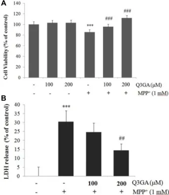
Fig. 1. Q3GA protectes SH-SY5Y cells against MPP+-induced cy- totoxicity. Cells were pretreated with different doses of Q3GA (100 or 200 μM) for 2 hr and then exposed to 1 mM MPP+ for 24 hr. (A) Cell viability was measured using MTT assay. (B) Cytotoxicity was determined by measuring LDH release into the media. Values are pre- sented relative to control as mean percentage change ± S.D. (n=3). Differences are statistically significant at ***p˂
0.001 vs the control group and ##p<0.01, ###p˂0.001 vs the MPP+ group.
세포 내 ROS 측정
세포 내 ROS는 DCFH-DA 형광 프로브(Sigma-Aldrich Chemicals)를 사용하여 평가하였다. 세포를 96 well 및 6 well plate에 동일하게 분주하고 Q3GA를 2시간 동안 처리 한 다음 MPP+(1 mM)로 24시간 동안 함께 처리 하였다. 이후 세포를 DCFH-DA(10 μM)와 함께 37℃에서 30분 동안 배양하고, 녹색 형광을 형광현미경(TS100-F, Nikon) 하에서 시각화하거나, Ex/Em : 485/535 nm에서 plate reader기(Wallac, Perkin Elmer, Waltham, MA, USA)로 형광을 측정하였다.
Western blot analysis
시료를 처리한 SH-SY5Y 세포에 RIPA buffer를 첨가한 다 음, 원심분리하여 상등액에서 Thermo Scientific 회사의 BCA 실험 키트(San Jose, California, USA)를 사용하여 단백질 농도 를 측정하였다. 동량의 단백질을 SDS-PAGE (10-12%)에서 영 동하고 nitrocellulose (NC) membrane으로 전사하였다. 전사 된 NC membrane을 5% 무지방유가 포함된 blocking buffer (0.1% Tween 20 in TBS)에서 blocking한 후 p-Akt, Akt, p- ERK, ERK, p-CREB, CREB, PARP, Bax, Bcl2 및 GAPDH 에 특이적인 항체(1: 1,000)와 함께 4℃에서 밤새 배양 하였다. 다 시 상응하는 2차 항체와 함께 1시간 배양하고 ECL 용액을 1:1로 잘 섞어서 NC membrane위에 가하여 발광시키고 암실 에서 X선 필름에 감광한 후 현상하였다. 특정 단백질 밴드의 상대적 강도는 ImageJ 소프트웨어를 사용하여 정량화하였다.
통계처리
각 실험군 간의 결과는 평균치와 표준오차로 나타내었으며, 3번 이상 독립적인 실험을 통하여 얻어진 실험 결과는 Graph Pad 소프트웨어를 사용하여 일원 분산 분석(one-way ANOVA) 후 Tukey’s post hoc multiple comparison으로 분석하였다.
실험군 간의 차이는 95% 수준(p<0.05)에서 유의적인 것으로 판단하였다.
결과 및 고찰
Q3GA의 MPP+ 유도 세포 독성에 대한 보호 효과 MPP+는 파킨슨병의 시험관 내 실험 모델로 사용되어온 신 경 독성 물질로서, 도파민성 신경세포를 비롯한 카테콜라민 신경세포를 선택적으로 손상시킨다[3, 35]. 먼저, SH-SY5Y 세 포에 MPP+를 처리하여 유도된 세포 독성에 대한 Q3GA의 영향을 확인하였다(Fig. 1). SH-SY5Y 세포에 Q3GA (100, 200 μM)로 2시간 동안 전처리 한 후 MPP+ (1 mM)로 24시간 배양 하였다. 세포의 생존율은 MTT 실험법을 이용하여 확인하였 다. MPP+에 노출된 SH-SY5Y 세포는 대조군 세포에 비해 세포 생존율이 유의적으로 감소됨을 관찰하였다. 그러나 Q3GA를 전처리하면 세포 생존율이 용량 의존적으로 유의적으로 증가
하였다(Fig. 1A). 세포막 손상에 의한 세포 사멸을 측정하는 LDH 실험 결과에서도, MPP+로 인한 세포 사멸이 Q3GA 처리 에 의해 농도의존적으로 억제되었다(Fig. 1B). 이 결과들로부 터 Q3GA가 MPP+에 의한 신경 세포 독성에 대해 보호 효과를 가지고 있음을 확인하였다.
MPP+에 의해 유도된 세포 내 ROS의 축적에 대한 Q3GA 의 영향
산화 스트레스는 MPP+에 의한 신경세포 사멸에서 중요한 역할을 하며[15], quercetin의 항산화 효과는 여러 논문을 통해 보고되어져 왔다[5]. 따라서, MPP+의 세포 독성에 대한 Q3GA 의 보호 효과가 항산화 기전에 의해 매개되는지 여부를 조사 하였다. 세포 내 활성산소종(ROS) 수준은 DCFH-DA 형광 분 석에 의해 측정하였다. 24시간 동안 MPP+에 노출된 SH-SY5Y 세포 내에서는 ROS 생성이 유의적으로 증가하였다(Fig. 2).
반면, 100 또는 200 μM의 Q3GA는 MPP+에 의해 유도된 세포 내 ROS의 축적을 유의적으로 억제하였다. 이 결과는 Q3GA가 MPP+에 의해 유도된 ROS의 축적을 억제하여 세포 보호 효과
A
B
Fig. 2. Q3GA suppresses MPP+-induced intracellular accumulation of ROS in SH-SY5Y cells. SH-SY5Y cells were pretreated with Q3GA (100 or 200 μM) for 2 hr, and then exposed to 1 mM MPP+ for 24 hr. Further, the cells were incubated for 30 min at 37°C with 10 μM 2,7-dichlorofluorescein diacetate (DCFH-DA). (A) Representative images of the cells under a fluorescence microscope are shown. Intracellular ROS evaluated by the oxidation of H2DCF-DA to green colour floursecence DCF. Scale bar = 50 μm. (B) DCFH-DA fluorescence intensities were measured by fluorimetry with a plate reader at excitation/emission wavelengths of 485/535 nm. Values are presented relative percentage to control as mean percentage change ± S.D. (n=3).
Differences are statistically significant at ***p<0.001 vs the control group and ###p<0.001 vs the MPP+ group.
를 나타낼 가능성을 보여준다.
Q3GA에 의한 Bax/Bcl-2 비율의 감소 효과
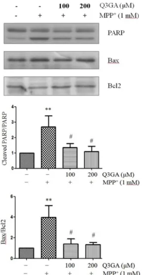
핵 내에 풍부하게 존재하며 DNA 수리 효소로서 기능하는 PARP단백질은 세포자멸사 과정에서는 활성화된 caspases에 의해 절단된다[9, 24]. 이 PARP 단백질의 절단은 세포자멸사 의 중요한 바이오마커이다. 세포자멸사 과정에서 세포질 내 cytochrome c는 caspase 9와 3의 활성화를 유도하며, PARP는 caspase 3의 하류 표적이다. 이러한 PARP 단백질의 절단은 소뇌 과립 신경세포에서 MPP+에 의한 세포 사멸 과정에서도 일어난다[29]. 따라서, Western blot 분석을 통하여 PARP 단백 질의 발현을 측정하였다. MPP+에 의해 PARP 단백질의 절단 이 증가하지만, Q3GA의 처리에 의해 감소함을 확인하였다 (Fig. 3).
MPP+에 의해 증가되는 Bax/Bcl-2 비율은 세포자멸사를 조 절하는 중요한 요소이다[12, 32]. Bcl-2, Bcl-xL 등은 세포자멸 사를 억제하며, Bax, Blk, Bad 등은 세포자멸사를 항진시킨다 [25]. Bax는 미토콘드리아 내로 이동하여 활성화된 mono- dimer가 되어 세포자멸사를 항진시키고, 이러한 Bax에 의한 세포사멸은 Bcl-2와의 heterodimer형성에 의해 억제된다.
Western blot 분석 결과, 1 mM MPP+의 24시간 처리는 Bax의 발현을 증가시켰으며, Bcl-2의 발현을 억제하였다. 반면,
Q3GA의 처리는 MPP+에 의한 Bcl-2의 단백질 발현을 유의적 으로 증가시켜, Bax/Bcl-2 비율을 감소시켰다(Fig. 3). 이 결과 는 Q3GA가 항세포사멸 단백질과 세포사멸 단백질 사이의 균형을 회복시켜 세포 생존을 촉진할 수 있다는 가능성을 보 여준다.
Q3GA에 의한Akt와 CREB 인산화 조절
Q3GA의 MPP+에 대한 세포 보호 효과의 기초가 되는 분자 메커니즘을 밝히기 위해, Western blot 분석을 통해 PI3K/Akt`, ERK 및 CREB 신호 전달 경로를 평가하였다. Fig. 4에서 MPP+ 는 Akt와 ERK, CREB의 인산화를 현저히 감소시켰다. 반면에, Q3GA로 전처리하면 MPP+에 의해 감소한 Akt와 CREB의 인 산화는 회복되었으나, ERK의 감소된 인산화는 회복되지 않았 다. 이러한 결과는 Q3GA가 ERK가 아니라, Akt 의 활성화를 통해 CREB의 활성화를 유도하고, 이를 통해 MPP+에 대한 보 호 효과를 나타낼 가능성을 시사한다.
파킨슨병의 발병 기전에 완전히 이해되지는 못했지만, 산화 적 스트레스와 신호전달체계의 조절 이상에 의한 도파민성 신경세포의 사멸은 중요한 유발 요인으로 보고되어졌다. 이전 의 연구들은 MPP+가 빠르고 가역적으로 Akt 의 인산화를 감 소시킨다는 것을 보여 주었으며, 이는 신경 세포의 사멸과 관 련되어 있다[6]. CREB은 Akt의 하류 표적으로 유전자 발현을
Fig. 3. Q3GA markedly attenuates MPP+ -induced PARP pro- teolysis and Bax/Bcl2 ratio. SH-SY5Y cells were pretreated with Q3GA for 2 hr and then treated with MPP+ (1 mM) for 24 hr. Protein levels of PARP, Bax, Bcl-2 and β-actin were measured by Western blot analysis. Representative blots and their densitometric quantification are shown.
β-actin was used as the internal control. Values are pre- sented relative to control as mean fold change ± S.D.
(n=3). Differences are statistically significant at **p<0.01 vs the control group and #p<0.05 vs the MPP+ group.
조절하며, MPP+ 유도 세포 손상에 관여하는 신호전달 분자 중 하나이다[11]. 본 연구에서도 MPP+는 SH-SY5Y 세포에서 ROS의 축적을 증가시키고, Akt의 Serine 473 위치의 인산화와 CREB의 Serine 133 위치의 인산화를 감소시켰다. Q3GA는 MPP+에 의해 증가된 ROS를 억제하였고, 감소된 Akt와 CREB 의 인산화를 회복시켰다. 반면에 CREB의 또 다른 상위 신호전 달 분자인 ERK의 경우, MPP+에 의해 그 인산화가 감소되었지 만, Q3GA에 의해 회복되지 못했다. 본 연구 결과로부터, Q3GA 는 ERK가 아니라, Akt의 신호전달 경로를 통하여 CREB의 인산화에 작용하고, 산화 스트레스를 완화시키는 것을 통해서
Fig. 4. MPP+ decreases Akt and CREB phosphorylation, whereas Q3GA reverses those effects. SH-SY5Y cells were pre- treated with Q3GA (100 or 200 μM) for 2 hr, and then exposed to MPP+ (1 mM) for 24 hr. After cell lysis, the extracted proteins were subjected to Western blot analysis.
Protein levels of p-Akt, Akt, p-ERK, ERK, p-CREB and GAPDH were determined. GAPDH was used as the in- ternal control. Representative blots and their densito- metric quantification are shown. Values are presented relative to control as mean fold change ± S.D. (n=3).
Differences are statistically significant at ***p<0.001 vs the control group and #p<0.05, ##p<0.01 vs the MPP+ group.
MPP+에 대한 신경세포 보호 작용을 나타낼 것으로 사료된 다. 본 연구는 MPP+로 유도된 세포 독성에 대한 Q3GA의 신경 보호 기작을 이해하는데 도움이 된다. 또한, quercetin 과 그 대사체인 Q3GA가 파킨슨병에 대한 치료제로 개발될 수 있는 가능성을 시사하였다. 이를 분명하게 밝히기 위하 여 앞으로 더 많은 연구가 필요할 것으로 생각된다.
감사의 글
논문은 2016년도 정부의 재원으로 한국연구재단의 지원을 받아 수행된 기초연구사업입니다(NRF-2016R1A2B4007588).
References
1. Baral, S., Pariyar, R., Kim, J., Lee, H. S. and Seo, J. 2017.
Quercetin-3-O-glucuronide promotes the proliferation and migration of neural stem cells. Neurobiol. Aging 52, 39-52.
2. Carlezon Jr, W. A., Duman, R. S. and Nestler, E. J. 2005.
The many faces of CREB. Trends Neurosci. 28, 436-445.
3. Chun, H. S., Gibson, G. E., DeGiorgio, L. A., Zhang, H., Kidd, V. J. and Son, J. H. 2001. Dopaminergic cell death induced by MPP+, oxidant and specific neurotoxicants shares the common molecular mechanism. J. Neurochem. 76, 1010-1021.
4. Comalada, M., Camuesco, D., Sierra, S., Ballester, I., Xaus, J., Galvez, J. and Zarzuelo, A. 2005. In vivo quercitrin anti-in- flammatory effect involves release of quercetin, which in- hibits inflammation through down-regulation of the NF- kB pathway. Eur. J. Immunol. 35, 584-592.
5. Costa, L. G., Garrick, J. M., Roque, P. J. and Pellacani, C.
2016. Mechanisms of neuroprotection by quercetin: counter- acting oxidative stress and more. Oxid. Med. Cell. Longev.
2016, 2986796.
6. Datta, S. R., Dudek, H., Tao, X., Masters, S., Fu, H., Gotoh, Y. and Greenberg, M. E. 1997. Akt phosphorylation of BAD couples survival signals to the cell-intrinsic death machinery.
Cell 91, 231-241.
7. Du, K. and Montminy, M. 1998. CREB is a regulatory target for the protein kinase Akt/PKB. J. Biol. Chem. 273, 32377- 32379.
8. Fearnley, J. M. and Lees, A. J. 1991. Ageing and Parkinson's disease: substantia nigra regional selectivity. Brain 114, 2283- 2301.
9. Fernandes-Alnemri, T., Litwack, G. and Alnemri, E. S. 1994.
CPP32, a novel human apoptotic protein with homology to Caenorhabditis elegans cell death protein Ced-3 and mam- malian interleukin-1 beta-converting enzyme. J. Biol. Chem.
269, 30761-30764.
10. Forno, L. S. 1996. Neuropathology of Parkinson's disease.
J. Neuropathol. Exp. Neurol. 55, 259-272.
11. Gu, X., Liu, L., Shen, Q. and Xing, D. 2017. Photoactivation of ERK/CREB/VMAT2 pathway attenuates MPP+-induced neuronal injury in a cellular model of Parkinson's disease.
Cell Signal. 37, 103-114.
12. Itano, Y. and Nomura, Y. 1995. 1-methyl-4-phenyl-pyr- idinium ion (MPP+) causes DNA fragmentation and in- creases the Bcl-2 expression in human neuroblastoma, SH- SY5Y cells, through different mechanisms. Brain Res. 704, 240-245.
13. Jantas, D., Greda, A., Golda, S., Korostynski, M., Grygier, B., Roman, A., Pilc, A. and Lason, W. 2014. Neuroprotective effects of metabotropic glutamate receptor group II and III activators against MPP+-induced cell death in human neuro- blastoma SH-SY5Y cells: the impact of cell differentiation state. Neuropharmacology 83, 36-53.
14. Lee, B., Cao, R., Choi, Y. I., Cho, H. E., Rhee, A. D., Hah, C. K., Hoyt, K. R. and Obrietan, K. 2009. The CREB/CRE transcriptional pathway: protection against oxidative stress- mediated neuronal cell death. J. Neurochem. 108, 1251-1265.
15. Li, J., O, W., Li, W., Jiang, Z. G. and Ghanbari, H. A. 2013.
Oxidative stress and neurodegenerative disorders. Int. J.
Mol. Sci. 14, 24438-24475.
16. Lin, Y. L., Wang, G. J., Huang, C. L., Lee, Y. C., Liao, W.
C., Lai, W. L., Lin, Y. J. and Huang, N. K. 2009. Ligusticum chuanxiong as a potential neuroprotectant for preventing serum deprivation-induced apoptosis in rat pheochromocy- toma cells: functional roles of mitogen-activated protein kinases. J. Ethnopharmacol. 122, 417-423.
17. Lonze, B. E. and Ginty, D. D. 2002. Function and regulation of CREB family transcription factors in the nervous system.
Neuron 35, 605-623.
18. Lonze, B. E., Riccio, A., Cohen, S. and Ginty, D. D. 2002.
Apoptosis, axonal growth defects, and degeneration of pe- ripheral neurons in mice lacking CREB. Neuron 34, 371-385.
19. Lu, S., Lu, C., Han, Q., Li, J., Du, Z., Liao, L. and Zhao, R.
C. 2011. Adipose-derived mesenchymal stem cells protect PC12 cells from glutamate excitotoxicity-induced apoptosis by upregulation of XIAP through PI3-K/Akt activation.
Toxicology 279, 189-195
20. Mattson, M. P. 2000. Apoptosis in neurodegenerative dis- orders. Nat. Rev. Mol. Cell. Biol. 1, 120-129.
21. Mu, X., Yuan, X., Du, L. D., He, G. R. and Du, G. H. 2016.
Antagonism of quercetin against tremor induced by unilat- eral striatal lesion of 6-OHDA in rats. J. Asian Nat. Prod.
Res. 18, 65-71.
22. Mukai, R., Kawabata, K., Otsuka, S., Ishisaka, A., Kawai, Y., Ji, Z. S., Tsuboi, H. and Terao, J. 2012. Effect of quercetin and its glucuronide metabolite upon 6-hydorxydopamine- induced oxidative damage in Neuro-2a cells. Free Radic. Res.
46, 1019-1028.
23. Murakami, A., Ashida, H. and Terao, J. 2008. Multitargeted cancer prevention by quercetin. Cancer Lett. 269, 315-325.
24. Oliver, F. J., de la Rubia, G., Rolli, V., Ruiz-Ruiz, M. C., de Murcia, G. and Mnissier-de Murcia, J. 1998. Importance of poly (ADP-ribose) polymerase and its cleavage in apoptosis Lesson from an uncleavable mutant. J. Biol. Chem. 273, 33533-33539.
25. Oltvai, Z. N. and Korsmeyer, S. J. 1994. Check points of dueling dimers foil death wishes. Cell 79, 189-192.
초록:1-methyl-4-phenylpyridinium으로 유도된 신경 손상에 대한 quercetin-3-O-glucuronide의 보호 효과
파리야르라메스․바스또라통킹․서정원*
(원광대학교 약학대학 약품연구소)
파킨슨병은 운동완서, 근육경직, 진전 및 비정상적인 자세 등을 임상적 특징으로 하는 주로 운동 신경계에 영향 을 주는 진행성 신경 퇴행성 질환이다. 파킨슨병은 산화 스트레스와 세포 내 신호 전달 경로의 조절 장애에 의한 뇌 흑색치밀부에서의 도파민성 신경세포의 사멸을 특징으로 한다. Quercetin의 주요 대사산물인 Quercetin-3-O- glucuronide (Q3GA)는 신경 보호 효과가 있는 것으로 보고 되어 왔다. 본 연구에서는 SH-SY5Y 세포에서 1-meth- yl-4-phenyl pyridinium (MPP+)에 의해 유도된 신경 독성에 대한 Q3GA의 신경 보호 효과와 그 분자 조절 기전을 조사하였다. Q3GA는 MPP+에 의해 유도된 세포 사멸을 유의적으로 감소시켰으며 PARP 절단을 감소시켰다. 또 한, Bax/Bcl-2 비율의 감소와 함께 MPP+에 의해 증가된 세포 내 ROS를 감소시켰다. Q3GA는 MPP+에 의해 감소 된 Akt와 CREB의 인산화를 유의적으로 회복시켰지만, ERK에는 영향을 미치지 않았다. 이 결과는 Q3GA가 ROS 생산 억제와 Akt/CREB 신호 전달 경로를 통해 MPP+ 에 의해 유도된 신경 독성을 억제시킬 수 있음을 시사한다.
본 연구는 Q3GA가 파킨슨병에 대한 예방제 또는 치료제로 개발될 수 있는 가능성을 제시한다.
26. Ossola, B., Kaariainen, T. M. and Mannisto, P. T. 2009. The multiple faces of quercetin in neuroprotection. Expert Opin.
Drug Saf. 8, 397-409.
27. Perez, A., Gonzalez-Manzano, S., Jimenez, R., Perez-Abud, R., Haro, J. M., Osuna, A., Santos-Buelga, C., Duarte, J. and Perez-Vizcaino, F. 2014. The flavonoid quercetin induces acute vasodilator effects in healthy volunteers: correlation with beta-glucuronidase activity. Pharmacol. Res. 89, 11-18.
28. Perez-Vizcaino, F., Duarte, J., Jimenez, R., Santos-Buelga, C.
and Osuna, A. 2009. Antihypertensive effects of the fla- vonoid quercetin. Pharmacol. Rep. 61, 67-75.
29. Pu, X., Song, Z., Li, Y., Tu, P. and Li, H. 2003. Acteoside from Cistanche salsa inhibits apoptosis by 1-methyl-4-phe- nylpyridinium ion in cerebellar granule neurons. Planta Med. 69, 65-66.
30. Saura, C. A. and Valero, J. 2011. The role of CREB signaling in Alzheimer's disease and other cognitive disorders. Rev.
Neurosci. 22, 153-169.
31. Wang, H., Xu, J., Lazarovici, P., Quirion, R. and Zheng, W.
2018. cAMP response element-binding protein (CREB): a
possible signaling molecule link in the pathophysiology of Schizophrenia. Front. Mol. Neurosci. 11, 255.
32. Wang, Y., Gao, J., Miao, Y., Cui, Q., Zhao, W., Zhang, J.
and Wang, H. 2014. Pinocembrin protects SH-SY5Y cells against MPP+-induced neurotoxicity through the mitochon- drial apoptotic pathway. J. Mol. Neurosci. 53, 537-545.
33. Weng, Z., Signore, A. P., Gao, Y., Wang, S., Zhang, F., Hastings, T., Yin, X. M. and Chen, J. 2007. Leptin protects against 6-hydroxydopamine-induced dopaminergic cell death via mitogen-activated protein kinase signaling. J. Biol. Chem.
282, 34479-34491.
34. Zhang, C., Yuan, X., Hu, Z., Liu, S., Li, H., Wu, M., Yuan, J., Zhao, Z., Su, J. and Wang, X. 2017. Valproic acid protects primary dopamine neurons from MPP+-induced neurotoxicity.
Biomed. Res. Int. 2017, 8124501.
35. Zhu, J. H., Horbinski, C., Guo, F., Watkins, S., Uchiyama, Y. and Chu, C. T. 2007. Regulation of autophagy by ex- tracellular signal-regulated protein kinases during 1-meth- yl-4-phenylpyridinium-induced cell death. Am. J. Pathol.
170, 75-86.