TFAP2C Promotes Cell Proliferation by Upregulating CDC20 and TRIB3 in Non-small Cell Lung Cancer Cells
Dain Kim1, Hyunhee Do1, JiHoon Kang2, BuHyun Youn2,3 and Wanyeon Kim1,4*
1Department of Science Education, Korea National University of Education, Cheongju-si, Chungbuk 28173, Korea
2Department of Integrated Biological Science, Pusan National University, Busan 46241, Korea
3Department of Biological Sciences, Pusan National University, Busan 46241, Korea
4Department of Biology Education, Korea National University of Education, Cheongju-si, Chungbuk 28173, Korea
Received April 4, 2019 /Revised June 19, 2019 /Accepted June 19, 2019
Non-small cell lung cancer (NSCLC) has the infamous distinction of being the leading cause of global cancer-related death over the past decade, and novel molecular targets are urgently required to change this status. We previously conducted a microarray analysis to investigate the association of transcription factor activating enhancer-binding protein 2C (TFAP2C) with NSCLC and revealed its oncogenic roles in NSCLC development. In this study, to identify new biomarkers for NSCLC, we fo- cused on several oncogenes from the microarray analysis that are transcriptionally regulated by TFAP2C. Here, the cell division cycle 20 (CDC20) and tribbles pseudokinase 3 (TRIB3) were sub- sequently found as potential potent oncogenes as they are positively regulated by TFAP2C. The re- sults showed that the mRNA and protein levels of CDC20 and TRIB3 were down-regulated in two NSCLC cell lines (NCI-H292 and NCI-H838), which were treated with TFAP2C siRNA, and that the overexpression of either CDC20 or TRIB3 was responsible for promoting cell viability in both NSCLC cell lines. In addition, apoptotic levels of NCI-H292 and NCI-H838 cells treated with TFAP2C siRNA were found to be suppressed by the overexpression of either CDC20 or TRIB3. Together, these results suggest that CDC20 and TRIB3 are positively related to NSCLC tumorigenesis and that they should be considered as potential prognostic markers for developing an NSCLC therapy.
Key words : CDC20, NSCLC tumorigenesis, oncogene, TFAP2C, TRIB3
*Corresponding author
*Tel : +82-43-230-3750, Fax : +82-43-232-7176
*E-mail : [email protected]
This is an Open-Access article distributed under the terms of the Creative Commons Attribution Non-Commercial License (http://creativecommons.org/licenses/by-nc/3.0) which permits unrestricted non-commercial use, distribution, and reproduction in any medium, provided the original work is properly cited.
Journal of Life Science 2019 Vol. 29. No. 6. 645~652 DOI : https://doi.org/10.5352/JLS.2019.29.6.645
Introduction
Though the incidence of lung cancer has started to gradu- ally decline, it is still the leading cause of cancer death worldwide [16]. Lung cancer may be classified as non-small cell lung cancer (NSCLC) accounting for approximately 85%
of cases, and small cell lung cancer. Despite advancements in the chemotherapy, radiotherapy, surgery, molecular tar- geted therapy and in other fields, the survival rate of lung cancer patients remains low. It is known that early diagnosis can improve NSCLC survival, and thus, concerted efforts are being made to identify biomarkers associated with its tumorigenesis and development.
Transcription factor activating enhancer-binding protein 2C (TFAP2C) is a member of the AP2 family and a se-
quence-specific DNA-binding transcription factor involved in the activations of several developmental genes that affect the embryonic developments of eyes, the face, body wall, limbs, and neural tube [11]. Studies on the topic demonstrate that TFAP2C can promote cell differentiation and pro- liferation and inhibit cell death by regulating the expressions of cancer-associated genes. For example, TFAP2C has been shown to bind to the promoter region of the EGFR (epidermal growth factor receptor) gene, increase EGFR lev- els, and promote tumorigenesis [2]. TFAP2C also plays crit- ical roles in cell cycle progression and cell survival in Her2-amplified breast cancer by regulating the transcription of EGFR [14]. In addition, TFAP2C has been shown to inter- act with several microRNAs and is considered essential for cancer development. In NSCLC cells, TFAP2C was found to be responsible for the induction of oncogenic miR-183 and for the down-regulation of tumor suppressive miR-33a, and thus, for cell cycle activation in NSCLC cells [5]. TFAP2C can also induce cancer development through interplay with long non-coding RNAs such as lncRNA UCA1 (urothelial cancer associated 1), which is expressed after TFAP2C binds to the upstream sequence of the UCA1 transcription start
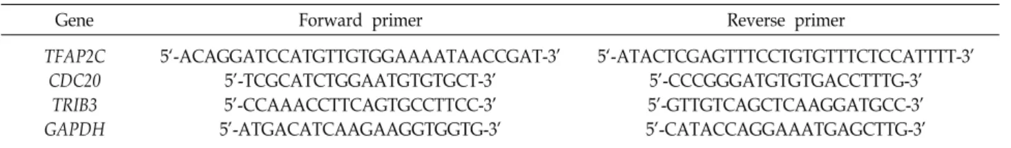
Table 1. Primers for determining expression levels of TFAP2C, CDC20 and TRIB3
Gene Forward primer Reverse primer
TFAP2C CDC20
TRIB3 GAPDH
5‘-ACAGGATCCATGTTGTGGAAAATAACCGAT-3’
5’-TCGCATCTGGAATGTGTGCT-3’
5’-CCAAACCTTCAGTGCCTTCC-3’
5’-ATGACATCAAGAAGGTGGTG-3’
5‘-ATACTCGAGTTTCCTGTGTTTCTCCATTTT-3’
5’-CCCGGGATGTGTGACCTTTG-3’
5’-GTTGTCAGCTCAAGGATGCC-3’
5’-CATACCAGGAAATGAGCTTG-3’
site and is required for cellular migration in colorectal cancer [1]. Accordingly, TFAP2C plays important regulatory roles for non-coding RNAs and functional coding-genes.
Recently, several studies have focused on finding novel oncogenes transcriptionally controlled by TFAP2C in NSCLC. We previously investigated several genes regulated by TFAP2C based on the analysis of a TFAP2C-related mi- croarray dataset [5, 9]. In the present study, among many genes screened, cell division cycle 20 (CDC20) and tribbles pseudokinase 3 (TRIB3) were selected as candidate onco- genes as they contribute to NSCLC cell viability and survival. Our results identify promising target genes for TFAP2C-mediated NSCLC tumorigenesis that may be useful prognostic biomarkers in NSCLC.
Materials and Methods
Antibodies and reagents
Antibodies specific for TFAP2C (sc-12762), a-tubulin (sc-23948), CDC20 (sc-13162) and TRIB3 (sc-365842) were purchased from Santa Cruz Biotechnology (Santa Cruz, CA, USA). Cell culture media (RPMI-1640), fetal bovine serum (FBS), penicillin and streptomycin were acquired from Gibco (Grand Island, NY, USA). Small interfering RNA (siRNA) specific for TFAP2C (sc-29696) and control siRNA (sc-37007) was purchased from Santa Cruz Biotechnology.
Cell lines and cell culture
The human NSCLC cell lines, NCI-H292 and NCI-H838 were acquired from the American Type Culture Collection (ATCC, Manassas, VA), authenticated, and then maintained in early passages for no more than 6 months after receipt from ATCC. Both were grown in RPMI-1640 supplemented with 10% FBS, 100 U/ml penicillin and 100 mg/ml strepto- mycin at 37℃ in a 5% CO2/95% air atmosphere.
Transient transfection and real-time quantitative RT-PCR
Plasmids containing a full-length CDC20 or TRIB3 con-
struct (pCMV-SPORT6-CDC20 and pCMV-SPORT6-TRIB3, respectively) were obtained from Dharmacon (Chicago, IL, USA). Cells were plated in 60 mm dishes before being tran- siently transfected with si-TFAP2C or control siRNA by us- ing Lipofectamine RNAiMAX (Invitrogen, Carlsbad, CA, USA). After 24 hr, cells were additionally transfected with plasmids containing CDC20 gene or TRIB3 gene by using Lipofectamine 3000 (Invitrogen). Cells were collected 48 hr after transfection and subjected to in vitro functional analyses.
To analyze mRNA levels [8], total RNA was isolated from cells using TRIzol (Invitrogen), and to obtain cDNA, the iso- lated RNA was converted using an MMLV Reverse Tran- scriptase system (Bioneer, Daejeon, Republic of Korea), ac- cording to the manufacturer’s protocol. The cycle parameters used for reverse transcription (RT) were 25℃ for 5 min, 37℃
for 60 min, 95℃ for 5 min, and then held at 4℃ until used.
Each RT product was used as a template for real-time quan- titative PCR, which was performed using the specific pri- mers detailed in Table 1. Aliquots of a master mix containing all reaction components and primers were dispensed into a real-time PCR plate (Applied Biosystems, Foster City, CA, USA). PCR was performed using reagents from a SYBR Green Core Reagent Kit (Applied Biosystems). mRNA levels were measured in triplicate in the reaction plate. Real-time qRT-PCR was performed using an Applied Biosystems-7900 HT qRT-PCR Instrument (Applied Biosystems) over 40 cy- cles of 15 s at 95℃ and 1 min at 60℃, after which samples were subjected to thermal denaturation. Gene expression levels were normalized versus GAPDH mRNA levels using the 2−ΔΔCT method [12]. To simplify data presentation, rela- tive expression values were multiplied by 102.
Western blot analysis
After experimental treatments, Western blotting was per- formed as previously described [6]. Briefly, whole-cell ly- sates and tissue lysates were obtained by using an EzRIPA Lysis kit (ATTO, Tokyo, Japan), which contained 20 mM HEPES, pH 7.5, 150 mM NaCl, 1% NP-40, 0.1% SDS, 0.5%
deoxycholic acid, and protease inhibitor and phosphatase in-
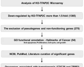
Fig. 1. Schematic of the microarray data (GSE79228)-based proc- ess used to identify TFAP2C-target genes that function as oncogenes.
hibitor cocktails. Protein concentrations in lysates were de- termined by using a BioRad protein assay kit (BioRad Laboratories, Hercules, CA, USA). Proteins were separated by SDS-PAGE and then transferred to nitrocellulose mem- branes, which were blocked with 5% bovine serum albumin in TBST (10 mM Tris, 100 mM NaCl, and 0.1% Tween 20) for 1 hr at room temperature. Membranes were incubated at 4℃ overnight with specific primary antibodies and then probed with peroxidase-conjugated secondary antibodies (Santa Cruz) for 1 hr at room temperature. Blots were vi- sualized by using an ECL detection system (Abfrontier, Seoul, Republic of Korea). Data acquisition and densito- metric analysis were performed using an iBright CL1000 imaging system (Thermo Fisher Scientific, Waltham, MA, USA).
Cell viability assay
Cells (5×10⁴) were cultured in 35-mm dishes for 24 hr, and treated with gene overexpression and/or siRNA transfection. They were detached with 10% trypsin-EDTA, washed with PBS, resuspended in PBS, and diluted 1:1 with trypan blue solution (Gibco). Cell viabilities were calculated by expressing the number of viable cells (not stained with trypan blue). Cell viability was also measured using thia- zolyl blue tetrazolium bromide solution (Sigma, St. Louis, MO) as previously described [10]. For experiments, treated cells were cultured in 96-well plates (1,000 cells per well) for 24 hr. Media were then removed, 0.05% thiazolyl blue tetrazolium bromide solution (Sigma) was added, and cells were incubated at 37℃ for 2 hr. The thiazolyl blue tetrazo- lium bromide solution was then replaced with dimethyl sulf- oxide and the cells were incubated for 10 min. After in- cubation, the solution was aliquoted into 96-well plates in triplicate, and absorbance was measured at 570 nm.
Apoptosis assay
A Caspase-Glo 3/7 assay kit (Promega, Madison, WI) was used to detect apoptosis [17]. Briefly, treated cells (104 cell per ml) in 100 μl of culture medium were transferred to a 96-well microplate, 100 μl of Caspase-Glo 3/7 reagent, which contained caspase 3/7 substrates, was added to each well.
Well contents were gently mixed at 300 to 500 rpm for 30 s, and incubated at room temperature for 1 hr. Sample lumi- nescence was measured using a Glomax multidetection sys- tem (Promega). Apoptosis was also assayed by measuring ATP levels using a CellTiter-Glo Assay Kit (Promega).
Briefly, treated cells (104 cells per ml) in 100 μl of culture medium were transferred to a 96-well microplate, 100 μl of CellTiter-Glo reagent (Promega) was added to each well.
Well contents were gently mixed at 300 to 500 rpm for 2 min, and the plate was incubated at room temperature for 10 min. The luminescence of each sample was measured us- ing a Glomax multidetection System (Promega).
Statistical analysis
Results are presented as the means ± standard deviations (SDs) at least three independent experiments and sample sizes were calculated to allow significance to be determined.
Experimental results were analyzed by one-way ANOVA for ranked data followed by Tukey's honestly significant differ- ence test. Prism 5 software (GraphPad Software, San Diego, CA, USA) was used to conduct the statistical analysis, and statistical significance was accepted for p values of <0.05.
Results and Discussion
Analysis of microarray gene profiling for TFAP2C- induced oncogenes
Based on the results of our previous studies on the roles played by TFAP2C in lung tumorigenesis [5, 9], we con- ducted gene profiling analysis to identify TFAP2C-induced oncogenes using a microarray dataset deposited in the Gene Expression Omnibus database (GEO Series accession num- ber GSE79228). Since we were interested in potential TFAP2C-
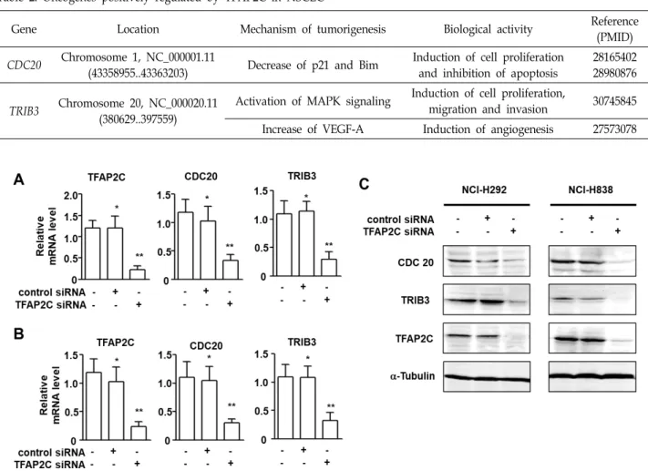
Table 2. Oncogenes positively regulated by TFAP2C in NSCLC
Gene Location Mechanism of tumorigenesis Biological activity Reference
(PMID) CDC20 Chromosome 1, NC_000001.11
(43358955..43363203) Decrease of p21 and Bim Induction of cell proliferation and inhibition of apoptosis
28165402 28980876
TRIB3 Chromosome 20, NC_000020.11 (380629..397559)
Activation of MAPK signaling Induction of cell proliferation,
migration and invasion 30745845 Increase of VEGF-A Induction of angiogenesis 27573078
Fig. 2. TFAP2C knockdown down-regulated the expression levels of CDC20 and TRIB3 in NSCLC cells. (A) Levels of CDC20 and TRIB3 mRNAs in TFAP2C siRNA-treated NCI-H292 cells were determined by real-time qRT-PCR. *p<0.05 compared with non-treated control NCI-H292 cells; **p<0.05 compared with NCI-H292 cells treated with control siRNA. (B) Levels of CDC20 and TRIB3 mRNAs in TFAP2C siRNA-treated NCI-H838 cells were determined by real-time qRT-PCR. *p<0.05 compared with non-treated control NCI-H838 cells; **p<0.05 compared with NCI-H838 cells treated with control siRNA. (C) Protein levels of CDC20 and TRIB3 in TFAP2C siRNA-treated NCI-H292 and NCI-H838 cells were assessed by Western blotting.
regulated oncogenes, we screened for genes down-regulated by TFAP2C knockdown (Fig. 1). Of the 1385 genes down- regulated by more than 1.5-fold, we excluded pseudogenes and non-functional genes and focused on the roles of func- tional genes based on hallmarks of cancer as indicated by GO annotation (e.g., proliferation, cell growth, cell cycle pro- motion, and anti-apoptosis). Of the remaining 54 genes, CDC20 and TRIB3 were selected for further study because they have been reported to play potent oncogenic roles in many types of solid cancers (Table 2).
Up-regulations of CDC20 and TRIB3 by TFAP2C in NSCLC cells
Based on a report of TFAP2C in NSCLC cells [5], we ob-
served that the mRNA levels of CDC20 and TRIB3 in TFAP2C siRNA-treated NSCLC cells (NCI-H292 and NCI-H838 cells) were assessed by real-time qRT-PCR (Fig. 2A, Fig. 2B). The mRNA levels of CDC20 and TRIB3 and that of TFAP2C were found to be significantly down-regulated after treating NCI-H292 and NCI-H838 cells with TFAP2C siRNA. Fur- thermore, the protein levels of CDC20 and TRIB3 were also down-regulated in both cell types (Fig. 2C). These results indicate that TFAP2C was directly responsible for the ex- pression of CDC20 and TRIB3 in NSCLC cells.
Oncogenic effects of the up-regulations of CDC20 and TRIB3 by TFAP2C on the cell viability of NSCLC cells
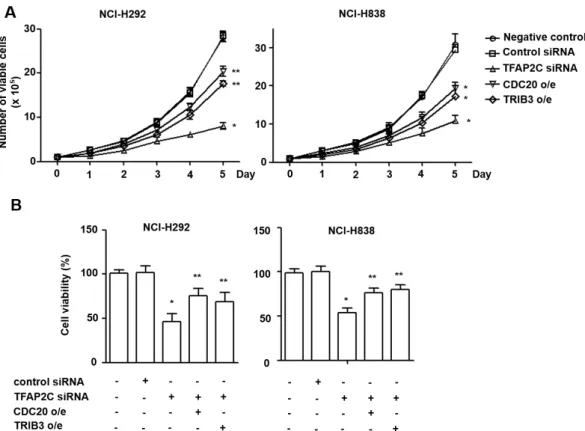
Fig. 3. The oncogenic effects of CDC20 and TRIB3 on NSCLC cell proliferation. (A-B) The effects of CDC20 or TRIB3 overexpression on the proliferation of NCI-H292 and NCI-H838 cells were determined using trypan blue (A) and thiazolyl blue tetrazolium bromide (B) cell viability assays. *p<0.05 compared with NCI-H292 or NCI-H838 cells treated with control siRNA; **p<0.05 compared with NCI-H292 or NCI-H838 cells treated with TFAP2C siRNA.
It has been reported on several occasions that CDC20 and TRIB3 play crucial roles in the survival and proliferation of many cancer cell types [3, 4, 13, 15, 18]. We analyzed the proliferative effects of CDC20 and TRIB3 up-regulations on NCI-H292 or NCI-H838 cells using cell viability assays.
Twenty four hours after transient transfection with TFAP2C siRNA, cells were transfected with plasmids containing CDC20 gene or TRIB3 gene. Trypan blue assays showed numbers of viable TFAP2C-knocked down NSCLC cells were significantly less than control cell numbers at day 5.
Furthermore, this reduction in TFAP2C-knocked down cell survival was prevented by overexpressing CDC20 or TRIB3 (Fig. 3A and B). In addition, the cell viability assay using thiazolyl blue tetrazolium bromide solution also showed that TFAP2C-knocked down cells were less viable than con- trol cells, and that this viability reduction was prevented by CDC20 or TRIB3 overexpression (Fig. 3C and D). Thus, these results indicate that CDC20 and TRIB3 are positively regu- lated by TFAP2C and are required for NSCLC cell pro- liferation.
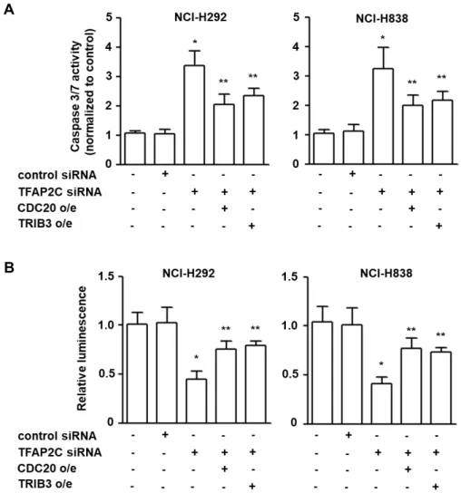
The inhibitory effects of CDC20 and TRIB3 up-reg- ulation by TFAP2C on the apoptosis of NSCLC cells To support the oncogenic roles of CDC20 and TRIB3 in NSCLC tumorigenesis, we investigated whether the expre- ssion levels of CDC20 and TRIB3 were associated with the inhibition of apoptosis by examining the activation of cas- pase 3/7. The activities of caspase 3/7 in NCI-H292 and NCI-H838 cells were increased by TFAP2C siRNA as com- pared with control cells (Fig. 4A and B). However, over- expression of CDC20 or TRIB3 significantly reduced caspase 3/7 activities in TFAP2C siRNA-treated NSCLC cells. In ad- dition, we measured ATP levels to support the apoptosis induced by caspase 3/7 activation [7], because the ATP pro- duction stops when apoptosis is initiated and ATP levels rapidly diminish. ATP levels were significantly lower in TFAP2C-knocked down NCI-H292 and NCI-H838 cells, but overexpression of CDC20 or TRIB3 in TFAP2C-knocked down cells inhibited these reductions in ATP levels (Fig. 4C and D). These findings show that the up-regulations of CDC20 and TRIB3 induced by TFAP2C activation are neg- atively associated with the apoptosis of NSCLC cells, which
A
B
Fig. 4. The inhibitory effects of CDC20 and TRIB3 on NSCLC cell apoptosis. (A) The effects of CDC20 or TRIB3 overexpression on NCI-H292 and NCI-H838 cell apoptosis were determined using a caspase 3/7 assay (A) and an ATP assay (B). *p<0.05 compared with NCI-H292 or NCI-H838 cells treated with control siRNA; **p<0.05 compared with NCI-H292 or NCI-H838 cells treated with TFAP2C siRNA.
potentially contribute to NSCLC tumorigenesis.
Many studies have been undertaken to identify promising target genes and to elucidate the molecular mechanisms un- derlying NSCLC tumorigenesis. In this study, we found that TFAP2C may positively participate in NSCLC tumorigenesis by up-regulating the oncogenes CDC20 and TRIB3. It has been reported that CDC20 and TRIB3 are direct targets of TFAP2C and that their expressions may be closely associated with cell proliferation and have anti-apoptotic effects in NSCLC although further studies for the apoptosis inhibitory pathway mediated by CDC20 and TRIB3 would be required.
We suggest that CDC20 and TRIB3 be considered putative oncogenes for NSCLC development and promising prog- nostic markers and therapeutic targets in NSCLC.
Acknowledgement
This work was supported by the 2017 New Professor Research Grant funded by Korea National University of Education.
References
1. Barbagallo, C., Brex, D., Caponnetto, A., Cirnigliaro, M., Scalia, M., Magnano, A., Caltabiano, R., Barbagallo, D., Biondi, A., Cappellani, A., Basile, F., Di Pietro, C., Purrello, M. and Ragusa, M. 2018. LncRNA UCA1, upregulated in CRC biopsies and downregulated in serum exosomes, con- trols mRNA expression by RNA-RNA interactions. Mol.
Ther. Nucleic Acids 12, 229-241.
2. De Andrade, J. P., Park, J. M., Gu, V. W., Woodfield, G.
W., Kulak, M. V., Lorenzen, A. W., Wu, V. T., Van Dorin, S. E., Spanheimer, P. M. and Weigel, R. J. 2016. EGFR is
regulated by TFAP2C in luminal breast cancer and is a tar- get for Vandetanib. Mol. Cancer Ther. 15, 503-511.
3. Dong, S., Xia, J., Wang, H., Sun, L., Wu, Z., Bin, J., Liao, Y., Li, N. and Liao, W. 2016. Overexpression of TRIB3 pro- motes angiogenesis in human gastric cancer. Oncol. Rep. 36, 2339-2348.
4. Hong, B., Zhou, J., Ma, K., Zhang, J., Xie, H., Zhang, K., Li, L., Cai, L., Zhang, N., Zhang, Z. and Gong, K. 2019.
TRIB3 promotes the proliferation and invasion of renal cell carcinoma cells via activating MAPK signaling pathway. Int.
J. Biol. Sci. 15, 587-597.
5. Kang, J., Kim, W., Lee, S., Kwon, D., Chun, J., Son, B., Kim, E., Lee, J. M., Youn, H. and Youn, B. 2017. TFAP2C pro- motes lung tumorigenesis and aggressiveness through miR- 183- and miR-33a-mediated cell cycle regulation. Oncogene 36, 1585-1596.
6. Kang, J., Kim, W., Seo, H., Kim, E., Son, B., Lee, S., Park, G., Jo, S., Moon, C., Youn, H. and Youn, B. 2018. Radiation- induced overexpression of transthyretin inhibits reti- nol-mediated hippocampal neurogenesis. Sci. Rep. 8, 8394.
7. Kangas, L., Gronroos, M. and Nieminen, A. L. 1984. Biolu- minescence of cellular ATP: a new method for evaluating cytotoxic agents in vitro. Med. Biol. 62, 338-343.
8. Kim, E., Kim, W., Lee, S., Chun, J., Kang, J., Park, G., Han, I., Yang, H. J., Youn, H. and Youn, B. 2017. TRAF4 promotes lung cancer aggressiveness by modulating tumor micro- environment in normal fibroblasts. Sci. Rep. 7, 8923.
9. Kim, W., Kim, E., Lee, S., Kim, D., Chun, J., Park, K. H., Youn, H. and Youn, B. 2016. TFAP2C-mediated upregula- tion of TGFBR1 promotes lung tumorigenesis and epi- thelial-mesenchymal transition. Exp. Mol. Med. 48, e273.
10. Kim, W., Youn, H., Lee, S., Kim, E., Kim, D., Sub Lee, J., Lee, J. M. and Youn, B. 2018. RNF138-mediated ubiquitina-
tion of rpS3 is required for resistance of glioblastoma cells to radiation-induced apoptosis. Exp. Mol. Med. 50, e434.
11. Kolat, D., Kaluzinska, Z., Bednarek, A. K. and Pluciennik, E. 2019. The biological characteristics of transcription factors AP-2alpha and AP-2gamma and their importance in various types of cancers. Biosci. Rep. 39, pii: BSR20181928.
12. Livak, K. J. and Schmittgen, T. D. 2001. Analysis of relative gene expression data using real-time quantitative PCR and the 2(-delta delta C(T)) method. Methods 25, 402-408.
13. Miyoshi, N., Ishii, H., Mimori, K., Takatsuno, Y., Kim, H., Hirose, H., Sekimoto, M., Doki, Y. and Mori, M. 2009.
Abnormal expression of TRIB3 in colorectal cancer: a novel marker for prognosis. Br. J. Cancer 101, 1664-1670.
14. Park, J. M., Wu, T., Cyr, A. R., Woodfield, G. W., De Andrade, J. P., Spanheimer, P. M., Li, T., Sugg, S. L., Lal, G., Domann, F. E., Zhang, W. and Weigel, R. J. 2015. The role of Tcfap2c in tumorigenesis and cancer growth in an activated Neu model of mammary carcinogenesis. Oncogene 34, 6105-6114.
15. Shang, G., Ma, X. and Lv, G. 2018. Cell division cycle 20 promotes cell proliferation and invasion and inhibits apop- tosis in osteosarcoma cells. Cell Cycle 17, 43-52.
16. Siegel, R. L., Miller, K. D. and Jemal, A. 2019. Cancer sta- tistics, 2019. CA Cancer J. Clin. 69, 7-34.
17. Tiwari, M., Prasad, S., Tripathi, A., Pandey, A. N., Ali, I., Singh, A. K., Shrivastav, T. G. and Chaube, S. K. 2015.
Apoptosis in mammalian oocytes: a review. Apoptosis 20, 1019-1025.
18. Zhang, Y., Xue, Y. B., Li, H., Qiu, D., Wang, Z. W. and Tan, S. S. 2017. Inhibition of cell survival by curcumin is associated with downregulation of cell division cycle 20 (Cdc20) in pancreatic cancer cells. Nutrients 9, E109.
초록:비소세포폐암 발달 과정에서 TFAP2C에 의해 발현되는 CDC20과 TRIB3의 원암유전자 기능에 관한 연구
김다인1․도현희1․강지훈2․윤부현2,3․김완연1,4*
(1한국교원대학교 일반대학원 과학교육학과, 2부산대학교 일반대학원 생명시스템학과, 3부산대학교 자연과학대학
생명과학과, 4한국교원대학교 제3대학 생물교육학과)
전세계적으로 폐암 발병율은 서서히 감소하는 추세이지만, 여전히 암 관련 사망의 주요 원인으로 지목되고 있 으며, 이에 따라 폐암 진단과 치료를 위한 새로운 분자적 지표를 발굴하는 연구가 활발히 이루어지고 있다. 본 연구진이 수행한 기존 연구에 따르면 폐암 환자에게서는 전사인자 중 하나인 TFAP2C가 높은 비율로 발현되며, 이 전사인자를 통해 폐암 발달에 상당한 영향을 끼치는 것을 확인할 수 있었다. TFAP2C는 다른 유전자들의 발현 을 조절하여 암 형성에 기여하게 된다. 마이크로어레이 분석을 통해 TFAP2C에 의해 발현양이 조절되는 잠재적 표적 유전자들을 확인하였고, 특히 TFAP2C siRNA를 처리하였을 때 발현이 감소되는 원암유전자들 중 CDC20과 TRIB3 유전자를 최종적으로 선별하였다. 리얼타임 qRT-PCR과 웨스턴블롯을 통하여 두 유전자가 TFAP2C에 의존 적으로 발현됨을 확인하였으며, 세포 생존 분석법을 통하여 CDC20과 TRIB3의 발현 증가가 폐암세포의 세포 증식 을 유의미하게 유도하는 것을 확인하였다. 이와 더불어, CDC20과 TRIB3의 과발현이 폐암세포의 세포사멸 수준을 감소시켜 폐암 형성에 관여함을 확인하였다. 본 연구를 통하여 CDC20과 TRIB3가 폐암 형성을 유도할 수 있는 잠재적인 원암유전자로 기능함을 밝힐 수 있었으며, 두 유전자가 폐암 진단을 위한 표적유전자로서의 역할을 수 행할 수 있을 것으로 기대한다.