Long Non-Coding RNA TUG1 Promotes Proliferation and Inhibits Apoptosis of Osteosarcoma Cells
by Sponging miR-132-3p and Upregulating SOX4 Expression
Gang Li, Keyu Liu, and Xinhui Du
Department of Orthopedics, The First Affiliated Hospital of the Medical College, Shihezi University, Shihezi, China.
Purpose: Long non-coding RNA taurine upregulated gene 1 (TUG1) is reported to be a vital regulator of the progression of various cancers. This study aimed to explore the exact roles and molecular mechanisms of TUG1 in osteosarcoma (OS) development.
Materials and Methods: Real-time quantitative PCR was applied to detect the expressions of TUG1 and microRNA-132-3p (miR- 132-3p) in OS tissues and cells. Western blot was performed to measure protein levels of sex determining region Y-box 4 (SOX4).
Cell viability was assessed using XTT assay. Cell apoptosis was evaluated using flow cytometry and caspase-3 activity detection as- says. Bioinformatics analysis and luciferase reporter experiments were employed to confirm relationships among TUG1, miR-132- 3p, and SOX4.
Results: TUG1 was highly expressed in human OS tissues, OS cell lines, and primary OS cells. TUG1 knockdown hindered prolif- eration and induced apoptosis in human OS cell lines and primary OS cells. Moreover, TUG1 inhibited miR-132-3p expression by direct interaction, and introduction of miR-132-3p inhibitor partly abrogated the effect of TUG1 knockdown on the proliferation and apoptosis of OS cells. Furthermore, SOX4 was validated as a target of miR-132-3p. Further functional analyses revealed that miR-132-3p inhibited proliferation and induced apoptosis of OS cells, while this effect was greatly abated following SOX4 overex- pression. Moreover, TUG1 knockdown suppressed proliferation and promoted apoptosis by upregulating miR-132-3p and down- regulating SOX4 in primary OS cells.
Conclusion: TUG1 facilitated proliferation and suppressed apoptosis by regulating the miR-132-3p/SOX4 axis in human OS cell lines and primary OS cells. This finding provides a potential target for OS therapy.
Key Words: Osteosarcoma, TUG1, miR-132-3p, SOX4
INTRODUCTION
Osteosarcoma (OS), a primary bone malignant tumor, is the
second leading cause of cancer-related death in children and young adults.1 Although advancements been made in the di- agnosis and treatment of OS, survival rates for metastatic or re- current OS patients are still very poor.2 Therefore, it is essential and urgent to further explore the mechanisms underlying OS development in order to find out novel diagnostic or prognos- tic biomarkers and effective therapeutic agents.
A growing amount of evidence indicates that aberrant ex- pression of long non-coding RNAs (lncRNAs) and microRNAs (miRNAs) is closely correlated with the development of vari- ous diseases, including OS.3-7 Some studies have also suggest- ed that lncRNAs could act as competing endogenous RNAs (ceRNAs) to modulate the expression of miRNAs and miRNAs target genes.8,9 These lncRNAs were found to exert their func- Received: September 25, 2017 Revised: November 29, 2017
Accepted: December 28, 2017
Corresponding author: Dr. Gang Li, Department of Orthopedics, The First Affiliated Hospital of the Medical College, Shihezi University, No.37 North Second Road, Shihezi 832008, China.
Tel: +86-0993-2858434, Fax: +86-0993-2850400, E-mail: [email protected]
•The authors have no financial conflicts of interest.
© Copyright: Yonsei University College of Medicine 2018
This is an Open Access article distributed under the terms of the Creative Com- mons Attribution Non-Commercial License (http://creativecommons.org/licenses/
by-nc/4.0) which permits unrestricted non-commercial use, distribution, and repro- duction in any medium, provided the original work is properly cited.
pISSN: 0513-5796 · eISSN: 1976-2437 Yonsei Med J 2018 Mar;59(2):226-235
https://doi.org/10.3349/ymj.2018.59.2.226
tions by miRNA response elements, which could absorb en- dogenous miRNAs like sponges, thereby relieving the repres- sion effect of miRNAs on their target messenger RNAs (mRNAs).8 Taurine upregulated gene 1 (TUG1), a lncRNA, could act as an oncogene or a tumor suppressor in the development and progression of various cancers. For example, TUG1 has been found to play carcinogenic roles, accompanied by a high-level expression, in some cancers, including esophageal squamous cell cancer and bladder urothelial cancer.10,11 However, in some cancers, such as non-small cell lung cancer, TUG1 has been shown to act as a tumor suppressor with low-level ex- pression.12 These studies indicate that TUG1 may be cancer type specific and that different tumor microenvironments might impact TUG1 activity.
In recent years, studies have revealed the critical roles of TUG1 in the progression of OS: Ma, et al.13 reported that TUG1 expression was up-regulated in OS and that high-level expres- sion of TUG1 was closely correlated with poor prognosis and disease status in OS. Moreover, Zhang, et al.14 demonstrated that down-regulation of TUG1 inhibited proliferation and in- duced apoptosis of OS cells, indicating that TUG1 acts as an oncogene in OS. However, the exact roles and molecular mech- anisms of TUG1 underlying OS progression have not been thoroughly elucidated.
In the present study, we identified that TUG1 is highly ex- pressed in human OS tumor tissues, cell lines, and primary OS cells. Moreover, TUG1 facilitated cell proliferation and sup- pressed apoptosis by sequestering miR-132-3p from its target gene sex determining region Y-box 4 (SOX4) in OS cell lines and primary OS cells.
MATERIALS AND METHODS
Patient tissue samples and OS cell culture
OS tumor tissue and matched adjacent normal tissue were col- lected from 22 patients diagnosed with primary OS at the First Affiliated Hospital of the Medical College, Shihezi University.
This study was performed with the approval of the Research Medical Ethics Committee of the First Affiliated Hospital of the Medical College, Shihezi University. Each patient signed writ- ten informed consent prior to enrolling in this medical study.
Human OS cell lines (U2OS, MG-63, Saos-2, and 143B) and the human normal osteoblastic cell line FOB1.19, together with Human Embryonic Kidney 293 cells (HEK293), were obtained from American Type Culture Collection (ATCC, Rockville, MD, USA). U2OS and 143B cells were cultured in RPMI-1640 (Gib- co Co., New York, NY, USA) medium supplemented with 10%
fetal bovine serum (FBS, Invitrogen, Carlsbad, CA, USA). MG- 63 were grown in MEM medium (Gibco) containing 10% FBS (Invitrogen). Saos-2 cells were cultured in McCoy’s 5A medium (Sigma-Aldrich, St. Louis, MO, USA) containing 15% FBS (Invit- rogen). The human normal osteoblastic cell line hFOB 1.19
was maintained in DMEM/F-12 medium (Gibco) supple- mented with 10% FBS (Invitrogen). HEK293 cells were main- tained in DMEM (Gibco) medium containing 10% FBS (Invit- rogen). All cells were maintained in humidified incubator containing 5% CO2 at 37°C.
Establishment of a primary OS cell line
Fresh OS tumor tissue obtained from patients with primary spontaneous OS was washed using sterile phosphate-buff- ered saline three times and then minced into small tumor pieces. Then, tumor samples were digested in digestion medi- um [DMEM/F-12 medium (Gibco) containing 0.1 mg/mL of hyaluronidase (Sigma-Aldrich), 2 mg/mL of collagenase A (Sig- ma-Aldrich), 60 U/mL of nystatin (Sigma-Aldrich), and 0.1 mg/
mL of gentamicin (Sigma-Aldrich)] for almost 50 min at 37°C.
Then, cells were cultured in DMEM/F12 medium (Gibco) sup- plemented with 10% FBS (Invitrogen) in a humidified incuba- tor containing 5% CO2 at 37°C.
Reagents and antibodies
Small interference RNA (siRNA) targeting TUG1 (si-TUG1) and its scramble control (si-NC), miR-132-3p mimic and its negative control (miR-NC), miR-132-3p inhibitor (anti-miR-132-3p) and its control anti-miR-NC, and SOX4 siRNA (si-SOX4) and its scramble control (si-NC) were obtained from GenePharma Co., Ltd. (Shanghai, China). TUG1 and SOX4 cDNA sequences were cloned into pcDNA3.1 (Invitrogen) to produce TUG1 and SOX4 overexpression vectors (pcDNA3.1-TUG1 and pcDNA3.1-SOX4), named TUG1 and SOX4.
Cell transfection
The MG-63, U2OS, and primary OS cells were transfected with miRNA, siRNA, miRNA inhibitors, and plasmids by Lipofe- ctamine 2000 (Invitrogen) according to the manufacturer’s in- structions, and the cells were harvested at the indicated time points after transfection.
RNA extraction and real time PCR analysis
Total RNA from the tissues and cells was obtained by TRIzol so- lution (Invitrogen) in accordance with the manufacturer’s pro- tocols. cDNA was synthesized by the PrimeScript RT reagent kit (TaKaRa, Tokyo, Japan). Quantitative PCR analysis of TUG1 was performed using the SYBR® Premix Ex TaqTM reagent (Ta- KaRa), with β-actin as an endogenous control. The reverse tr- anscription and quantification of miR-132-3p was carried out using the miDETECTA TrackTM miRNA qRT-PCR Starter kit (Ri- boBio, Guangzhou, China), referring to the manufacturer’s pro- tocols, with U6 snRNA as an internal normalization reference.
The real-time quantitative PCR (RT-qPCR) primers sequenc- es were as follows: TUG1, 5'-CTGAAGAAAGGCAACATC-3' (forward) and 5'-GTAGGCTACTACAGGATTTG-3' (reverse);
β-actin, 5'-AGTGTGACGTGGACATCCGCAAAG-3' (forward) and 5'-ATCCACATCTGCTGGAAGGTGGAC-3' (reverse). Re-
verse transcription primers, 5'-GTCGTATCCAGTGCAG GGTCCGAGGTATTCGCACTGGATACGACCGACCATG-3' (miR-132-3p); 5'-AAAATATGGAACGCTTCACGAATTTG-3' (U6). Quantification primers, miR-132-3p, 5'-GCGCGCGTAA CAGTCTACAGC-3' (forward) and 5'-GTCGTATCCAGTGCAG GGTCC-3' (reverse); U6, 5'-CTCGCTTCGGCAGCACATA TACT-3' (forward) and 5'-CGCTTCACGAATTTGCGTGT-3' (reverse).
Western blot assays
Cells from different treatment conditions were obtained 48 hours after transfection, and western blot assays were carried out with the SOX4 and β-actin antibodies (Santa Cruz Bio- technology, Dallas, TX, USA) according to manufacturer’s pro- tocols. Briefly, MG-63 and U2OS cells were lysed using RIPA lysis buffer (Beyotime, Shanghai, China) containing cocktail (Roche, Mannheim, Germany) to obtain the whole protein. The protein concentration was measured using PierceTM BCA Pro- tein Assay Kit (Thermo scientific, Rockford, IL, USA). Then, equal weights (50 μg) of proteins were separated using SDS- PAGE gel, followed by being transferred to PVDF membranes (Millipore, Billerica, MA, USA). Next, the membranes were blocked for 1 h with 5% skimmed milk and incubated with ap- propriate concentrations of antibodies against SOX4 and β-actin antibodies (Santa Cruz Biotechnology) overnight at 4°C. On the next day, the membranes were incubated with horserad- ish peroxidase-conjugated secondary antibody for another 1 h.
Finally, protein signals were detected using the BeyoECL Plus kit (Beyotime) and quantified with Image J software (National Institutes of Health, Bethesda, MD, USA).
XTT assays
MG-63, U2OS, and primary OS cells were seeded into 96-well plates and transfected using Lipofectamine 2000 reagent.
Then, cell proliferation was assessed using the XTT Cell Prolif- eration Assay Kit (Abnova, Taipei, Taiwan) following the man- ufacturer’s protocols. Briefly, 10 μL of XTT was added into 96- well plates at indicated time points (0, 24, 48, and 72 h) after transfection and then incubated for another 3 h at 37°C incu- bator. Finally, absorbance was measured at the wavelength of 450 nm.
Luciferase assays
Partial DNA sequences of TUG1 and SOX4 3’-UTR containing wild-type (WT) or mutant (MUT) miR-132-3p binding sites were amplified by PCR and then cloned into pmiR-RB-RE- PORTTM vectors (RiboBio) to produce WT-TUG1, MUT-TUG1, WT-SOX4-3’UTR, and MUT-SOX4-3’UTR reporter plasmids.
Then, constructed reporter plasmids were respectively co- transfected with miR-132-3p or miR-NC into 293T cells using Lipofectamine 2000 (Invitrogen). Then, the luciferase activity in cells from different treatment conditions was measured by the Dual-Luciferase® Reporter Assay kit (Promega, Madison, WI,
USA) according to manufacturer’s instructions.
Apoptosis rate analysis
After transfection for 48 h, transfected cells were treated using the AnnexinV-FITC/PI Apoptosis Detection Kit (BD Bioscienc- es, Franklin Lakes, NJ, USA), referring to manufacturer’s instruc- tions, and then, flow cytometry (FACScan; BD Biosciences) was used to determine cell apoptosis rates.
Caspase-3 activity assays
MG-63 and U2OS cells were collected 48 h after transfection, and caspase-3 activity in these cells were measured using the Caspase 3 Activity Assay Kit (Beyotime), referring to the man- ufacturer’s protocols.
Statistical analysis
All data were obtained from at least three independent experi- ments and are reported as means±standard deviations. One- way analysis of variance or Student’s t-test was conducted to analyze difference therein. Differences were regarded as sta- tistically significant when p<0.05.
RESULTS
TUG1 expression is upregulated in OS tumors and cells
To determine the expression pattern of TUG1 in OS tissues, 22 pairs of OS tumor tissues and correspondingly adjacent nor- mal tissues were collected, followed by the detection of TUG1 levels using RT-qPCR assays. TUG1 expression was remark- ably upregulated in 17 (77.3%) OS tumor tissues, compared to correspondingly normal tissues, together with a slight down- regulation in the remaining (22.7%) OS tumor tissues (Fig. 1A).
The overall expression of TUG1 was upregulated in OS tumor tissues, compared with normal tissues (Fig. 1B). RT-qPCR re- sults revealed that TUG1 was highly expressed in OS cell lines (MG-63, U2OS, Saos-2, and 143B), compared with the human osteoblast cell line hFOB 1.19 (Fig. 1C). All these results indi- cated that the expression of TUG1 in OS tumors and cells is sig- nificantly upregulated, which is in accordance with previous results.14,15
TUG1 knockdown suppresses proliferation and
induces apoptosis in OS cells (MG-63 and U2OS)
To investigate the roles of TUG1 in OS cell lines, TUG1 overex- pression plasmid (pcDNA3.1-TUG1, TUG1) and siRNA of TUG1 (si-TUG1) were constructed or synthesized. Then, the transfection efficiency of TUG1 and si-TUG1 was respectively assessed by RT-qPCR assays. As shown in Fig. 1D, the trans- fection of pcDNA3.1-TUG1 resulted in a noticeable upregula- tion of TUG1 expression, compared with the negative control (vector), in MG-63 cells. We also observed that TUG1 expres-sion was strikingly downregulated in U2OS cells transfected with si-TUG1 in comparison with the transfection of non-spe- cific siRNA (si-NC) (Fig. 1E). These results revealed that the TUG1 overexpression plasmid and TUG1 siRNA could exert
its function in the following experiments.
Next, the effects of TUG1 on the proliferation and apoptosis of MG-63 and U2OS cells were further investigated. TUG1 and si-TUG1 were respectively transfected into MG-63 and U2OS
Fold changes of TUG1 level in OS/N, log2
4
3
2
1
0
-1
−
−
−
−
−
−
1 2 3 4 5 6 7 8 9 10 11 12 13 14 15 16 17 18 19 20 21 22
*
Relative TUG1 expression
Vector TUG1
MG-63
1
0 25 20 15 10
−
−
−
−
−
−
Relative TUG1 expression
FOB1.19 MG-63 U2OS Saos-2 143B
3
2
1
0
−
−
−
−
*
*
* *
Relative TUG1 expression
si-NC si-TUG1 1.2 U2OS
1.0 0.8 0.6 0.4 0.2 0.0
−
−
−
−
−
−
− A
C
F G
D
H
E
I
Apoptosis rate (%)
si-NC si-TUG1 20 U2OS
15
10
5
0
−
−
−
−
−
*
Relative caspase-3 activity
si-NC si-TUG1 5 U2OS
4
3
2
1
0
−
−
−
−
−
−
*
Relative cell viability
0 24 h 48 h 72 h 6 U2OS
5 4 3 2 1 0
−
−
−
−
−
−
−
* si-NC
si-TUG1
Relative cell viability
0 24 h 48 h 72 h MG-63 8
7 6 5 4 3 2 1 0
−
−
−
−
−
−
−
−
−
* Vector
TUG1
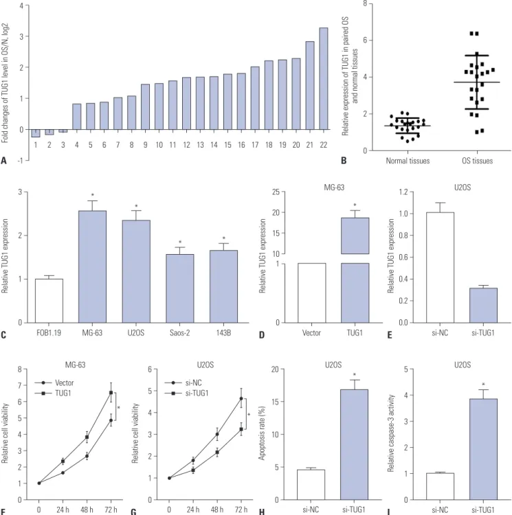
Fig. 1. TUG1 expression is up-regulated in OS tissues and cells, and TUG1 depletion hampers proliferation and induces apoptosis of OS cells. (A-E) Real-time quantitative PCR assays were employed to measure the expression of TUG1 in tissues and cells. (A) Expression of TUG1 in 22 cases of OS tissues with the expression in correspondingly adjacent normal tissues as a normalization standard. (B) Overall expression analysis of TUG1 in 22 pairs of OS tissues and adjacent normal tissues with β-actin as an internal control. (C) TUG1 expression in human normal osteoblastic cell line FOB1.19 and OS cell lines (U2OS, MG-63, Saos-2, 143B) with β-actin as an internal control. (D and E) The transfection efficiency of pcDNA3.1-TUG1 in MG-63 cells (D) and the knockdown efficiency of TUG1 siRNA in U2OS cells (E). (F and G) The effect of TUG1 overexpression (F) and knockdown (G) on OS cell proliferation was assessed by XTT assays at the indicated time point (0, 24, 48, and 72 h) after transfection. (H and I) The effect of TUG1 knockdown on U2OS cell apoptosis was evaluated by flow cytometry (H) and caspase-3 activity detection (I) after transfection 48 h. *p<0.05. TUG1, taurine upregulated gene 1; OS, osteosarcoma.
Relative expression of TUG1 in paired OS and normal tissues 8
6
4
2
0
−
−
−
−
−
Normal tissues OS tissues B
cells, and cells proliferation was assessed at 0, 24, 48, and 72 h by XTT assays (Fig. 1F and G). The results revealed that TUG1 overexpression significantly promoted MG-63 cell prolifera- tion (Fig. 1F) and that TUG1 knockdown markedly suppressed U2OS cell proliferation (Fig. 1G). The effect of TUG1 on U2OS cell apoptosis was confirmed by the flow cytometry and cas- pase-3 activity detection. As expected, the depletion of TUG1 by si-TUG1 induced increases in apoptosis (Fig. 1H) and cas- pase-3 activity (Fig. 1I) in U2OS cells.
Si-TUG1 exerts its anti-proliferation and pro-apoptosis effects partly by targeting miR-132-3p in OS cells
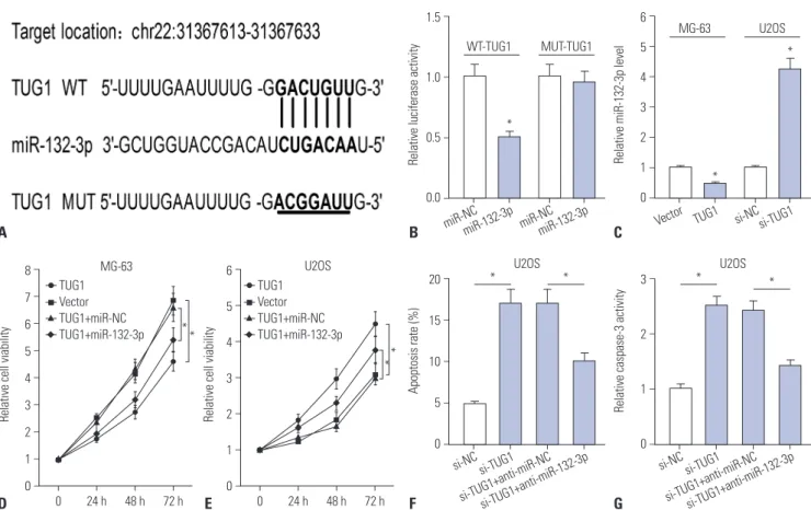
Emerging evidence indicates that lncRNAs act as ceRNAs of miRNAs to regulate target mRNAs expression.8,9 Therefore, bio- informatics prediction analysis was performed by the miRcode online website to search for potential target miRNAs of TUG1.Among candidate miRNAs, miR-132-3p was chosen due to its anti-tumor effect in OS (Fig. 2A).16,17 For validation thereof, WT- TUG1 (containing the putative miR-132-3p binding sites) and MUT-TUG1 (putative miR-132-3p binding sites mutated to the indicated sequences) luciferase reporter plasmids were constructed. Then, the effect of miR-132-3p on the luciferase
activity of the WT-TUG1 or MUT-TUG1 reporter plasmid was detected, and the results indicated that the introduction of miR-132-3p mimic significantly inhibits the luciferase activity of WT-TUG1 reporter, compared with miR-NC, whereas miR- 132-3p had no effect on the luciferase activity of MUT-TUG1 reporter in HEK293 cells (Fig. 2B). These finding suggested that the putative binding sites were indeed essential for the di- rect interaction of miR-132-3p and TUG1. RT-qPCR assays further demonstrated that enforced expression of TUG1 mark- edly suppressed miR-132-3p expression in MG-63 cells, while TUG1 knockdown facilitated miR-132-3p expression in U2OS cells (Fig. 2C). Thus, TUG1 inhibits the expression of miR-132- 3p by direct interaction in MG-63 and U2OS cells.
Next, we further explored whether miR-132-3p could influ- ence the effect of TUG1 on proliferation and apoptosis in OS cells. XTT assay results indicated that the restoration of miR- 132-3p partly abolished the promotion effect of TUG1 overex- pression on proliferation in MG-63 cells (Fig. 2D). Also, the in- troduction of miR-132-3p inhibitor partially abrogated si- TUG1-mediated anti-proliferative effects in U2OS cells (Fig.
2E). Moreover, the transfection of miR-132-3p inhibitor resulted in a marked decrease in apoptosis rate (Fig. 2F) and caspase-3
A B C
D E F G
Relative cell viability Relative cell viability
0 24 h 48 h 72 h 0 24 h 48 h 72 h
MG-63 U2OS
8 7 6 5 4 3 2 1 0
6 5 4 3 2 1 0
−
−
−
−
−
−
−
−
−
−
−
−
−
−
−
−
**
**
Fig. 2. si-TUG1 exerts its anti-proliferation and pro-apoptosis effect by targeting miR-132-3p in OS cells. (A) The putative binding sites between TUG1 and miR-132-3p and the mutation sites of TUG1 in the MUT-TUG1 reporter. (B) The effect of miR-132-3p on luciferase activity of WT-TUG1 or MUT- TUG1 reporter. (C) The effect of TUG1 overexpression and knockdown on miR-132-3p expression in MG-63 and U2OS cells. (D) The effect of miR-132- 3p on TUG1-mediated pro-proliferation effect in MG-63 cells. (E-G) The influence of miR-132-3p inhibitor on si-TUG1-mediated anti-proliferation (E) and pro-apoptosis (F and G) effect in U2OS cells. *p<0.05. TUG1, taurine upregulated gene 1; OS, osteosarcoma; WT-TUG1, wild-type TUG1; MUT- TUG1, mutant TUG1.
Relative luciferase activity
miR-NCmiR-132-3p miR-NCmiR-132-3p WT-TUG1 MUT-TUG1 1.5
1.0
0.5
0.0
−
−
−
−
*
Relative miR-132-3p level
Vector TUG1 si-NCsi-TUG1
MG-63 U2OS
6 5 4 3 2 1 0
−
−
−
−
−
−
−
*
*
Apoptosis rate (%)
si-NC
si-TUG1+anti-miR-NC si-TUG1
si-TUG1+anti-miR-132-3p U2OS 20
15
10
5
0
−
−
−
−
−
* *
Relative caspase-3 activity
si-NC
si-TUG1+anti-miR-NC si-TUG1
si-TUG1+anti-miR-132-3p U2OS 3
2
1
0
−
−
−
−
* *
TUG1 Vector TUG1+miR-NC TUG1+miR-132-3p
TUG1 Vector TUG1+miR-NC TUG1+miR-132-3p
activity (Fig. 2G) in U2OS cells upon transfection of si-TUG1, compared to U2OS cells transfected with si-TUG1 and control (anti-miR-NC). Collectively, si-TUG1 exerted its anti-prolifer- ation and pro-apoptosis effects partly by targeting miR-132- 3p in OS cells.
SOX4 is a target of miR-132-3p
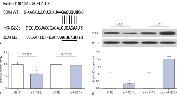
As is known, miRNAs can exert regulatory roles by affecting target gene expression. Hence, TargetScan online was em- ployed to search for genes that had a chance to interact with miR-132-3p. Among candidate genes, SOX4 has been identi- fied as an oncogene in OS (Fig. 3A),16,18,19 and was chosen for further study. To validate whether the putative binding sites of SOX4-3’UTR and miR-132-3p are important for the interac- tion between miR-132-3p and SOX4, dual luciferase reporter assays and mutation assays were performed. Then, constructed WT-SOX4-3’UTR or MUT-SOX4-3’UTR reporter was co-trans- fected with miR-132-3p or miR-NC into HEK293 cells, and their luciferase activities were measured 48 h after transfec- tion. As shown in Fig. 3B, the introduction of miR-132-3p mim- ic brought about a notable reduction in luciferase activity of the WT-SOX4-3’UTR reporter, compared with negative con- trol (miR-NC). However, miR-132-3p had no effect on the lu- ciferase activity of MUT-SOX4-3’UTR reporter (Fig. 3B). These findings indicated that miR-132-3p interacts with SOX4- 3’UTR via the putative binding sites. Next, western blot assays further suggested that the ectopic expression of miR-132-3p
decreases SOX4 protein levels, compared with the negative control miR-NC, in MG-63 cells (Fig. 3C). Conversely, miR-132- 3p deficiency resulted in a marked increase in SOX4 protein expression, compared with control (anti-miR-NC), in U2OS cells (Fig. 3C). Overall, these results suggest that SOX4 is a tar- get of miR-132-3p in OS cells.
MiR-132-3p inhibits proliferation and induces apoptosis by targeting SOX4 in OS cells
Then, the effects of miR-132-3p and SOX4 on proliferation and apoptosis were further investigated in MG-63 and U2OS cells. XTT assays showed that the introduction of miR-132-3p mimic remarkably suppressed MG-63 cell proliferation, com- pared with negative control (miR-NC) (Fig. 4A). As expected, the transfection of miR-132-3p inhibitor increased U2OS cell proliferation, compared with negative control (anti-miR-NC) (Fig. 4B). Moreover, the overexpression of miR-132-3p gave rise to a notable increase in apoptosis rate (Fig. 4C) and caspase-3 activity (Fig. 4D) in MG-63 cells. In restoration assays, the re- introduction of SOX4 partly reversed miR-132-3p-mediated anti-proliferation (Fig. 4A) and pro-apoptosis (Fig. 4C and D) effects in MG-63 cells. Also, the introduction of si-SOX4 weak- ened the promotive effect of miR-132-3p inhibitor on prolifer- ation in U2OS cells (Fig. 4B). Collectively, miR-132-3p was deemed to inhibit proliferation and induce apoptosis by tar- geting SOX4 in OS cells.
A
B C
Fig. 3. SOX4 is a target of miR-132-3p. (A) The putative binding sites between SOX4-3’UTR and miR-132-3p and the mutation sites of SOX4-3’UTR in the MUT-SOX4-3’UTR reporter. (B) The effect of miR-132-3p on luciferase activity of WT-SOX4-3’UTR or MUT-SOX4-3’UTR reporter. (C) SOX4 protein ex- pression in miR-132-3p-transfected MG-63 cells and anti-miR-132-3p-transfected U2OS cells. The lower histogram presented the gray values. *p<
0.05. SOX4, sex determining region Y-box 4; WT, wild-type; MUT, mutant.
Relative luciferase activity
miR-132-3p miR-NC
miR-132-3p miR-NC
MUT-SOX4 WT-SOX4
1.5
1.0
0.5
0.0
−
−
−
−
*
Relative SOX4 expression
anti-miR-132-3p anti-miR-NC
miR-132-3p miR-NC
U2OS MG-63
SOX4
β-actin
2.5 2.0 1.5 1.0 0.5 0.0
−
−
−
−
−
−
*
TUG1 enhances SOX4 expression by acting as a sponge of miR-132-3p
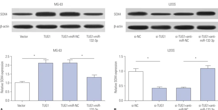
Interestingly, TUG1 and SOX4 shared the same binding sites in the process of interacting with miR-132-3p, indicating that TUG1 might act as a ceRNA to regulate the expression of miR- 132-3p and SOX4. Western blot assays demonstrated that TUG1 overexpression markedly improved SOX4 protein levels, while this effect of TUG1 on SOX4 expression was dramatical- ly abated by miR-132-3p in MG-63 cells (Fig. 5A). On the con- trary, TUG1 knockdown resulted in a significant reduction in SOX4 protein expression and miR-132-3p inhibitor reversed the inhibition effect of si-TUG1 on SOX4 expression in U2OS cells (Fig. 5B). Taken together, TUG1 might serve as a ceRNA of miR-132-3p to increase SOX4 expression in OS cells.
The knockdown of TUG1 suppresses proliferation and promotes apoptosis by regulating miR-132-3p/SOX4 axis in primary OS cells
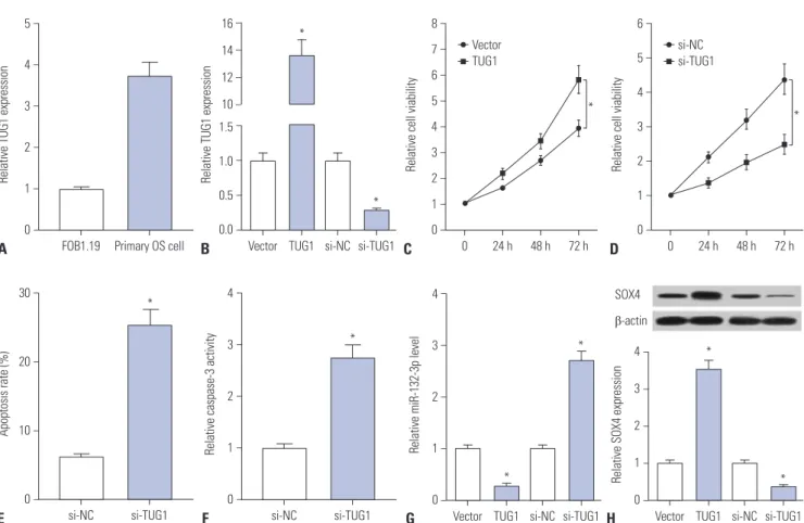
Primary OS cells were isolated from OS patients to further ex- plore whether TUG1 exerts its carcinogenic effect by regulat- ing miR-132-3p/SOX4 in human OS patients. First, we dem- onstrated that TUG1 is highly expressed in human primary OS cells, compared with the human normal osteoblastic cell line FOB1.19 (Fig. 6A). Next, the transfection efficiency of pcDNA- TUG1 and si-TUG1 was detected in human primary OS cells.
As shown in Fig. 6B, the transfection of pcDNA-TUG1 overex-
pression plasmid strikingly promoted TUG1 expression and the introduction of si-TUG1 markedly inhibited TUG1 expres- sion in human primary OS cells. Then, the effects of TUG1 over- expression and silencing on cell proliferation were further ex- amined in human primary OS cells. The results showed that cell proliferation capacity was elevated in TUG1-overexpressed primary OS cells, which was markedly reduced in TUG1-de- pleted primary OS cells (Fig. 6C and D). Moreover, we further demonstrated that TUG1 knockdown facilitated primary OS cell apoptosis, reflected as increases in apoptosis rate and cas- pase-3 activity in si-TUG1-transfected cells (Fig. 6E and F). As demonstrated above, TUG1 exerted its carcinogenic effect by downregulating miR-132-3p expression and upregulating SOX4 expression in OS cell lines. Thus, the effects of TUG1 on miR-132-3p and SOX4 expression were explored in primary OS cells. The results revealed that enforced expression of TUG1 suppresses miR-132-3p expression and facilitates SOX4 ex- pression in primary OS cells (Fig. 6G and H). Conversely, TUG1 silencing induced a notable increase in miR-132-3p level and a marked reduction in SOX4 expression in primary OS cells (Fig. 6G and H). Overall, these data indicated that the knock- down of TUG1 suppresses proliferation and promotes apopto- sis by regulating the miR-132-3p/SOX4 axis in primary OS cells.
A B
Relative cell viability Relative cell viability
0 24 h 48 h 72 h 0 24 h 48 h 72 h
MG-63 U2OS
6 5 4 3 2 1 0
8 7 6 5 4 3 2 1 0
−
−
−
−
−
−
−
−
−
−
−
−
−
−
−
−
** miR-NC
miR-132-3p miR-132-3p+vector miR-132-3p+SOX4
anti-miR-NC anti-miR-132-3p anti-miR-132-3p+si-NC anti-miR-132-3p+si-SOX4
*
*
C D
Fig. 4. MiR-132-3p inhibits proliferation and induces apoptosis of MG-63 and U2OS cells partly by targeting SOX4. (A, C, D) MG-63 cells were trans- fected with miR-NC, miR-132-3p, miR-132-3p+pcDNA3.1vector, or miR-132-3p+pcDNA3.1-SOX4, followed by the detection of cell proliferation (A), apoptosis rate (C) and caspase-3 activity (D). (B) U2OS cells were transfected with anti-miR-NC, anti-miR-132-3p inhibitor, anti-miR-132-3p+si-NC scramble control, or anti-miR-132-3p+si-SOX4, followed by cell proliferation by XTT assays. *p<0.05. SOX4, sex determining region Y-box 4.
Apoptosis rate (%) Relative caspase-3 activity
miR-132-3p+SOX4 miR-132-3p+SOX4
miR-132-3p+vector miR-132-3p+vector
miR-132-3p miR-132-3p
miR-NC miR-NC
* *
* *
25 20 15 10 5 0
4
3
2
1
−
−
−
−
−
−
−
−
−
−
MG-63 MG-63
DISCUSSION
OS, a frequent malignant bone tumor derived from primitive mesenchymal cells, seriously threatens the health of humans, especially madolescents.20 LncRNAs have been identified as critical mediators in the progression and prognosis of OS:7,21 for instance, lncRNA MALAT1 facilitated metastasis and prolifer- ation of OS cells by the PI3K/AKT axis,22 while lncRNA HIF- 2PUT hindered proliferation and invasion of OS cells.23 In the present study, the roles and molecular mechanisms of TUG1 underlying OS development were further investigated. Firstly, RT-qPCR results showed that TUG1 expression is upregulated in OS tumor tissues and cell lines, in accordance with previous reports.13,15 Functional analyses further revealed that TUG1 knockdown suppresses proliferation and facilitates apoptosis of MG-63 and U2OS cells, which was also consistent with pre- ceding studies.14,15
Herein, bioinformatics predict analysis, luciferase reporter assays, and RT-qPCR assays revealed that TUG1 inhibits miR- 132-3p expression by direct interaction. Previous studies have suggested that miR-132-3p might act as a tumor suppressor in OS. For example, Liu, et al.16 demonstrated that miR-132-3p hampered proliferation and metastasis by downregulating target gene SOX4 expression in OS cells. Wang, et al.17 demon- strated that miR-132-3p inhibited OS cell proliferation by tar- geting cyclin E1. Moreover, Yang, et al.24 reported that low-level expression of miR-132 is associated with poor survival and prog- nosis in OS patients. Hence, we supposed that TUG1 might
exert its carcinogenic effect by downregulating miR-132-3p expression in OS. Indeed, subsequent assays further demon- strated that introduction of miR-132-3p inhibitor partially re- versed the effect of TUG1 depletion on proliferation and apop- tosis of OS cells. In other words, TUG1 exerted its pro-prolifer- ation and anti-apoptosis effect by targeting miR-132-3p in OS cells.
Mounting evidence suggests that miRNAs exert functions by regulating target mRNAs expression.25 Hence, TargetScan online was employed to search for genes that possibly interact with miR-132-3p. The results indicated that there are some complementary sites between miR-132-3p and SOX4 3’UTR region. SOX4 has been identified as an oncogene in various can- cers, including hepatocarcinoma, prostate cancer, and OS.16,26,27 For example, SOX4, as a target of microRNA-212 and microR- NA-25-3p together with miR-132, could weaken these miRNAs- mediated inhibition effects on progression of OS cells.16,18,19 Consequently, we further demonstrated that SOX4 was a tar- get gene of miR-132-3p by bioinformatics analysis, luciferase reporter assays, and western blot assays, the results of which were consistent with those in a previous study.16 Additionally, miR-132-3p repressed proliferation and induced apoptosis of OS cells, while this effect of miR-132-3p was partly abrogated by SOX4 overexpression.
Previous studies suggested that TUG1 could act as ceRNA to influence the expression of miRNA target genes. For exam- ple, Xie, et al.28 demonstrated that TUG1 promotes the occur- rence and progression of OS by acting as an endogenous
A B
Fig. 5. TUG1 acts as a ceRNA of miR-132-3p to facilitate SOX4 expression in OS cells. (A) MG-63 cells were transfected with pcDNA3.1 vector, pcDNA3.1-TUG1, pcDNA3.1-TUG1+miR-NC, or pcDNA3.1-TUG1+miR-132-3p. (B) U2OS cells were transfected with si-NC, si-TUG1, si-TUG1+anti-miR- NC, si-TUG1+ anti-miR-132-3p. After transfection 48 h, SOX4 protein levels in MG-63 (A) and U2OS (B) cells were detected by western blot assays. *p<
0.05. TUG1, taurine upregulated gene 1; SOX4, sex determining region Y-box 4.
Relative SOX4 expression Relative SOX4 expression
TUG1+miR-
132-3p si-TUG1+anti-
miR-132-3p
TUG1+miR-NC si-TUG1+anti-
miR-NC
TUG1 si-TUG1
Vector si-NC
* *
* *
MG-63 U2OS
2.5 2.0 1.5 1.0 0.5 0.0
1.5
1.0
0.5
0.0
−
−
−
−
−
−
−
−
−
−
MG-63 U2OS
Vector TUG1+miR-NC si-NC si-TUG1+anti-
miR-NC
TUG1 TUG1+miR- si-TUG1
132-3p si-TUG1+anti-
miR-132-3p
SOX4 SOX4
β-actin β-actin
sponge of miR-9-5p and regulating the expression of the miR- 9-5p target gene POU2F1. In addition, Wang, et al.29 also dem- onstrated that TUG1 promotes the migration and invasion of OS cells by acting as an endogenous sponge of miR-335-5p and by regulating the expression of the miR-335-5p target gene ROCK1. Therefore, we aimed to further explore whether TUG1 could act as a ceRNA to sequester miR-132-3p from its target gene SOX4 in OS. Interestingly, we noticed that TUG1 and SOX4 shared the same binding sites in their interactions with miR-132-3p, indicating that TUG1 might regulate SOX4 ex- pression. Then, we confirmed that TUG1 facilitated SOX4 ex- pression in OS cells. Furthermore, the reintroduction of miR- 132-3p partially abrogated TUG1-induced SOX4 up-regulation in OS cells. In other words, our study demonstrated that TUG1 acted as a ceRNA of miR-132-3p to promote SOX4 expression and inhibit miR-132-3p expression, which resulted in an en- hancement of proliferation and a reduction of apoptosis in OS cell lines. Additionally, we showed that TUG1 knockdown sup- presses proliferation and promotes apoptosis by upregulating miR-132-3p and downregulating SOX4 in human primary OS
cells, indicating that TUG1 might exert its carcinogenic effect in OS patients.
Previous studies reported that the promoter of TUG1 en- compasses an abundance of highly conserved binding sites of p53 and that its expression is regulated by p53.30 Due to the important roles of p53 in various respects of cancer progres- sion, our study is vitally important and meaningful for investi- gating therapy and prognosis values of TUG1, miR-132-3p, and SOX4 in multiple cancers, including OS.
ORCID
Gang Li https://orcid.org/0000-0003-2033-5628
REFERENCES
1. Mirabello L, Troisi RJ, Savage SA. Osteosarcoma incidence and survival rates from 1973 to 2004: data from the Surveillance, Epi- demiology, and End Results Program. Cancer 2009;115:1531-43.
2. Meyers PA, Schwartz CL, Krailo MD, Healey JH, Bernstein ML, Betcher D, et al. Osteosarcoma: the addition of muramyl tripep-
Relative TUG1 expression
FOB1.19 Primary OS cell 5
4
3
2
1
0
−
−
−
−
−
−
D A
E
B
F G H
Apoptosis rate (%)
si-NC si-TUG1 30
20
10
0
−
−
−
−
*
Relative caspase-3 activity
si-NC si-TUG1 4
3
2
1
0
−
−
−
−
−
*
Relative cell viability
0 24 h 48 h 72 h 6
5 4 3 2 1 0
−
−
−
−
−
−
−
* si-NC
si-TUG1
Relative cell viability
0 24 h 48 h 72 h 8
7 6 5 4 3 2 1 0
−
−
−
−
−
−
−
−
−
* Vector
TUG1
Fig. 6. Knockdown of TUG1 suppresses proliferation and promotes apoptosis by regulating miR-132-3p/SOX4 axis in primary OS cells. (A) RT-qPCR as- says were performed to measure TUG1 expression in FOB1.19 and primary OS cells. (B) Primary OS cells were transfected with pcDNA3.1 empty vector, pcDNA3.1-TUG1, si-NC, or si-TUG1 for 48 h, followed by the detection of TUG1 level using RT-qPCR assays. (C and D) Primary OS cells were transfected with pcDNA3.1 empty vector, pcDNA3.1-TUG1, si-NC, or si-TUG1. At the indicated time points (0, 24, 48, 72 h) after transfection, cell viabili- ty was determined using XTT assays. (E and F) The effects of TUG1 knockdown on apoptosis rate and caspase-3 activity were measured in primary OS cells at 48 h following transfection. (G and H) Primary OS cells were transfected with pcDNA3.1 empty vector, pcDNA3.1-TUG1, si-NC, or si-TUG1 for 48 h, followed by the detection of miR-132-3p and SOX4 expressions. *p<0.05. TUG1, taurine upregulated gene 1; SOX4, sex determining region Y- box 4; OS, osteosarcoma; RT-qPCR, real-time quantitative PCR.
Relative TUG1 expression
Vector TUG1 si-NC si-TUG1 16
14 12 10 1.5 1.0 0.5 0.0
−
−
−
−
−
−
−
−
*
*
C
Relative miR-132-3p level
Vector TUG1 si-NC si-TUG1 4
3
2
1
0
−
−
−
−
−
*
*
Relative SOX4 expression
Vector TUG1 si-NC si-TUG1 4
3
2 1 0
−
−
−
−
−
*
* SOX4
β-actin
tide to chemotherapy improves overall survival--a report from the Children’s Oncology Group. J Clin Oncol 2008;26:633-8.
3. Esteller M. Non-coding RNAs in human disease. Nat Rev Genet 2011;12:861-74.
4. Catto JW, Alcaraz A, Bjartell AS, De Vere White R, Evans CP, Fussel S, et al. MicroRNA in prostate, bladder, and kidney cancer: a sys- tematic review. Eur Urol 2011;59:671-81.
5. Yang G, Lu X, Yuan L. LncRNA: a link between RNA and cancer.
Biochim Biophys Acta 2014;1839:1097-109.
6. Zhou G, Shi X, Zhang J, Wu S, Zhao J. MicroRNAs in osteosarco- ma: from biological players to clinical contributors, a review. J Int Med Res 2013;41:1-12.
7. Li Z, Yu X, Shen J. Long non-coding RNAs: emerging players in osteosarcoma. Tumour Biol 2016;37:2811-6.
8. Tay Y, Rinn J, Pandolfi PP. The multilayered complexity of ceRNA crosstalk and competition. Nature 2014;505:344-52.
9. Salmena L, Poliseno L, Tay Y, Kats L, Pandolfi PP. A ceRNA hy- pothesis: the Rosetta Stone of a hidden RNA language? Cell 2011;
146:353-8.
10. Han Y, Liu Y, Gui Y, Cai Z. Long intergenic non-coding RNA TUG1 is overexpressed in urothelial carcinoma of the bladder. J Surg Oncol 2013;107:555-9.
11. Xu Y, Wang J, Qiu M, Xu L, Li M, Jiang F, et al. Upregulation of the long noncoding RNA TUG1 promotes proliferation and migration of esophageal squamous cell carcinoma. Tumour Biol 2015;36:
1643-51.
12. Zhang EB, Yin DD, Sun M, Kong R, Liu XH, You LH, et al. P53-regu- lated long non-coding RNA TUG1 affects cell proliferation in hu- man non-small cell lung cancer, partly through epigenetically regulating HOXB7 expression. Cell Death Dis 2014;5:e1243.
13. Ma B, Li M, Zhang L, Huang M, Lei JB, Fu GH, et al. Upregulation of long non-coding RNA TUG1 correlates with poor prognosis and disease status in osteosarcoma. Tumour Biol 2016;37:4445-55.
14. Zhang Q, Geng PL, Yin P, Wang XL, Jia JP, Yao J. Down-regulation of long non-coding RNA TUG1 inhibits osteosarcoma cell prolifera- tion and promotes apoptosis. Asian Pac J Cancer Prev 2013;14:
2311-5.
15. Yun-Bo F, Xiao-Po L, Xiao-Li L, Guo-Long C, Pei Z, Fa-Ming T. Ln- cRNA TUG1 is upregulated and promotes cell proliferation in os- teosarcoma. Open Med (Wars) 2016;11:163-7.
16. Liu Y, Li Y, Liu J, Wu Y, Zhu Q. MicroRNA-132 inhibits cell growth and metastasis in osteosarcoma cell lines possibly by targeting Sox4. Int J Oncol 2015;47:1672-84.
17. Wang J, Xu G, Shen F, Kang Y. miR-132 targeting cyclin E1 sup- presses cell proliferation in osteosarcoma cells. Tumour Biol 2014;
35:4859-65.
18. Luo XJ, Tang DG, Gao TL, Zhang YL, Wang M, Quan ZX, et al. Mi-
croRNA-212 inhibits osteosarcoma cells proliferation and inva- sion by down-regulation of Sox4. Cell Physiol Biochem 2014;34:
2180-8.
19. Wu X, Zhou H, Yue B, Li M, Liu F, Qiu C, et al. Upregulation of mi- croRNA-25-3p inhibits proliferation, migration and invasion of osteosarcoma cells in vitro by directly targeting SOX4. Mol Med Rep 2017;16:4293-300.
20. Ritter J, Bielack SS. Osteosarcoma. Ann Oncol 2010;21 Suppl 7:
vii320-5.
21. Yang Z, Li X, Yang Y, He Z, Qu X, Zhang Y. Long noncoding RNAs in the progression, metastasis, and prognosis of osteosarcoma.
Cell Death Dis 2016;7:e2389.
22. Dong Y, Liang G, Yuan B, Yang C, Gao R, Zhou X. MALAT1 pro- motes the proliferation and metastasis of osteosarcoma cells by activating the PI3K/Akt pathway. Tumour Biol 2015;36:1477-86.
23. Wang Y, Yao J, Meng H, Yu Z, Wang Z, Yuan X, et al. A novel long non-coding RNA, hypoxia-inducible factor-2α promoter up- stream transcript, functions as an inhibitor of osteosarcoma stem cells in vitro. Mol Med Rep 2015;11:2534-40.
24. Yang J, Gao T, Tang J, Cai H, Lin L, Fu S. Loss of microRNA-132 pre- dicts poor prognosis in patients with primary osteosarcoma. Mol Cell Biochem 2013;381:9-15.
25. Vlachos IS, Paraskevopoulou MD, Karagkouni D, Georgakilas G, Vergoulis T, Kanellos I, et al. DIANA-TarBase v7.0: indexing more than half a million experimentally supported miRNA:mRNA in- teractions. Nucleic Acids Res 2015;43:D153-9.
26. Liao YL, Sun YM, Chau GY, Chau YP, Lai TC, Wang JL, et al. Iden- tification of SOX4 target genes using phylogenetic footprinting- based prediction from expression microarrays suggests that over- expression of SOX4 potentiates metastasis in hepatocellular carcinoma. Oncogene 2008;27:5578-89.
27. Liu P, Ramachandran S, Ali Seyed M, Scharer CD, Laycock N, Dal- ton WB, et al. Sex-determining region Y box 4 is a transforming oncogene in human prostate cancer cells. Cancer Res 2006;66:
4011-9.
28. Xie CH, Cao YM, Huang Y, Shi QW, Guo JH, Fan ZW, et al. Long non-coding RNA TUG1 contributes to tumorigenesis of human os- teosarcoma by sponging miR-9-5p and regulating POU2F1 ex- pression. Tumour Biol 2016;37:15031-41.
29. Wang Y, Yang T, Zhang Z, Lu M, Zhao W, Zeng X, et al. Long non- coding RNA TUG1 promotes migration and invasion by acting as a ceRNA of miR-335-5p in osteosarcoma cells. Cancer Sci 2017;
108:859-67.
30. Khalil AM, Guttman M, Huarte M, Garber M, Raj A, Rivea Mo- rales D, et al. Many human large intergenic noncoding RNAs as- sociate with chromatin-modifying complexes and affect gene ex- pression. Proc Natl Acad Sci U S A 2009;106:11667-72.