Effects of Olanzapine on the Pacemaker Potentials of Interstitial Cells of Cajal in Murine Small Intestine
Byung Joo Kim*
Division of Longevity and Biofunctional Medicine, School of Korean Medicine, Pusan National University, Yangsan 50612, Korea Received May 27, 2019 /Revised August 20, 2019 /Accepted September 6, 2019
The interstitial cells of Cajal (ICCs) are the pacemaker cells in the gastrointestinal (GI) tract. In the present study, the effects of olanzapine, an atypical antipsychotic agent, on pacemaker potentials in cultured ICCs from the small intestine of the mouse were investigated. The whole-cell patch-clamp configuration was used to record pacemaker potentials from cultured ICCs. Olanzapine produced pacemaker depolarizations in a concentration-dependent manner in current clamp mode. Methoctr- amine, a muscarinic M2 receptor antagonist, did not inhibit olanzapine-induced pacemaker depolariza- tions, whereas 4-diphenylacetoxy-N-methylpiperidine methiodide (4-DAMP) muscarinic M3 receptor antagonist did inhibit it. When guanosine 5'-[β-thio] diphosphate (GDP-β-S; 1 mM) was in the pipette solution, olanzapine-induced pacemaker depolarization was blocked. Also, low Na+ solution externally eliminated the generation of pacemaker potentials and inhibited the olanzapine-induced pacemaker depolarizations. Additionally, the nonselective cation channel blocker, flufenamic acid, inhibited the olanzapine-induced pacemaker depolarizations. Pretreatment with U-73122, an active phospholipase C (PLC) inhibitor, also eliminated the generation of pacemaker potentials and suppressed the olanza- pine-induced pacemaker depolarizations. These results suggested that olanzapine modulates the pace- maker potentials through muscarinic M3 receptor activation by G protein–dependent external Na+ and PLC pathway in the ICCs. Therefore, olanzapine could affect intestinal motility through ICCs.
Key words : Gastrointestinal tract, ICCs, interstitial cells of Cajal, olanzapine, pacemaker potentials
*Corresponding author
*Tel : +82-51-510-8469, Fax : +82-51-510-8420
*E-mail : [email protected]
This is an Open-Access article distributed under the terms of the Creative Commons Attribution Non-Commercial License (http://creativecommons.org/licenses/by-nc/3.0) which permits unrestricted non-commercial use, distribution, and reproduction in any medium, provided the original work is properly cited.
Journal of Life Science 2019 Vol. 29. No. 9. 1010~1015 DOI : https://doi.org/10.5352/JLS.2019.29.9.1010
서 론
위장관 운동은 음식이 입을 통해 식도, 위, 소장, 대장을 지 나 항문을 통해 몸 밖으로 배출되는 이동을 말한다. 위장관 운동은 교감신경, 부교감 신경 등과 같은 외인성 신경계, 위장 관 자체내 내인성 신경계, 자체내 내인성 인자 및 약물, 또는 평활근 등에 의해 조절된다[12]. 위장관 운동은 특수한 간질세 포중 하나인 카할세포(interstitial cells of Cajal; ICCs)에 의해 서 발생되어 향도잡이 세포(pacemaker cell; 향도(嚮導)는 “길 을 인도한다”는 의미로 이곳에서 향도잡이는 위장관운동의 리듬을 조절한다는 의미임)라고 불리우고 있다[8, 19]. 따라서 이 세포는 위장관 연동운동에서 중요한 역할을 하고 있다. 카 할세포의 중요기능은 향도잡이 세포 기능이외에 다양한 위장 관에서의 신경전달물질의 작용 및 기계적 자극의 조절기능도 가지고 있다[8, 19]. 카할세포는 평활근과 함께 간충직(mesen- chyme)의 전구세포에서 분화한 세포들로 위장관 평활근 및
신경망과 간극연접(gap junction)을 통해 연결되어 있다[11].
카할세포는 세포막의 티로신 인산화효소(receptor tyrosine kinase)인 키트(Kit)에 작용하는 c-kit 유전자를 가지고 있다 [18]. 평활근과 장관신경계는 키트 수용체(Kit receptor)가 존 재하지 않아 c-Kit 항체를 이용한 세포면역학적방법이 카할세 포를 확인하는데 결정적 역할을 하고 있다[18]. 여러 위장관 운동성 질환이 카할세포와 크게 관련되어 있다는 많은 실험결 과들이 보고 되었다[13, 18]. 즉 카할세포의 수가 감소하거나 형태학적 이상이 정상적인 위장관 운동성의 리듬을 유지하지 못함으로 인하여 운동성 장애를 일으킨다는 것이다. 이와 관 련된 질환들로는 염증성 장 질환(inflammatory bowel dis- ease), 만성특발성장가짜폐색(chronic idiopathic intestinal pseudo-obstruction), 식도이완불능증(achalasia), 선천성 거대 결장증(Hirschsprug's disease), 유문협착(pyloric stenosis), 장 폐색(intestinal obstruction), 항문직장기형(anorectal malfor- mations) 등이 있다[13, 18].
한편 올란자핀(Olanzapine)은 디에나벤조다이아제핀(thie- nobenzodiazepine) 계 약물로 비벤조다이아제핀(dibenzodia- zepine) 구조를 가지고 있고, 특히 조현병(schizophrenia)치료 에 효과적인 약물로 잘 알려져 있다[1, 16]. 올란자핀은 5-HT2, 5-HT3, 5-HT6 등의 세로토닌 수용체, D2, D3, D4 등의 도파민 수용체, M1에서 M5에 이르는 무스카린 수용체, α1 수용체 그리 고 H1 수용체 등 뇌 의 여러 수용체에 대해 강한 친화력을 가지
고 있다[1]. 올란자핀은 높은 항정신병 치료 효과와 치료 반응 률을 보였고, 안전성 측면에서도 이상반응 호소와 추체외로 증상의 발생 빈도도 낮았다. 또한 정신병에 동반된 우울증상 과 불안증상에도 효과적임이 확인되었다[1]. 하지만 피로감 (tiredness), 몸무게 증가(weight increase), 변비(constipation) 등의 부작용등이 보고 되고 있고[16], 다른 조현병 치료제들과 비교해보면 변비가 생길 확률이 더 높은 것으로 알려지고 있 다[16]. 변비는 삶의 질을 감소시키는 대표적인 부작용으로 다 양한 합병증을 또한 발생시킬 수 있다. 하지만 이러한 위장관 운동성에 대한 올란자핀의 효능은 잘 알려져 있지 않다.
따라서, 본 실험에서는 위장관 운동 조절 기전에 관한 연구 에 중요한 향도잡이 세포인 카할세포를 이용하여 올란자핀의 카할세포에 대한 효과와 작용기전을 알아보고 장운동성 조절 가능성을 확인하고자 한다.
재료 및 방법
재료
올란자핀 및 이 논문에 사용된 모든 시약은 sigma (Sigma Chemical Co., St. Louis, MO, USA)에서 구입하였고 사용직전 에 원하는 농도로 만든 후 세포에 투여하였다.
카할세포 배양
카할세포의 배양을 위해 주로 이용되는 마우스는 3-5일령 Balb/C를 사용하고 에테르(ether)로 마취 시킨 후 개복하여 유문륜(pyloric ring)에서부터 회장에 해당하는 소장부위를 적 출하였다. 실온에서 크렙스 링거 중탄산염(Krebs-Ringer bi- carbonate)용액으로 채워진 준비 용기 속에서 창자간막 가장 자리를 따라 절개하여 점막층을 제거하고 윤상근을 노출시킨 후 분리된 소장 근육조직을 교원질 분해 효소(collagenase) (Worthington Biochemical Co., Lakewood, NJ, USA) 1.3 mg/
mL, 소혈청알부민(bovine serum albumin) (Sigma Chemical Co., St. Louis, MO, USA) 2 mg/ml, 트립신 저해제(trypsin inhibitor) (Sigma Chemical Co., St. Louis, MO, USA) 2 mg/
mL등이 들어 있고, Ca2+이 들어 있지 않은 행크(Hank's) 용액 에 옮긴 다음 37℃에서 20분간 항온 소화시킨 후 진탕시켜 세포를 분리하였다. 분리된 세포들을 유리 커버 글라스 위에 분주하고, 10분 후에 간상세포인자(stem cell factor) (Sigma Chemical Co., St. Louis, MO, USA) 5 ng/ml와 2% 항생/항진 균제(antibiotic/antimycotic) (Gibco, Grand Island, NY, USA) 가 들어 있는 smooth muscle growth medium (Clonetics Corp., San Diego, CA, USA) 용액을 분주한 후, 37℃(95% O2
- 5% CO2) 배양기에서 배양 시켰다. 배양된 다음날 전날 배양 된 용액에서 항생/항진균제(antibiotic/antimycotic)만 제외시 켜 영양액을 바꾸어 주고 실험은 배양 1일째 후부터 시행하였 다. 모든 동물 실험 과정은 부산대학교 동물실험윤리 위원회
의 승인을 받아 실시 하였다(PNU-2017-1754).
전기생리학적 실험
배양한 카할세포에서 막전압을 기록하기 위해 패치클램프 (patch clamp) 실험기법 중 전 세포(whole cell patch)방법을 이용하였다. 전압은 패치클램프 앰프(standard patch clamp amplifiers) (Axon Instruments, Foster, CA, USA)를 통해 증 폭시키며, 나오는 신호는 컴퓨터 모니터 및 생리적 기록기 (Gould 2200, Gould, Valley View, OH, USA)를 통해서 관찰 하였다. 막전압을 기록하는 동안 세포외 관류용액의 조성은 다음과 같다(각 수치는 mM 단위임): KCl 5, NaCl 135, CaCl2
1.2, glucose 10, HEPES 10이며 Tris를 첨가하여 pH가 7.4가 되도록 하고, 전극내 용액의 조성은 다음과 같이 하였다(각 수치는 mM 단위임): KCl 140, MgCl2 5, K2ATP 2.7, Na2GTP 0.1, creatinine phosphate disodium 2.5, HEPES 5, EGTA 0.1 이며 Tris를 첨가하여 pH가 7.2가 되도록 하였다. 배양된 카할 세포에서 세포막 전류 고정법(Current clamp)을 시행하여 자 발적으로 발생되는 내향성 향도잡이 전압을 기록한 후 올란자 핀의 효능을 알아 보았다.
통계 분석
대조군과 실험군 사이의 통계학적 유의성 검정은 Student’s t-test를 사용하였으며 유의수준 p<0.05를 사용하였다. 실험결 과는 mean ± SD로 기재하였다.
결 과
올란자핀의 카할세포에 대한 효과
소장 카할세포의 향도잡이 기능(pacemaking activity)에서 올란자핀에 의한 조절능을 알아 보았다. 올란자핀은 농도 의 존적으로 향도잡이 기능(pacemaking activity)의 탈분극(de- polarization)을 증가시킴을 알 수 있었다(Fig. 1A - Fig. 1C).
올란자핀 50 μM에서는 5.0±0.8 mV (n=6), 100 μM에서는 11.7±1.1 mV (n=6), 300 μM에서는 24.2±1.5 mV (n=7)정도로 탈분극 정도가 증가되었다(Fig. 1D).
올란자핀의 카할세포 효과에 무스카린성 수용체(muscarinic receptor)의 관련성
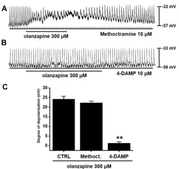
올란자핀에 의한 카할세포 조절에 무스카린성 수용체 (muscarinic receptor)의 관련성을 알아보기 위해서 M2 수용체 억제제인 methoctramine과 M3 수용체 억제제인 4-DAMP를 사용하여 보았다[7]. 일단 카할세포에 의한 향도잡이 전압에 methoctramine과 4-DAMP 자체는 아무런 영향을 미치지 않 았다(Fig. 2A, Fig. 2B). Methoctramine을 투여한 후에 올란자 핀을 투여한 경우에는 정상적인 향도잡이 전압의 탈분극이 나타났지만(Fig. 2A), 4-DAMP를 투여한 후 올란자핀을 투여
A
B
C
D
Fig. 1. Effects of olanzapine on pacemaker potentials in cultured ICCs from the murine small intestine. (A-C) Pacemaker potentials of ICCs exposed to olanzapine (50-300 μM) in current clamping mode (I=0). (D) Summary of responses to olanzapine. The values were expressed as the mean
± S.D. *p<0.05. **p<0.01. CTRL, control.
A
B
C
Fig. 2. Effects of muscarinic receptor subtype antagonists on olanzapineinduced pacemaker potential depolarizations in cultured ICCs. (A) Pacemaker potentials of ICCs ex- posed to olanzapine in the presence of methoctramine muscarinic M2 receptor antagonist (10 μM). Pacemaker potential depolarizations were depolarized. (B) Pace- maker potentials of ICCs exposed to olanzapine in the presence of 4-DAMP muscarinic M3 receptor antagonist (10 μM). Pacemaker potential depolarizations were not altered. (C) Responses to olanzapine in the presence of different receptor antagonists. The values were expre- ssed as the mean ± S.D. *p<0.05. **p<0.01. CTRL, control.
CTRL, control; Methoct, methoctramine; 4-DAMP, 4-di- phenylacetoxy-N-methylpiperidine methiodide.
한 경우에는 향도잡이 전압의 탈분극이 억제됨을 알 수 있었다 (Fig. 2B). Methoctramine을 투여한 경우 올란자핀에 의한 탈 분극 정도는 22.3±0.8 mV (n=7), 4-DAMP를 투여한 경우 올란 자핀에 의한 탈분극 정도는 1.3±0.7 mV (n=5) 이다(Fig. 2C).
따라서, 이 실험으로 올란자핀은 무스카린성 3번 수용체(M3
muscarinic receptor)를 통해서 카할세포의 향도잡이 기능을 억제 시킴을 알 수 있었다.
올란자핀의 카할세포 효과에 G protein의 관련성
올란자핀에 의한 카할세포 조절에 G protein의 관련성을 알아보기 위해서 세포내 G protein의 기능을 억제 시키는 것 으로 알려지고 있는 GDPβS를 사용하였다[15]. 올란자핀의 카 할세포의 향도잡이 탈분극이 GDPβS (1 mM)의 세포내 존재로 억제되었다(Fig. 3). 따라서 G protein이 올란자핀에 의한 카할 세포 향도잡이 탈분극에 관여함을 알 수 있다.
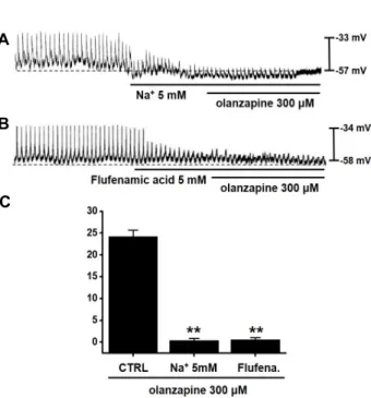
올란자핀의 카할세포 효과에 세포밖 Na+ 농도 및 비선택성 양이온 통로의 관련성
올란자핀에 의한 카할세포 조절에 세포밖 Na+ 농도 및 비선 택성 양이온 통로의 관련성을 알아보기 위해서 세포밖 Na+을 같은 농도의 NmethylDglucamine (NMDG; 생리학적 삼투압 을 유지하면서 사용가능한 Na+ 대체물질)로 바꾸어 세포밖 Na+ 농도를 5 mM로 만들었다[5]. 이런 경우 카할세포에 의한
향도잡이 발생이 억제되고, 이때 올란자핀 투여시 향도잡이 탈분극은 발생되지 않았다(Fig. 4A). 비선택성 양이온 통로 억 제제인 flufenamic acid [22]의 존재에서도 카할세포에 의한 향도잡이 발생이 억제되고, 이때 올린자핀 투여서 향도잡이 탈분극 역시 발생되지 않았다(Fig. 4B). 세포밖 Na+ 5 mM을 투여한 경우 올란자핀에 의한 탈분극 정도는 0.4±0.4 mV (n=
5), flufenamic acid를 투여한 경우 올란자핀에 의한 탈분극 정도는 0.6±0.5 mV (n=5)이다(Fig. 4C). 따라서, 이 실험으로 올란자핀에 의한 카할세포 향도잡이 탈분극은 세포밖 Na+과 비선택성 양이온 통로가 관여함을 알 수 있다.
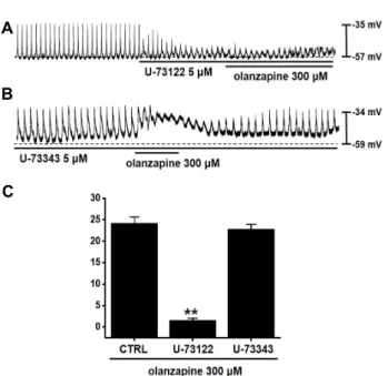
올란자핀의 카할세포 효과에 phospholipase C (PLC) 관련성
PLC기전의 관련성을 알아보기 위해서 활성형 PLC 억제제 인 U-73122을 이용하였다[6]. U-73122 존재할때, 카할세포의 향도잡이 기능은 억제가 되고, 이때 올란자핀에 의한 카할세 포 향도잡이 탈분극은 억제 되었다(Fig. 5A). U-73122의 기능 을 확실히 하기 위해서 U-73122의 비활성형인 U-73343을 처치
A
B
Fig. 3. Effects of GDPβS on the response to on olanzapinein- duced pacemaker potential depolarizations in cultured ICCs. (A) Pacemaker potentials of ICCs exposed to olan- zapine in the presence of GDPβS (1 mM) in the pipette.
Under these conditions, olanzapine-induced pacemaker potential depolarization was blocked. (B) Responses to olanzapine in the presence of GDP-β-S in the pipette. The values were expressed as the mean ± S.D. **p<0.01. CTRL, control; GDP-β-S, guanosine 5'[β-thio] diphosphate.
A
B
C
Fig. 4. Effects of external low Na+ solution or flufenamic acid, a nonselective cation channel blocker, on olanzapine-in- duced pacemaker potential depolarizations in cultured ICCs. (A) External low Na+ solution eliminated the gen- eration of pacemaker potentials. Under these conditions, olanzapine did not depolarize pacemaker potentials. (B) Flufenamic acid (10 μM) eliminated the generation of pacemaker potentials. In these conditions, flufenamic acid inhibited the olanzapine-induced pacemaker poten- tial depolarizations. (C) Responses to olanzapine in the external low Na+ solution and in the presence of flufe- namic acid. The values were expressed as the mean ± S.D. **p<0.01. CTRL, control.
해 보니[6] 올란자핀에 의한 카할세포 향도잡이 탈분극에는 아무 효능이 없음을 확인 하였다(Fig. 5B). U-73122를 투여한 경우 올란자핀에 의한 탈분극 정도는 1.6±0.5 mV (n=6), U- 73343을 투여한 경우 올란자핀에 의한 탈분극 정도는 22.8±1.1 mV (n=6) 이다(Fig. 5C). 따라서, 이 실험으로 올란자핀에 의한 카할세포 향도잡이 탈분극은 PLC 기전이 관여함을 알 수 있다.
고 찰
본 연구에서 올란자핀은 위장관 소장 카할세포에서 향도잡 이 전압을 탈분극 시키고 무스카린성 3번 수용체를 통해서 G protein-PLC-세포밖 Na+ 의존 기전이 관여하여 향도잡이 기능을 조절하고 있다.
카할세포가 위장관에서 자발적 운동과 관련된 향도잡이 세 포라는 사실은 많은 연구에서 증명되고 있다[8, 19]. 카할세포 는 방추형 모양으로 세포막에 많은 가지를 가지면서 다른 카 할세포나 평활근 또는 신경등과 간극연접(gap junction)으로 연결되어 있다[17, 20]. 또한 세포내에 많은 미토콘드리아를 가지고 있다[17, 20]. 많은 연구에서 카할세포는 자발적인 향도 잡이 전압을 일으켜서 탈분극을 일으키고 이 자극이 평활근의 탈분극을 일으켜 평활근 세포막에 있는 전압의존성 칼슘채널 을 활성화 시켜 세포내 칼슘농도를 증가시킨다. 그래서 결국 엔 평활근의 수축을 일으켜 위장관의 운동성을 조절하는 것으 로 알려지고 있다[17, 20]. 따라서 카할세포는 호르몬, 신경전
달물질, 또는 다양한 물질들에 의해서 작용이 조절받아 위장 관 운동성을 조절할 수 있게 된다. 하지만 이러한 위장관 운동 성은 카할세포 뿐만 아니라 신경이나 위장관에 존재하는 수많 은 다양한 세포에 의해서도 조절 받기 때문에 카할세포의 반 응이 반드시 위장관 운동 조절에 바로 적용 할 수 있는 것은 아닌 것이다. 기존엔 카할세포만의 정확한 생리학적 기능들을 이해하는데 어려움이 많았다. 이런 어려움을 극복하기 위하여 효소를 이용한 조직에서 단일세포를 분리한 후 형태학적으로 카할세포를 확인 한 다음 전기생리학적 방법을 이용하여 조직 과 동일한 서파에 관련한 이온전류들을 기록 분석하고 있다 [14]. 단일 카할세포에서 세포막 전류 고정법(Current-clamp method)을 시행하면 자발적으로 향도잡이 전압(pacemaker potentials)이 기록된다[9]. 향도잡이 전압(Pacemaker poten- tials)의 발생기전은 세포내 Ca2+ 저장고인 내형질세망에서 IP3
를 통한 주기적 Ca2+분비가 미토콘드리아로 Ca2+를 유입시켜 발생한다[17, 20]. 이때 세포내 Ca2+ 농도의 감소로 세포밖에서 비선택성 양이온 통로인 일과성 수용체 전압(TRPM7; tran- sient receptor potential melastatin 7) 이온통로나 칼슘에 의해
A
B
C
Fig. 5. Effects of PLC inhibitors on olanzapineinduced pace- maker potential depolarizations in cultured ICCs. (A) U-73122 (5 μM), a PLC inhibitor, stopped the gen- eration of pacemaker potentials. U-73122 blocked the olanzapine-induced pacemaker potential depolariza- tions. (B) The application of U-73343 (5 μM), an in- active PLC inhibitor did not show any influence on the generation of pacemaker potentials. Also, U-73343 did not block the olanzapine-induced pacemaker po- tential depolarizations. (C) Responses to olanzapine in PLC inhibitors are summarized. The values were expressed as the mean ± S.D. **p<0.01. CTRL, control.
활성화되는 염소(Ca2+-activated Cl-) 이온통로로 Ca2+이 세포 내로 들어오면서 향도잡이 전압이 발생하는 것으로 알려지고 있다[10, 23].
올란자핀은 조현병치료제로 널리 사용되고 있지만 다양한 부작용들이 보고 되고 있다[16]. 그중에서 위장관 운동성과 관 련된 부작용도 일으키는 것으로 알려지고 있다. 본 연구에서 는 카할세포의 향도잡이 전압을 탈분극을 일으키는 것을 알 수 있고, 따라서 올란자핀에 의한 위장관 이상 증상인 변비 등은 카할세포를 조절해서 나타나는 증상이라고 생각된다. 하 지만 위장관 운동은 카할세포뿐 아니라 다양한 위장관 신경이 나 세포의 반응에 나타나는 결과이기 때문에 앞으로 신경 및 평활근 세포에서의 반응 등 종합적인 결과를 보고 판단해야 할 것으로 생각된다. 또한 올라자핀은 다양한 수용체와의 작 용에 관여하며, 특히 5-HT 수용체나 무스카린성 수용체를 조 절하는 것으로 알려지고 있다[2, 3]. 또한 myosin light chain kinase (MLCK) 의 작용으로 반응이 나타나는 것으로 알려지 고 있다[21]. 본 연구에서는 MLCK의 작용 관련성에 대한 연구 는 아직 진행하지 못하였는데, 앞으로 평활근에 대한 연구와 함께 MLCK의 관련성에 대한 심도있는 연구를 진행할 예정이
다. 또한 올란자핀이 카할세포의 칼슘 이온통로에는 어떤 효 과를 보이는지를 확인하고 실제로 올란자핀이 카할세포의 기 능조절에 이들 이온통로를 조절하고 있는지를 연구할 예정이 다. 카할세포의 바이오 마커로는 c-kit과 칼슘에 의해 활성화되 는 비선택성 양이온 통로 및 염소 이온 통로가 알려지고 있는 데 올란자핀에 의한 in vivo 실험 시 이러한 바이오마커의 카할 세포 세포막 발현에 미치는 영향도 큰 의미 있는 실험 주제가 될 것으로 생각된다[10, 18, 23].
현재까지 알려진 위장관 운동과 관련된 질환은 카할세포의 수가 감소하거나 기능장애에 의한 향도잡이 기능의 손실로 인한 위장관 운동 장애로 추정하고 있다. 따라서 카할세포에 의한 다양한 물질들에 의한 약리학적 연구는 작용기전에 관한 이해뿐만 아니라 위장관 운동 관련 치료제의 개발과 연관하여 특히 중요할 것으로 사료된다.
감사의 글
이 성과는 2017년도 정부(과학기술정보통신부)의 재원으로 한국연구재단의 지원을 받아 수행된 연구임(No. 2017R1A2B 2003764).
References
1. Ahn, Y. M., Kang, D., Kwon, J. S., Kim, C. Y., Kim, C. E., Bahn, G. , Shin, Y., Lee, G., Lee, D. W., Yi, J. S., Cho, H., Chae, J. and Kim, Y. S. 2001. Efficacy and Safety of Olanza- pine in the Treatment of Korean Patients with Schizophrenia and Schizophreniform Disorder. J. Kor. Neuropsychiatr. Asso.
40, 693-707.
2. Bhana, N., Foster, R. H., Olney, R. and Plosker, G. L. 2001.
Olanzapine: an updated review of its use in the manage- ment of schizophrenia. Drugs 61, 111-161.
3. Bymaster, F. P., Nelson, D. L., DeLapp, N. W., Falcone, J.
F., Eckols, K., Truex, L. L., Foreman, M. M., Lucaites, V.
L. and Calligaro, D. O. 1999. Antagonism by olanzapine of dopamine D1, serotonin2, muscarinic, histamine H1 and al- pha 1-adrenergic receptors in vitro. Schizophr. Res. 37, 107- 122.
4. Bymaster, F., Perry, K. W., Nelson, D. L., Wong, D. T., Rasmussen, K., Moore, N. A. and Calligaro, D. O. 1999.
Olanzapine; a basic science update. Br. J. Psychiatry 174, 36- 40.
5. Chromý, V., Zahradnícek, L. and Voznícek, J. 1981. Use of N-methyl-D-glucamine as buffer in the determination of se- rum alkaline phosphatase activity. Clin. Chem. 27, 1729-1732.
6. D, MacMillan. and J. G., McCarron. 2010. The phospholipase C inhibitor U-73122 inhibits Ca2+ release from the intra- cellular sarcoplasmic reticulum Ca2+ store by inhibiting Ca2+
pumps in smooth muscle. Br. J. Pharmacol. 160, 1295-1301.
7. Ehlert, F. J., Sawyer, G. W. and Esqueda, E. E. 1999. Contrac- tile role of M2 and M3 muscarinic receptors in gastro-
초록:생쥐 소장 카할세포에서 발생하는 향도잡이 기능의 올란자핀 효능에 관한 연구
김병주*
(부산대학교 한의학전문대학원)
카할세포는 위장관에서 향도잡이 역할을 한다. 본연구에서는 생쥐 소장 카할세포에서 발생되는 향도잡이 기능 에서 올란자핀의 역할을 연구하였다. 패치클램프 방법을 사용하여 향도잡이 전압을 측정하였다. 올란자핀에 의해 서 카할세포 향도잡이 전압이 탈분극 되었으며, 이 탈분극은 무스카린성 3번 수용체 억제제에 의해서 억제 되었 다. 세포내 GDPβS을 넣어주니 올란자핀에 의해 향도잡이 전압 탈분극이 억제되었다. 또한, 세포밖 Na+ 농도 감소 와 비선택성 양이온 통로 억제제에 의해서 올란자핀에 의한 향도잡이 전압 탈분극이 억제 되었다. 세포내 PLC기 전의 억제제인 U-73122에 의해서 올란자핀에 의한 향도잡이 전압 탈분극이 억제 되었다. 이러한 결과로 올란자핀 은 무스카린성 3번 수용체를 통해서 세포내 G 단백질과 PLC기전 및 세포밖 Na+이 관여함을 알 수 있었다. 따라 서 올란자핀은 카할세포를 통해서 장운동성을 조절 할 수 있을 것으로 생각된다.
intestinal smooth muscle. Life Sci. 64, 387-394.
8. Huizinga, J. D., Thuneberg, L., Kluppel, M. and Malysz, H.
B. 1995. The W/kit gene required for interstitial cells of Cajal and for intestinal pacemaker activity. Nature 373, 347-349.
9. Kho, S. D., Sanders, K. M. and Ward, S. M. 1998. Spontaneous electrical rhythmicity in cultured interstitial cells of Cajal from the murine small intestine. J. Physiol. 513, 203-213.
10. Kim, B. J., Lim, H. H., Yang, D. K., Jun, J. Y., Chang, I. Y., Park, C. S., So, I., Stanfield, P. R. and Kim, K. W. 2005.
Melastatin-type transient receptor potential channel 7 is re- quired for intestinal pacemaking activity. Gastroenterology 129, 1504-1517.
11. Koh, S. D., Ward, S. M., Ordög, T., Sanders, K. M. and Horowitz, B. 2003. Conductances responsible for slow wave generation and propagation in interstitial cells of Cajal. Curr.
Opin. Pharmacol. 3, 579-582.
12. Koutouratsas, T., Kalli, T., Karamanolis, G. and Gazouli, M.
2019. Contribution of ghrelin to functional gastrointestinal disorders' pathogenesis. World J. Gastroenterol. 25, 539-551.
13. Kubota, M., Kanda, E., Ida, K., Sakakihara, Y. and Hayashi, M. 2005. Severe gastrointestinal dysmotility in a patient with congenital myopathy: causal relationship to decrease of in- terstitial cells of Cajal. Brain Dev. 27, 447-450.
14. Lee, H. K. and Sanders, K. M. 1993. Comparison of the elec- tric currents from interstitial cells and smooth muscle cells of canine proximal colon. J. Physiol. 460, 135-152.
15. Ogata, R., Inoue, Y., Nakano, H., Ito, Y. and Kitamura, K.
1996. Oestradiol-induced relaxation of rabbit basilar artery by inhibition of voltage-dependent Ca channels through
GTP-binding protein. Br. J. Pharmacol. 117, 351-359.
16. Prommer, E. 2013. Olanzapine: palliative medicine update.
Am. J. Hosp. Palliat. Care. 30, 75-82.
17. Sanders, K. M., Koh, S. D., Ro, S. and Ward, S. M. 2012.
Regulation of gastrointestinal motility--insights from smooth muscle biology. Nat. Rev. Gastroenterol. Hepatol. 9, 633-645.
18. Sanders, K. M., Ordög, T. and Ward, S. M. 2002. Physiology and pathophysiology of the interstitial cells of Cajal: from bench to bedside. IV. Genetic and animal models of GI mo- tility disorders caused by loss of interstitial cells of Cajal.
Am. J. Physiol. Gastrointest. Liver Physiol. 282, G747-756.
19. Sanders, K. M., Tamas, O., Kho, S. D., Torihashi, S. and Ward, S. M. 1999. Deveolpment of interstitial cells of cajal.
Neurogastroenterol. Motil. 11, 3-11.
20. Sanders, K. M., Ward, S. M. and Koh, S. D. 2014. Interstitial cells: regulators of smooth muscle function. Physiol. Rev. 94, 859-907.
21. Zhang, J., Qiao, Y., Le, J., Sun, D., Guan, Y. and Li, Z. 2016.
Olanzapine may inhibit colonic motility associated with the 5-HT receptor and myosin lightchain kinase. Psychiatry Investig. 13, 232-238.
22. Zhang, Y. H. and Hancox, J. C. 2003. A novel, voltage-de- pendent nonselective cation current activated by insulin in guinea pig isolated ventricular myocytes. Circ. Res. 92, 765- 768.
23. Zhu, M. H., Kim, T. W., Ro, S., Yan, W., Ward, S. M., Koh, S. D. and Sanders, K. M. 2009. A Ca2+-activated Cl-con- ductance in interstitial cells of Cajal linked to slow wave currents and pacemaker activity. J. Physiol. 587, 4905-4918.