Original Article
원고 접수일 2011년 10월 21일, 원고 수정일 2011년 11월 9일, 게재 확정일 2011년 11월 22일
책임저자 김수관
(501-825) 광주시 동구 서석동 421, 조선대학교 치의학전문대학원 구강악안면외과 학교실
Tel: 062-220-3815, Fax: 062-228-7316, E-mail: [email protected]
RECEIVED October 21, 2011, REVISED November 9, 2011, ACCEPTED November 22, 2011
Correspondence to Su-Gwan Kim
Department of Oral and Maxillofacial Surgery, College of Dentistry, Chosun University
421, Seoseok-dong, Dong-gu, Gwangju 501-825, Korea
Tel: 82-62-220-3815, Fax: 82-62-228-7316, E-mail: [email protected]
CC This is an open access article distributed under the terms of the Creative Commons Attribution Non-Commercial License (http://creativecommons.org/licenses/
by-nc/3.0) which permits unrestricted non-commercial use, distribution, and reproduction in any medium, provided the original work is properly cited.
의도적으로 연조직에 노출시킨 수산화인회석 코팅 임플란트의 골유착과 안정성에 대한 실험적 연구
정경인ㆍ김수관ㆍ문성용ㆍ오지수ㆍ조지호ㆍ임형섭ㆍ김정선ㆍ임성철1ㆍ정미애2
조선대학교 치의학전문대학원 구강악안면외과학교실, 1조선대학교 의학전문대학원 병리학교실, 2강원대학교 치위생과
Abstract
Experimental Study of Osseointegration and Stability of Intentionally Exposed Hydroxyapatite Coating Implants
Kyung-In Jeong, Su-Gwan Kim, Seong-Yong Moon, Ji-Su Oh, Ji-Ho Jo, Hyoung-Sup Lim, Jeong-Sun Kim, Sung-Chul Lim
1, Mi-Ae Jeong
2Department of Oral and Maxillofacial Surgery, School of Dentistry, Chosun University,
1Department of Pathology, School of Medicine, Chosun University,
2Department of Dental Hygiene, Kangwon National University
Purpose: The purpose of this study is to evaluate the effect of exposure of hydroxyapatite coating on the amount of bone formation and stability in the dogs.
Methods: In this study, hydroxyapatite coated implants (HAPTITE) was placed over the femur bone surface of four dogs about 1 mm. The experimental group was divided into 4 weeks group of 8 implants and 8 weeks group of 8 implants, and then they were sacrificed. The stability of implants was evaluated twice with OsstellTM mentor (Osstell AB, Goteborg, Sweden) at right after placement and sacrifice. The amount of bone formation was evaluated through histomorphometric examination.
Results: The stability of implants was in normal range, and tended to increase as time goes by. Mean percentages of new bone formation rates were 90.5±6.6 at uppermost 1 mm bone level adjacent to soft tissues (level 1) and 92.9±4.1 at next 1 mm bone level (level 2) in 4 weeks group, 90.1±11.5 at level 1 and 95.9±2.3 at level 2 in 8 weeks group. Mean percentages of bone-implant contact rates were 85.1±10.8 at level 1 and 88.1±13.8 at level 2 in 4 weeks group, 88.5±14.4 at level 1 and 95.3±3.1 at level 2 in 8 weeks group. There was no statistically significant difference of new bone formation rate and bone-implant contact rate between uppermost 1 mm bone level adjacent to soft tissues and next 1 mm bone level.
However, there was no significant difference in bone formation between 4 and 8 weeks groups.
Conclusion: These results suggested that exposed hydroxyapatite coated implants to soft tissue showed favorable bone formation and implant stability.
Key words: Dental implants, Coated materials, Hydroxyapatite
Table 1. The number of placed implants in the dogs
Group n (4 wk) n (8 wk)
Experimental group 8 8
서 론
임플란트 표면구조는 초기 골유착에 중요한 요소로 알려져 있으며 골유착 시기를 단축시키고 조기하중을 위하여 여러 표면처 리 방식이 개발되어 왔다[1]. 특히 생체친화성을 가져 골유착 기간 을 단축시킬 수 있다고 알려진 hydroxyapatite (HA)를 이용한 HA 코팅 임플란트가 1990년대에 개발되었다[2]. HA의 장점에 대하여 HA-골계면의 강도가 티타늄-골계면의 강도보다 더 크다 고 보고한 연구가 있으며, 골의 성숙을 활성화시킬 수 있다는 여러 연구가 발표된 바 있다[3]. 그러나 HA 코팅 임플란트는 식립 시 코팅층이 벗겨지거나 흡수되어 골유착이 파괴될 가능성을 가지며 HA 표면은 감염이 쉽고 이로 인한 임플란트 조기탈락의 위험성이 제기되기도 하였다[4,5].
최근의 HA 코팅 임플란트는 코팅 기술이 발전하여 코팅층의 소실이 줄고 양호한 골-임플란트 접촉을 보인다고 보고되고 있다 [6]. 특히 최근 국내에서 시판중인 HA 코팅 임플란트(hydroxyapa- tite coated implants, HAPTITE)는 상온에서 초박막코팅기법을 이용하여 표면처리한 임플란트로, 기존의 고온 플라스마 기법의 단점과 두꺼운 HA 코팅 두께로 인한 HA의 용해의 문제점을 최소화한다고 알려져 있다. 그러나, 현재까지 HA 코팅 임플란트 에서 노출로 인한 임플란트의 안정성과 임플란트와 골과의 접촉률 등에 대한 연구가 없어 본 연구에서는 HA 코팅 임플란트의 최상 부 1 mm를 의도적으로 노출시켜 조직형태계측학적 분석을 통하 여 이를 평가해보고자 하였다.
연구방법
1. 임플란트
실험군 임플란트로 직경 3.7 mm, 길이 10 mm의 HA coated implant (HAPTITE, Dentis Implant co., Daegu, Korea)를 사용하였다.
2. 동물실험 1) 마취
마취 유도를 위해 Zoletil 50 (Virbac Lab., Carros, France)과 2% Rumpun (Bayer Korea Ltd., Seongnam, Korea)을 동량으 로 섞은 용액을 0.2 mL/kg으로 근주하여 전신마취를 유도하였다.
대퇴골 부위에는 1:100,000 에피네프린이 함유된 2% 염산리도카 인(Yuhan Co. Ltd., Seoul, Korea)으로 침윤마취를 시행하였 다.
2) 임플란트 식립
성견의 대퇴골에 직경 3.7 mm, 길이 10 mm인 HAPTITE
(Dentis Co., Ltd., Daegu, Korea)를 골 표면보다 1 mm 노출되 도록 식립하고 커버 나사를 연결하였다. HAPTITE의 나사날부분 은 모두 HA 코팅 처리방식으로 구성되어 있으며 이 실험에서는 골 상방 연조직으로 HA 코팅이 노출되도록 계획하였다. 4주군 임플란트는 좌측 대퇴골에 각각 2개씩 총 8개, 8주군 임플란트는 우측 대퇴골에 각각 2개씩 네 마리의 성견에 나누어 식립하였다 (Table 1).
골막은 4-0 vicryl로 피부는 2-0 black-silk를 이용하여 층별 봉합을 시행하였다. 수술 부위의 감염 예방을 위해 술후 genta- mycin sulfate (0.1 mL/kg, Deasung Gentamycin inj., Deasung Microbiological Labs. Co. Ltd., Uiwang, Korea)를 근주하고 1일 1회 5일간 주사하였다.
3) 실험 동물의 희생
임플란트를 식립한 후 4주와 8주에 ketamine (Ketalar, Yuhan Co. Ltd.)의 과량 투여로 동물을 희생시키고, 대퇴골에 식립된 임플란트와 주변골 일부가 포함되도록 조직을 채취하였 다.
3. 실험 평가 1) 안정성 평가
임플란트 식립 직후 및 4주와 8주의 치유기간을 부여한 후 Osstell
TMmentor (Osstell AB, Goteborg, Sweden)를 이용하여 implant stability quotient (ISQ) 계수를 측정하여 임플란트 안정성을 평가하였다.
2) 조직학적 및 조직형태계측학적 분석을 위한 표본 제작
임플란트 식립 후 골형성 정도를 평가하기 위해 조직학적 및
조직형태계측학적 분석을 시행하였다. 임플란트 표본을 주변골과
함께 채득하여 즉시 10% 포르말린 용액에 넣어 고정하였다. 이후
70% 알코올에 6일간 담가 다시 고정한 후 24시간 동안 상온의
흐르는 물에서 세정하였다. 탈수를 위해 알코올로 세척한 다음
glycomethacrylate resin (spur low-viscosity embedding me-
dia, Polyscience, PA, USA)에 포매하였다. 이 시편은 임플란트
장축 방향으로 약 200 μm 두께로 연마하여 표본을 제작하여
조직형태계측학적 분석을 위해 Villanueva osteochrome bone
stain (SBT, San clemente, CA, USA)을 시행한 후 광학현미경
(Olympus BX51, Olympus, Tokyo, Japan)을 이용하여 골-임
플란트 접촉률(Bone-implant contact, BIC)을 분석하였다. BIC
Table 2. The assessment of implant stability quotient (ISQ) value with OsstellTMmentor between initial and sacrifice period at 4 and 8 weeks group
4 wk group ISQ 8 wk group ISQ
Initial After
sacrifice Initial After sacrifice Experimental
group 64.3±5.71 79.2±5.1* 67.4±5.7 80.3±4.0*
*Statistically significant difference relative to initial group (P
<0.05).
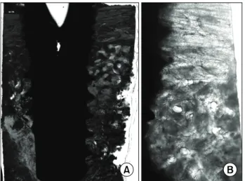
Fig. 1. (A) Low power view showed absence of new bone formation above the first thread level (asterisk) and some peri-implant spaces devoid of new bone formation (arrows). (B) Higher power view of the right half of the left figure showed new bone formation above the first thread level. There were some peri-implant spaces devoid of new bone formation (arrows). Villanueva osteochrome bone stain, A: ×12.5, B: ×40.
Fig. 2. (A) Low power view showed almost complete new bone formation above the first thread level and peri-implant spaces.
(B) Higher power reverse view of the left half of the left figure showed almost complete new bone formation around the implant.
Villanueva osteochrome bone stain, A: ×12.5, B: ×40.
는 골이 접촉되어 있는 길이/전체 길이×100 (%)을 이용하여 구하였다. 더불어 임플란트 나사선 사이에 형성된 신생골의 면적 을 백분율로 산출하여 신생골 충전율(New bone formation rate, NBFR)을 얻었다. 통계적 평가를 위해 임플란트-골 접촉률(BIC) 과 신생골 충전율(NBFR)을 평가하였다. 연조직에 인접한 피질골 최상방 1 mm 부분을 level 1로 표기하였고, level 1 하방의 1 mm 층을 level 2라고 표기하였다.
3) 통계학적 분석
본 연구에서 측정한 ISQ, BIC, NBFR의 통계적 분석을 위하여 SPSS version 17 (SPSS Inc., Chicago, IL, USA)을 이용한 ANOVA 검정을 사용하였으며, P 값이 0.05보다 작은 경우에 통계학적으로 유의성이 있는 것으로 간주하였다.
결 과
1. 안정성 평가
1) 4주 경과 시 ISQ 계수
각 군당 2번의 ISQ를 측정하였고 평균값을 Table 2에 표기하 였다. 4주 경과 후의 ISQ는 79.2±5.1로 임플란트 식립 직후보다 유의적으로 증가하였다.
2) 8주 경과 시 ISQ 계수
8주 경과 후 ISQ 계수는 80.3±4.0으로 식립 직후보다 유의적 으로 임플란트 안정성이 증가하였다. ISQ 계수는 4주군보다 평균 값은 증가하였으나 4주군과 8주군 간 유의적인 차이는 관찰되지 않았다(Table 2).
2. 조직학적 소견 1) 4주군
임플란트 계면을 따라 신생골 형성이 관찰되었으며 소주골의 두께가 두꺼워진 소견이 관찰되었다. 수질골 부위의 골형성은 제한적이었다(Fig. 1).
2) 8주군
매식 후 8주에서는 임플란트 계면을 따라 피질골과 수질골
부분 모두에서 증가된 신생골 형성 및 골성숙 소견이 관찰되었다
(Fig. 2).
Table 4. Mean percentages of bone-implant contact rate in the experimental groups at 4 and 8 weeks after placement of implants (%)
Experimental group 4 wk group
8 wk group
Level 1 Level 2 Level 1 Level 2
85.1±10.8 88.1±13.8 88.5±14.4 95.3±3.1 Level 1: at uppermost 1 mm bone level adjacent to soft tissues, level 2: at next 1 mm bone level.
Table 3. Mean percentages of new bone formation rate in the experimental groups at 4 and 8 weeks after placement of implants (%)
Experimental group 4 wk group
8 wk group
Level 1 Level 2 Level 1 Level 2
90.5±6.6 92.9±4.1 90.1±11.5
95.9±2.3 Level 1: at uppermost 1 mm bone level adjacent to soft tissues, level 2: at next 1 mm bone level.
3. 조직형태계측학적 분석
4주군과 8주군에서 실험군의 평균값과 표준편차는 다음과 같 다(Table 3, 4). 4주군과 8주군 모두 NBFR은 level 2가 level 1보다 평균값은 높았으나 모두 유의적 차이는 관찰되지 않았다 ( P >0.05). 또한 level 1에서 4주군과 8주군, level 2에서 4주군 과 8주군의 유의적 차이도 관찰되지 않았다. 4주군 BIC에서도 마찬가지로 level 2가 level 1보다 평균값은 높았으나 모두 유의적 차이는 관찰되지 않았다( P >0.05). 또한 4주군과 8주군의 유의 적 차이도 관찰되지 않았다.
고 찰
HA 코팅 임플란트는 우수한 생체적합성을 보이며, HA가 생체 친화성을 가지는 이유로 HA는 serum protein과 성장인자의 결 합력이 우수하며 따라서 골모세포의 부착과 증식을 촉진시킬 수 있다는 근거도 제시되었다[7,8]. 이후 HA 코팅 임플란트는 임플란 트와 골 접촉 강도가 증가하며 특히 골질이 불량한 부위에서 부하 시기를 단축시킬 수 있다는 장점으로 여러 임플란트 표면처 리 방식이 개발되었다[9,10]. 그러나 과거의 HA 코팅 임플란트는 제품에 따라 코팅의 질과 두께에 많은 차이가 있었으며 임플란트 식립 시 코팅층이 벗겨진다는 단점이 있었고, 특히 치밀한 골에 식립 시 이는 자주 나타났다. 탈락된 HA는 골유착을 파괴시키며 염증반응을 나타낼 수 있다는 가능성이 있다[11]. 또한 노출된 HA 표면은 감염의 위험성이 높아 임플란트 조기탈락의 원인이 될 수 있다고 하였다[4].
그러나 최근의 HA 코팅 임플란트는 이 같은 단점을 보완하였 으며 HA 코팅 임플란트의 생존율은 점차 늘어나고 있다[12,13].
HAPTITE는 상온에서 코팅하는 방법을 통해 기존의 고온 플라스 마 기법으로 HA 코팅 시 열팽창에 의해 일어나는 미세균열의 문제점을 해결하고자 하였다. 또한 Heimann[14]의 연구에서 밝 힌 바와 같이 저온 코팅기법은 고온에서의 HA 입자가 탈수산화되 며 β-TCP 등의 상이 변한다는 한계를 극복할 수 있다. 또한 코팅 두께에서도 변화가 있었다. 두꺼운 HA코팅은 금속 이온이 용출되는 것을 막아줄 수 있으나 취성이 크고 잔류응력을 내재하
여 후에 금균열이 생길 수 있는 가능성이 있다[15]. 기존의 분사형 HA 코팅 기법이 30∼200 μm였다면 최근의 임플란트는 코팅 두께를 점점 줄여가고 있으며, HAPTITE는 2 μm 이하의 두께로 HA가 코팅되는 초박막코팅기법을 통하여 불균일한 코팅과 박리 현상을 최소화하고자 하였다[16].
본 실험은 임플란트를 골높이보다 1 mm 이상 높게 식립하여 의도적으로 HA 코팅층이 연조직에 노출되도록 계획하였다. 실험 결과상 HA 코팅의 연조직 노출에 의한 감염은 관찰되지 않았다.
ISQ 계수는 1 mm 이상 노출되게 식립되었음에도 불구하고 기존 의 연구와 유사한 정도의 안정성을 관찰할 수 있었다[17-19].
임플란트 식립 시 가장 골과 접촉이 잦고 연조직에 인접한 피질골의 최상방 1 mm 구간에서는 NBFR과 BIC 측면에서 모두 우수한 골형성 소견을 관찰할 수 있었으며 이는 다음 1 mm 구간과 유의적인 차이가 없어, HA 코팅이 연조직에 노출되어도 감염의 위험성이 낮을 뿐 아니라 HA의 박리 측면에서 안전하다는 결과를 얻을 수 있었다. 아울러 4주와 8주간 골형성의 차이가 적어 기존의 연구보다 빠른 골유도 소견이 관찰되었음을 확인할 수 있었다[20,21]. 다만 본 연구는 샘플의 수가 적어 통계학적으로 유의한 차이를 발견하기 어려웠을 가능성이 있으며, 피질골의 BIC를 관찰하였기 때문에 기존의 연구에서 밝힌 임플란트 표면처 리에 따른 평균적 BIC와 객관적인 비교를 하기는 어렵다[22].
또한, 본 연구에서는 실제 임플란트가 노출되는 구강 내 환경이 아닌 대퇴골에 식립하여 노출된 임플란트 주변이 치은이 아닌 근육층이었던 점과, 노출의 정도를 1 mm로 국한시킨 한계점을 가지고 있어 추후 이에 대한 추가적인 연구가 필요하리라 생각한 다.
결 론
본 연구에서는 상온초박막 코팅기법의 HA 코팅 임플란트를
의도적으로 연조직에 노출시켜 식립 후 임플란트 안정성과 골유착
정도를 평가해보고자 하였으며, 노출된 HA 코팅 임플란트도 안정
적이며 우수한 골유착 소견을 관찰할 수 있었다. 이번 연구에서는
샘플의 수가 적었음에도 불구하고 불구하고 연조직에 노출된 HA
코팅 임플란트는 일관된 임플란트 안정성과 우수한 골형성 소견을
보여 HA 코팅 임플란트의 단점에 대한 우려를 줄일 수 있을 것으로 생각한다.
References
![Table 1. The number of placed implants in the dogs Group n (4 wk) n (8 wk) Experimental group 8 8서 론임플란트 표면구조는 초기 골유착에 중요한 요소로 알려져 있으며 골유착 시기를 단축시키고 조기하중을 위하여 여러 표면처 리 방식이 개발되어 왔다[1]](https://thumb-ap.123doks.com/thumbv2/123dokinfo/5114377.330986/2.892.463.815.178.227/experimental-론임플란트-표면구조는-골유착에-중요한-단축시키고-조기하중을-개발되어.webp)