Background and Purpose Cerebral visual impairment (CVI) is an underdiagnosed condi- tion in children, and its assessment tools have focused on older children. We aimed to devel- op a parental questionnaire for cerebral visual impairment (PQCVI) for screening CVI in young children.
Methods The PQCVI comprised 23 questions based on a modified version of Houliston and Dutton’s questionnaire for older children. The PQCVI with neurocognitive function tests was applied to 201 child–parent pairs with typically developing children younger than 72 months (age 32.4±20.1 months, mean±standard deviation). The children were classified into six age groups. The normative data, cutoff scores, and internal reliability were assessed and item anal- ysis was performed. We referred to the total score for all questions as the cerebral visual func- tion (CVF) score.
Results The normative data showed that the CVF score and the scores corresponding to ventral-stream and dorsal-stream visual functions plausibly increased with age. The scores rapidly reached 90% of their maximum values up to the age of 36 months, after which they in- creased slowly. Cronbach’s alpha for all questions across all age groups was 0.97, showing ex- cellent consistency. The item difficulty and item discrimination coefficients showed that the questions were generally adequate for this age stage.
Conclusions The PQCVI items produced reliable responses in children younger than 72 months. The rapid increase in scores before the age of 3 years supports the importance of early identification of CVI. Following additional clinical verification, the PQCVI may be use- ful for CVI screening.
Key Words vision disorders, development, neurodevelopmental disorders, preschool children, early diagnosis.
Development of the Parental Questionnaire for Cerebral Visual Impairment in Children Younger than 72 Months
INTRODUCTION
Cerebral visual impairment (CVI) encompasses various conditions related to problems in higher-order visual processing caused by injury to the retrogeniculate pathways and brain structures.1,2 Children with cerebral or cortical visual problems have difficulties in recog- nizing people, objects, depth, and movement, simultaneous perception, orientation and nav- igation, and in performing visual field tests.1,3,4 Problems with higher visual functions may be outstanding or unnoticed, and may lead to the underdevelopment of motor coordina- tion and cognitive abilities, possibly even affecting socioemotional development.5,6 Thus, early detection and intervention of CVI is important for preventing negative consequences.
There are various causes of CVI, including periventricular leukomalacia (PVL) or hypoxic ischemic encephalopathy in premature infants, hydrocephalus, meningoencephalitis, trau- ma, and genetic disorders. CVI is an important cause of visual impairment in children with Jin-Hwa Moona, Gun-Ha Kimb
Sung Koo Kimc, Seunghyo Kimd Young-Hoon Kime, JoonSik Kimf Jin-Kyung Kimg, Byoungho H. Nohh Jung Hye Byeoni, Jung Sook Yeomj Baik-Lin Euni, So Hee Euni Jieun Choik, Hee Jung Chungl
a Department of Pediatrics, Hanyang University College of Medicine, Seoul, Korea
b Department of Pediatrics,
Korea Cancer Center Hospital, Seoul, Korea
c Department of Pediatrics, Dongtan Sacred Heart Hospital, Hallym University College of Medicine, Hwasung, Korea
d Department of Pediatrics,
Jeju National University School of Medicine, Jeju, Korea
e Department of Pediatrics, College of Medicine,
The Catholic University of Korea, Seoul, Korea
f Department of Pediatrics,
Keimyung University Dongsan Hospital, Daegu, Korea
g Department of Pediatrics, Daegu Catholic University School of Medicine, Daegu, Korea
h Department of Pediatrics,
Kangwon National University Hospital, Chuncheon, Korea
i Department of Pediatrics, Korea University College of Medicine, Seoul, Korea
j Department of Pediatrics, Gyeongsang National University School of Medicine, Gyeongsang Institute of Health Science, Jinju, Korea
k Department of Pediatrics, Seoul National University College of Medicine,
SMC-SNU Boramae Medical Center, Seoul, Korea
l Department of Pediatrics, National Health Insurance Service Ilsan Hospital, Goyang, Korea
pISSN 1738-6586 / eISSN 2005-5013 / J Clin Neurol 2021;17(3):354-362 / https://doi.org/10.3988/jcn.2021.17.3.354
Received September 29, 2020 Revised February 10, 2021 Accepted February 10, 2021 Correspondence
Hee Jung Chung, MD, PhD Department of Pediatrics, National Health Insurance Service Ilsan Hospital, 100 Ilsan-ro, Ilsandong-gu, Goyang 10444, Korea Tel +82-31-900-0530
Fax +82-31-900-0343 E-mail [email protected]
cc This is an Open Access article distributed under the terms of the Creative Commons Attribution Non-Com- mercial License (https://creativecommons.org/licenses/by-nc/4.0) which permits unrestricted non-commercial use, distribution, and reproduction in any medium, provided the original work is properly cited.
JCN
Open Access ORIGINAL ARTICLEMoon JH et al.
JCN
JCN
Open Accessvarious disabilities. It is found in 40–50% of children with ce- rebral palsy and in 21–47% of children born prematurely.7 CVI may exist in patients without organic disease and with normal brain imaging findings.8,9
CVI remains an underdiagnosed condition in children. Rou- tine eye examinations and cognitive function tests might not detect abnormalities in higher visual functions. Undiagnosed patients may be considered clumsy and have delayed visuo- spatial learning. Therefore, CVI-specific screening tools for the early detection of such children are required.5,8
Typical currently used diagnostic tools for visuospatial dis- ability in children and adults are the Developmental Test of Visual Perception (DTVP), Beery–Buktenica Developmental Test of Visual–Motor Integration (Beery VMI), and Motor- Free Visual Perception Test (MVPT). Most tests require the motor performance of patients (DTVP and VMI) and are aimed at subjects older than 4 years (DTVP and MVPT). The scores on these tests may be more affected by general intel- ligence than pure visual perception ability. Considering that it is not easy to examine young children, structural interviews of parents or caregivers who observe children over a long time can also be helpful.
There are few CVI-specific tests for younger children. The ABCDEFV (A test Battery of Child Development for Exam- ining Functional Vision) for children aged 0–36 months and computerized assessment tools for visual perception deficits in preschool children, such as the L94 and CVIT 3-6 (Chil- dren’s Visual Impairment Test for 3- to 6-year-olds), are used both clinically and in research.9-12 For clinical ease of use, short- ened screening tools such as Five Questions, CVI Inventory, and CVI Questionnaire have been developed.13-15 These are parental questionnaires with adequate reliability and validi- ty, but the target age of most surveys is over 5 years, and the available normative data are insufficient.
In Korea, the National Health Screening Program for In- fants and Children (NHSPIC) of the National Health Insur- ance Service provides a periodic health screening program for children younger than 72 months from across the country.16 Although the national birth rate is decreasing in Korea, the survival rate of low-birthweight infants has increased more than tenfold since the 1960s.17 For children who have a high risk of developing CVI, such as those with PVL caused by pre- maturity or cerebral palsy, additional screening for CVI could be helpful in its early detection.18-20
With this background, a taskforce of the Korean Child Neu- rology Society (KCNS) was convened to develop a CVI screen- ing tool for use in children younger than 72 months. The task- force developed a parental questionnaire for cerebral visual impairment (PQCVI) based on Houliston and Dutton’s ques- tionnaire that was previously developed for cognitive visual
problems in children with hydrocephalus.14 In the present study, the newly developed PQCVI was administered to chil- dren with typical development who were younger than 72 months. The normative data, cutoff scores, internal reliabil- ity, and item analysis of the PQCVI were assessed.
METHODS
Development of the PQCVI
The KCNS taskforce for the development of a CVI screening tool had met periodically and critically reviewed the origi- nal questionnaire. The important considerations addressed in the reviews were whether the tool was appropriate for eval- uating CVI in young children and the cultural suitability of the questions. The original questionnaire used in Houliston and Dutton’s research was targeted at children aged 5–12 years.14 The KCNS taskforce sorted the questions by age and modi- fied them for use in infants and younger children, which re- sulted in the questionnaire being modified to 23 questions.
Some questions were combined (e.g., questions 10 and 11, and 13 and 14 of Houliston and Dutton’s study were combined into questions 12 and 23, respectively, of our study) or split (from question 17 of Houliston and Dutton’s study were split into questions 3 and 8 of our study).14 New questions for younger children were added (questions 6 and 7 of our study). The fi- nal version used in the present study was written in Korean.
Table 1 lists the PQCVI questions translated into English in the order from that with the highest mean score to the low- est mean score. Nine questions (questions 1, 4, 5, 10, 11, 13, and 19–21) were assumed to mainly reflect ventral-stream function, and 13 questions (questions 2, 3, 6–8, 12, 14–18, 22, and 23) were assumed to mainly reflect dorsal-stream func- tion. Question 9 was related to binocular vision.
Parents were asked to choose an answer for each question that corresponded to their child’s behavior on the following scale: never, 1; occasionally, 2; most of the time, 3; always, 4;
and don’t know, 5. Each answer from 1 to 4 was scored as the corresponding points, while answer 5 (“don’t know”) was ex- cluded from the scoring. We designated the total score for the 23 questions as the cerebral visual function (CVF) score in this questionnaire (range, 23–92). The scores for 9 questions related to ventral-stream cerebral visual function (vCVF) (range, 9–36) and 13 questions related to dorsal-stream ce- rebral visual function (dCVF) (range, 13–52) were analyzed separately.
Subjects
The PQCVI was developed and then administered to subjects from December 2016 to March 2018. Volunteer child–parent pairs who visited for the NHSPIC were enrolled. They were
Cerebral Visual Impairment Screening in Children
JCN
recruited from 11 hospitals across the country. The inclusion criteria were children younger than 72 months within the nor- mal development ranges on the Korean Developmental Screen- ing Test (K-DST) for infants and children, and having normal visual acuity in vision screening.21 The exclusion criteria were children already diagnosed with a neurodevelopmental dis- order, development quotient (DQ) <70 or intelligence quotient (IQ) <70, or ophthalmological, visuospatial, hearing, or mo- tor problems, or other disabilities. The subjects were divided into six age groups: 1) <12 months, 2) 12–23 months, 3) 24–
35 months, 4) 36–47 months, 5) 48–59 months, and 6) 60–71 months. All research protocols were approved by the Insti- tutional Review Board of Ilsan Hospital (NHIMC 2017-05- 004). Written informed consent was obtained from parents for both themselves and on behalf of their children.
Administration of PQCVI and neuropsychological tests
The PQCVI and developmental screening using the K-DST were administered to all of the included children. DQ and IQ were determined using the Korean version of Bayley Scales of Infant Development-II (age <42 months), the Korean ver-
sion of the Wechsler Preschool and Primary Scale of Intelli- gence (K-WPPSI), and K-WPPSI-IV (age ≥42 months). K- Beery VMI-6 (age 30–48 months) and K-DTVP-II (age ≥48 months) were applied to assess visuospatial function. The sub- jects with abnormal results on K-DTVP-II or K- Beery VMI-6, a visuospatial index (VSI) of <70 on K-WPPSI-IV, or statisti- cally significant differences in verbal IQ (VIQ) and perfor- mance IQ (PIQ) on the K-WPPSI test (VIQ exceeding PIQ by >12, and PIQ <90) were excluded from the analysis due to the likely presence of visuospatial dysfunction. All neuro- psychological tests were conducted by child psychologists at each hospital.
Statistical analyses
Demographic and normative data for the study population are presented as mean±standard deviation (SD) values for continuous variables and as frequencies with percentages for categorical variables. Normative outcomes were compared between age groups using the Kruskal–Wallis test, and Bon- ferroni correction was applied to the post-hoc comparison.
Cutoffs for the CVI scores were obtained by estimating the scores corresponding to mean minus 2 SDs and mean minus Table 1. Parental questionnaire for cerebral visual impairment (PQCVI)
The questions below are related to the visual perception-related behavior of your child. Read these questions and choose the best answer for your child:
never, 1; occasionally, 2; most of the time, 3; always, 4; or don’t know, 5. The items in this list are ordered from the highest to the lowest mean score.
Question Answer
1. Does your child recognize mother/father’ face before you speak? (1) (2) (3) (4) (5)
2. Does your child reach out and grasp objects? (1) (2) (3) (4) (5)
3. Is your child able to see slow-moving objects (e.g., a rolling ball)? (1) (2) (3) (4) (5)
4. Does your child recognize the faces of other family members? (1) (2) (3) (4) (5)
5. Does your child recognize familiar objects (e.g., cup, shoes, or doll)? (1) (2) (3) (4) (5)
6. Does your child find objects covered by a blanket or paper? (1) (2) (3) (4) (5)
7. Does your child pick up a small object with their thumb and index finger? (1) (2) (3) (4) (5) 8. Is your child able to see fast-moving objects (e.g., a moving car)? (1) (2) (3) (4) (5) 9. Does your child eat food from parts of a large plate rather than only from one part? (1) (2) (3) (4) (5)
10. Does your child recognize other people in photographs? (1) (2) (3) (4) (5)
11. Does your child recognize themself in photographs? (1) (2) (3) (4) (5)
12. Does your child find their way well to rooms or the toilet at home? (1) (2) (3) (4) (5)
13. Does your child recognize friends’ faces? (1) (2) (3) (4) (5)
14. Can your child easily find their way to doorways or along corridors? (1) (2) (3) (4) (5) 15. Does your child judge the height of steps without missing their footing? (1) (2) (3) (4) (5) 16. Does your child recognize objects when they themselves are moving quickly? (1) (2) (3) (4) (5) 17. Can your child find objects on a blanket with a complex pattern? (1) (2) (3) (4) (5)
18. Does your child remember well where they put things at home? (1) (2) (3) (4) (5)
19. Can your child differentiate shapes (e.g., triangles, rectangles, and circles)? (1) (2) (3) (4) (5)
20. Can your child gather or match colors? (1) (2) (3) (4) (5)
21. Can your child name colors? (1) (2) (3) (4) (5)
22. Can your child find objects in a complex picture? (1) (2) (3) (4) (5)
23. Does your child easily find their way in new surroundings? (1) (2) (3) (4) (5)
Moon JH et al.
JCN
1 SD for each age group. As a measure of the item internal consistency, Cronbach’s alpha for items was calculated accord- ing to the range of ages.
The item response theory (IRT) was adapted to measure the reliability of the questionnaire.22,23 For each item, the unique item characteristic curve (ICC), which represents the proba- bility of answering correctly according to the subject’s ability, was constructed for all age groups together and for each age group separately. The ICC of an item was analyzed to estimate the item difficulty coefficient (β) and the item discrimination coefficient (α). β measures the difficulty of an item, and it gen- erally ranges from -2 to +2, with a larger value indicating that the item is more difficult. α measures the discrimination ca- pability of an item, and it generally ranges from 0 to +2, with a larger value indicating that the item has a better discrimi- nation capability.
The IRT analysis was conducted using R software (version 3.4.0, R Foundation for Statistical Computing, Vienna, Aus- tria), while all other statistical analyses were performed using the Statistical Package for the Social Sciences (version 24.0, IBM Corp., Armonk, NY, USA). All p values <0.05 were con- sidered statistically significant.
RESULTS
Subject characteristics
Initially 205 children were enrolled. Four children were ex- cluded because they did not meet the inclusion criteria (VSI on K-WPPSI-IV of 67 in one child, VIQ and PIQ mismatch of >12 with PIQ <90 on K-WPPSI in two children, and K- Beery VMI score <70 on K-DTVP-II in one child). Finally, the data of 201 children aged 32.4±20.1 months (89 males and 112 females) were analyzed. Detailed demographic data and neurocognitive function test results are presented in Ta- ble 2, Supplementary Table 1 (in the online-only Data Sup- plement).
Normative data
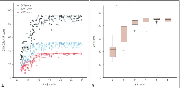
The scores for each of the 23 questions and the CVF scores according to age group are listed in Table 3. The CVF scores increased with the children’s age, being 41.2±10.5, 66.2±14.3, 85.1±5.0, 87.2±5.5, 89.8±2.7, and 89.1±2.9 in Groups A–F, respectively, and 74.0±20.2 for all subjects. The mean CVF scores increased rapidly from Group A to Group C, approach- ing 90% of their maximum values (Fig. 1A, Table 3). The mean CVF score was highest in Group E, which was higher than Group F, but the difference was not statistically significant. The mean CVF score differed significantly between age groups in the overall test (p<0.001). Post-hoc analysis showed signif- icant increases from Group A to Group B (p<0.001) and from
Group B to Group C (p=0.001) (Fig. 1B).
The mean scores for each question (range, 1.0–4.0 points) increased as age increased. When the maximal score of each question was 4.0 points, the score corresponding to 90% (near- mastered) was 3.6. After 36 months, the mean score of each question was >3.6 points for almost all questions (Table 3).
As for the CVF scores, both the vCVF and dCVF scores increased rapidly from Group A to Group C, and then reached a near plateau [Fig. 1A, Supplementary Table 2 (in the online- only Data Supplement)]. The vCVF and dCVF scores also dif- fered significantly between the overall age groups (p<0.001 for both). Post-hoc analyses of the vCVF and dCVF scores showed that there were considerable increases from Group A to Group B (p=0.066 and p=0.002, respectively) and Group B to Group C (p<0.001 and p=0.008).
There were no significant differences between male and fe- male children in total CVF scores or vCVF and dCVF scores.
Cutoff scores for CVI
In each age group, cutoffs for CVI scores were set at the mean minus 2 SDs and the mean minus 1 SD (Table 3). In-depth evaluation was recommended for subjects with a score of the less than the mean minus 2 SDs. For subjects with a score be- tween the mean minus 2 SDs and the mean minus 1 SD, follow up evaluation was recommended. Detailed cutoffs for the to- tal CVF, vCVF, and dCVF scores are presented in Table 3 and Supplementary Table 2 (in the online-only Data Supplement).
Item internal consistency
Cronbach’s alpha for assessing internal consistency of the questionnaire was calculated for all subjects and according to the different age groups. For all subjects, Cronbach’s alpha for all 23 questions was 0.97, which indicated excellent consisten- cy. Cronbach’s alpha values for all subjects for the 9 questions related to vCVF and the 13 questions related to dCVF showed good consistency (0.84) and excellent consistency (0.96), re- spectively. The Cronbach’s alpha values in each age group are presented in Table 4.
Table 2. Demographic data of the subjects Age
group
Age range, months
Number of subjects
Sex (male:female)
Age, months (mean±SD)
A <12 38 18:20 7.7±2.2
B 12–23 46 19:27 17.0±3.4
C 24–35 31 11:20 29.0±3.2
D 36–47 29 17:12 41.1±4.0
E 48–59 29 14:15 52.8±3.5
F 60–71 28 10:18 65.1±3.2
Total ≤71 201 89:112 32.4±20.1
SD: standard deviation.
Cerebral Visual Impairment Screening in Children
JCN
Item response analysis
Item response analysis was performed for all subjects and for each age group (Table 5). For all subjects, the β values for the 23 items were between -2.05 and 1.11, and the α values were between 1.78 and >2.00. In Groups A to F, each item corre- sponding to its age stage generally showed β values between -4.67×10⁸ and +2.00 and α values between 0.0 and >2.00 (Ta- ble 5). These β and α values were considered to be adequate given that items required different levels of development and were implemented at different ages. The overall ICC varied with age according to a sigmoid pattern.
DISCUSSION
After the first descriptions of visual perception disorders in children with brain injuries in the 1980s, CVI has been de- fined differently by authors in several conditions.1-3,8,24,25 In a recent systematic review, Sakki et al.25 proposed that CVI could be defined as “A verifiable visual dysfunction which cannot be attributed to disorders of the anterior visual path- ways or any potentially co-occurring ocular impairment.”8 Children with CVI can present with difficulties in recogni- tion, text reading, spatial memory, handwriting, object dis- crimination, spatial exploration, sequential movement, at- tention, motor planning, and spatial reasoning. CVI could Table 3. Normative scores on the PQCVI according to age group
Age group*
A B C D E F Total
PQCVI score Question
1 3.7±0.6 4.0±0.0 4.0±0.0 4.0±0.0 4.0±0.0 4.0±0.0 4.0±0.3
2 3.7±0.7 4.0±0.2 4.0±0.0 4.0±0.0 4.0±0.0 4.0±0.0 3.9±0.3
3 3.7±0.7 3.9±0.4 4.0±0.0 4.0±0.0 4.0±0.0 4.0±0.0 3.9±0.4
4 3.3±1.0 3.8±0.4 4.0±0.0 4.0±0.0 4.0±0.2 4.0±0.2 3.8±0.5
5 2.9±1.2 3.9±0.4 4.0±0.0 4.0±0.0 4.0±0.0 4.0±0.0 3.8±0.7
6 2.7±1.1 3.8±0.4 4.0±0.2 3.9±0.3 4.0±0.0 3.9±0.3 3.7±0.7
7 2.7±1.1 3.8±0.4 4.0±0.0 3.9±0.3 4.0±0.0 4.0±0.0 3.7±0.7
8 3.1±1.0 3.7±0.5 3.9±0.3 4.0±0.2 4.0±0.2 4.0±0.0 3.7±0.6
9 2.8±1.2 3.7±0.5 4.0±0.2 4.0±0.2 4.0±0.2 3.9±0.4 3.7±0.7
10 1.5±0.9 3.8±0.5 4.0±0.2 4.0±0.2 4.0±0.0 4.0±0.2 3.6±1.0
11 1.3±0.9 3.5±0.8 3.9±0.3 4.0±0.2 4.0±0.2 4.0±0.0 3.5±1.1
12 1.5±1.0 3.3±1.1 4.0±0.2 4.0±0.2 3.9±0.3 4.0±0.2 3.4±1.1
13 1.5±0.9 3.0±1.1 3.8±0.5 3.9±0.3 4.0±0.2 4.0±0.0 3.3±1.1
14 1.3±0.8 3.0±1.2 3.8±0.5 3.9±0.3 3.8±0.4 3.9±0.3 3.2±1.1
15 1.0±0.2 2.5±1.0 3.7±0.4 3.8±0.4 4.0±0.0 4.0±0.2 3.1±1.2
16 1.1±0.4 2.5±1.1 3.5±0.6 3.8±0.5 4.0±0.2 3.9±0.3 3.1±1.2
17 1.5±0.9 2.9±1.2 3.5±0.6 3.7±0.5 3.9±0.4 3.9±0.4 3.1±1.1
18 1.1±0.6 2.9±1.1 3.5±0.6 3.8±0.4 3.7±0.5 3.6±0.6 3.1±1.2
19 1.0±0.0 2.1±1.1 3.6±0.7 4.0±0.2 4.0±0.0 4.0±0.0 3.0±1.3
20 1.0±0.0 1.8±1.0 3.6±0.7 3.9±0.3 3.9±0.4 4.0±0.0 2.9±1.3
21 1.0±0.0 1.4±0.7 3.3±0.9 3.8±0.6 4.0±0.0 4.0±0.0 2.7±1.4
22 1.0±0.0 1.9±1.0 3.2±0.8 3.6±0.6 3.6±0.5 3.7±0.5 2.7±1.2
23 1.0±0.2 1.7±1.0 2.8±0.9 3.4±0.7 3.4±0.7 3.4±0.7 2.5±1.2
Total 2.0±1.1 3.1±0.8 3.7±0.3 3.9±0.2 3.9±0.2 3.9±0.2 3.4±0.4
CVF score† 41.2±10.5 66.2±14.3 85.1±5.0 87.2±5.5 89.8±2.7 89.1±2.9 74.0±20.2
Cutoff CVF scores
Mean minus 1 SD 31 52 80 82 87 86 54
Mean minus 2 SDs 23 38 75 76 84 83 34
Normative data are mean±SD values.
*Age group: A, <12 months; B, 12–23 months; C, 24–35 months; D, 36–47 months; E, 48–59 months; F, 60–71 months, †CVF score is the sum for the 23 questions.
CVF: cerebral visual function, PQCVI: parental questionnaire for cerebral visual impairment, SD: standard deviation.
Moon JH et al.
JCN
influence the future development in various domains, learn- ing, and social interaction.1,3-6
Several screening tools for CVI have been developed. The CVI Inventory reported by Macintyre-Beon et al.26 comprises 51 questions tested in children with CVI (aged 5–16.5 years) and school children. The subsections of the CVI Inventory include visual fields, perception of movement, search, guid-
ance of movement, attention, crowded scenes, recognition, and navigation.26 The CVI Questionnaire reported by Orbi- tus et al.15 comprises 46 questions answered by parents. This questionnaire estimates the visual attitude, ventral stream, dorsal stream, and other factors in children aged 3–17 years.
Houliston and Dutton’s questionnaire comprises 22 questions and was applied to children older than 5 years with hydro- cephalus.14 That questionnaire includes questions about dif- ficulties related to visual perception that parents usually com- plain of and questions about visual field defects and visual inattention. A questionnaire for children younger than 24 months was developed by Pueyo et al.27 However, it is not a screening tool and was developed to help in ophthalmologi- cal assessments of visual behavior. The CVI screening ques- tionnaires described above were mostly developed for older children, whereas the present PQCVI was designed to assess the usefulness of CVI screening in children younger than those covered by the existing tools.
The present study found that the PQCVI scores increased with age. The mean CVF score already approached nearly half (44.8%) of the maximum score by the age of 12 months, over two-thirds (72.0%) at 12–23 months, 92.5% at 24–35 months, and approached the maximum score up to 71 months (Ta- ble 3, Fig. 1). The patterns in vCVF and dCVF scores were similar. Thus, our data show that there is significant develop- ment of higher visual functions until 5–6 years old, and the Table 4. Cronbach’s alpha values for the PQCVI questions according
to age group
Age group* Cronbach’s alpha
CVF score† vCVF score‡ dCVF score§
A 0.903 0.800 0.737
B 0.920 0.919 0.829
C 0.808 0.746 0.756
D 0.849 0.773 0.787
E 0.776 0.671 0.494
F 0.675 0.590 -0.043
Total 0.974 0.840 0.955
*Age group: A, <12 months; B, 12–23 months; C, 24–35 months; D, 36–47 months; E, 48–59 months; F, 60–71 months, †CVF score is the sum for the 23 questions, ‡vCVF score is the sum score for the nine questions related to vCVF (questions 1, 4, 5, 10, 11, 13, and 19–21),
§dCVF score is the sum score for the 13 questions related to dCVF (questions 2, 3, 6–8, 12, 14–18, 22, and 23).
CVF: cerebral visual function, dCVF: dorsal-stream cerebral visual function, PQCVI: parental questionnaire for cerebral visual impair- ment, vCVF: ventral-stream cerebral visual function.
100
80
60
40
20
0
100
80
60
40
20
0
CVF/dCVF/vCVF scores CVF scores
0 A
CVF score dCVF score vCVF score
Age (months) Age group
12 24 36 48 60 72 B C D E F
Fig. 1. Graphs of scores on the parental questionnaire for cerebral visual impairment according to age and age group. A: Scatter plots of scores for cerebral visual function (CVF) (black crosses), dorsal-stream cerebral visual function (dCVF) (blue diamonds), and ventral-stream cerebral visual function (vCVF) (red triangles) according to age. B: Box plots of the CVF scores according to age group. The mean CVF scores differed significantly between age groups in the overall test (p<0.001). Post-hoc analysis showed significant increases from Group A to Group B (*p<0.001), and from Group B to Group C (†p=0.001). Age group: A, <12 months; B, 12–23 months; C, 24–35 months; D, 36–47 months; E, 48–59 months; F, 60–71 months.
A B
* †
^
Cerebral Visual Impairment Screening in Children
JCN
basis of CVF is formed before the age of 3 years.
In the dual-stream hypothesis of visual information pro- cessing, the ventral stream comprises the occipitotemporal pathway to the anterior temporal cortex. Ventral-stream func- tion is responsible for visual recognition and visual memory, and is related to face and object recognition (also called the
“what” pathway).28,29 It works in cooperation with the dorsal stream when solving complex visual problems. Integrating the ventral and dorsal streams in terms of color, speed, and form provide the information necessary for intermediate ob- ject representations in the dorsal stream.30 In addition, direct evidence of the involvement of crosstalk networks between ventral and dorsal streams in skilled hand actions has been demonstrated by white-matter tractography.31
The recognition of faces is examined by questions 1 (par- ent), 4 (family), and 13 (friends) of the PQCVI. The average scores for questions related to the recognition of parents, fam- ily, and friends increased sequentially with age, and the recog- nition of friends was nearly mastered by 24–36 months. The sequential recognition of faces may be the basis of pretend play with other people emerging at approximately 18 months
and parallel play with friends appearing at approximately 24 months.32 Recognition of photographs is reviewed by ques- tions 10 (other people) and 11 (themself). The sequential de- velopment of face and photograph recognition may also be related to the development of socioemotional behavior and self-concept.
The recognition of shape and objects is examined by ques- tions 5 (object) and 19 (shape). The ability to recognize famil- iar objects developed earlier, and was accomplished before 24 months. However, shape recognition developed later, and was not nearly mastered until 36 months. This shows that mental representations began with individual objects and then pro- gressed to abstract forms.
The matching and naming of colors are examined by ques- tions 20 and 21, respectively. The score was slightly lower for color naming than for color matching, but both abilities were nearly mastered after 36 months. Color preferences are known to develop as early as 3 months,33 while the ability to match colors develops at approximately 28 months.32 Delayed suc- cess with matching and naming colors relative to the acqui- sition of color vision is related to cognition and language de- Table 5. Item difficulty (β) and item discrimination (α) coefficients of the PQCVI according to age group
Question Age group*
A B C D E F Total
1 -1.204/2.671 5.998×10⁷/0.000 -1.133×10⁸/0.000 -2.045×10⁸/0.000 -4.678×10⁸/0.000 -2.124×10⁸/0.000 -2.045/22.149 2 -0.375/2.447 -1.365/1.546 -1.133×10⁸/0.000 -2.045×10⁸/0.000 -1.048/54.359 -3.029/1.177 -1.511/2.263 3 -0.971/5.201 -2.346/1.638 -1.133×10⁸/0.000 -2.045×10⁸/0.000 -4.678×10⁸/0.000 -2.124×10⁸/0.000 -1.949/3.972 4 0.604/1.514 -1.540/1.274 -2.926/1.409 -3.104/0.775 -4.678×10⁸/0.000 -2.181/1.041 -1.131/1.775 5 0.311/3.687 -1.046/3.194 -1.133×10⁸/0.000 -1.808/2.433 -4.678×10⁸/0.000 -2.124×10⁸/0.000 -1.100/3.476 6 -0.901/3.866 -1.345/29.238 -1.133×10⁸/0.000 -2.045/0.000 -4.678×10⁸/0.000 -2.124×10⁸/0.000 -1.807/3.761 7 0.115/4.013 -0.631/1.704 -1.334/2.211 -4.656/0.776 -1.734/2.193 -2.124×10⁸/0.000 -0.997/2.352 8 0.289/4.636 -0.457/1.639 -4.502/0.807 2.446/-0.136 -1.048/54.359 -1.412/1.492 -0.876/2.145 9 0.159/5.049 -1.328/1.625 -1.133×10⁸/0.000 -2.045×10⁸/0.000 -4.678×10⁸/0.000 -2.124×10⁸/0.000 -1.190/4.319 10 1.453/3.074 -0.440/2.628 -1.723/48.314 -1.724/50.591 -4.678×10⁸/0.000 -4.563/0.739 -0.629/4.724 11 1.479/2.948 0.189/3.667 -1.780/1.678 -1.724/50.591 -1.048/54.359 -2.124×10⁸/0.000 -0.399/5.858 12 3.088/1.265 0.774/4.056 -1.888/1.206 -1.382/30.129 -1.048/54.359 -2.124×10⁸/0.000 -0.154/6.616 13 6.115×10⁶/0.000 1.449/3.231 -9.650×10⁻¹/1.363 -1.724/50.591 -4.678×10⁸/0.000 -2.124×10⁸/0.000 0.000/198.004 14 6.115×10⁶/0.000 1.877/45.932 -2.160×10⁻¹/0.834 -7.570/24.728 -4.678×10⁸/0.000 -2.124×10⁸/0.000 0.207/5.177 15 6.115×10⁶/0.000 2.027/1.802 -8.560×10⁻¹/1.259 -1.173/2.498 -9.050×10⁻¹/2.208 -2.124×10⁸/0.000 0.150/4.919 16 6.115×10⁶/0.000 1.355/14.385 -6.620×10⁻¹/35.012 -1.386/1.666 -4.678×10⁸/0.000 -1.360/29.921 0.064/4.872 17 6.115×10⁶/0.000 1.203/3.228 -5.700×10⁻²/31.172 -1.167/4.275 -1.734/2.193 -1.019/40.799 0.093/5.221 18 3.088/1.265 0.654/3.035 -1.800×10⁻²/1.649 -6.420/12.578 -6.610×10⁻¹/35.510 -0.977/2.179 0.143/2.681 19 6.115×10⁶/0.000 2.073/1.721 3.140×10⁻¹/3.741 -1.400/2.166 7.400×10⁻²/10.720 -0.460/2.154 0.690/3.360 20 1.383/42.794 0.521/2.654 -1.490×10⁻¹/1.324 -8.330/2.036 -4.700×10⁻²/26.860 -0.771/1.283 0.179/2.089 21 1.041/59.869 -0.357/2.654 -3.750/1.005 -1.724/50.591 -1.358/1.635 -1.360/30.801 -0.571/3.533 22 1.041/59.869 0.381/2.452 -2.856/0.713 -1.382/29.946 -6.710×10⁻¹/2.614 -1.233/2.576 -0.249/2.935 23 6.115×10⁶/0.000 3.417/0.920 1.610/1.394 1.880/2.216 2.250×10⁻¹/1.062 0.548/22.210 1.109/2.294 Data are β/α values.
*Age group: A, <12 months; B, 12–23 months; C, 24–35 months; D, 36–47 months; E, 48–59 months; F, 60–71 months.
PQCVI: parental questionnaire for cerebral visual impairment.
Moon JH et al.
JCN
velopment.
The dorsal stream comprises the occipitoparietal pathway and is responsible for spatial working memory, visually guid- ed action, and navigation (also called the “where” or “how”
pathway).28 The dorsal stream is more vulnerable in children with periventricular white-matter injuries, autistic spectrum disorders, and Williams syndrome.7,29,34 Questions 2 (reach- ing and grasping) and 7 (pincer grasping) examine visually guided action and depth perception, both of which relate to fine motor development. Parietopremotor pathways mediate reaching and grasping and visually guided actions.28 In healthy children, reaching and grasping is possible at approximately 4 months, and pincer grasping is possible at 9–12 months. Our results show that reaching and grasping is nearly mastered before 12 months, while pincer grasping is nearly mastered after 12 months.
Motion perception is examined by questions 3 (slow-mov- ing object), 8 (fast-moving object), and 16 (while self-mov- ing). Motion perception is controlled by interactions between the visual and vestibular systems. The motion perception pro- cess is attributed to both ventral-stream function (stimuli du- ration) and dorsal-stream function (perceived vection).35 In our study, the abilities to perceive the motions of slow-mov- ing and fast-moving objects were achieved before 12 months and after 12 months, respectively, while self-moving was per- ceived after 36 months.
Orientation and navigation are examined by questions 12 (finding the way around the home), 14 (finding the way to doorways), and 23 (finding the way in new surroundings). Sev- eral reports have classified navigation as a ventral-stream func- tion.15,29 A recent study aimed at identifying a neural frame- work suggested that ventral-stream information and dorsal- stream information converged and were integrated in the medial temporal lobe. The dorsal stream is crucial for navi- gation using encoded landmarks.28 Finding the way around the home was nearly mastered before 36 months, and finding the way in new surroundings developed latest, with the full score not being approached until 71 months.
Figure-ground and simultaneous perceptions are examined by questions 17 (finding an object in a complex pattern) and 22 (finding an object in a complex picture). Finding an object in a complex pattern was nearly mastered by 36–47 months, whereas finding an object in a complex picture was more dif- ficult, not being almost mastered until 60–71 months.
Visuospatial memory is examined by questions 6 (finding covered objects) and 18 (finding an object in the home). The retrosplenial cortex in the dorsomedial parietal area is im- portant for learning landmark locations and spatial memo- ry,28,36 and is related to object permanence. The milestone of remembering covered objects usually occurs at approximate-
ly 10 months.32 In the present study, finding covered objects was nearly mastered after 12 months. The ability to find ob- jects in the home was the best at 36–47 months, after which this ability slightly, but not significantly, decreased. However, it is unclear whether this function truly deteriorated, and so this function should be further evaluated.
Cognitive visual function has been considered difficult to evaluate before the age of 5 years. Higher visual functions might not be distinguished from general cognitive functions at this age. The behavior lists of the PQCVI include visual percep- tion and practical abilities in everyday life. These questions may differ qualitatively from the items in traditional visuo- spatial function tests, which are more relevant to general cog- nitive functions or problem-solving. Most of the PQCVI items are easily observed behaviors but are practically related to cog- nitive visual function. Our results suggest that CVF can be as- sessed in young children using a parental questionnaire.
The main limitation of this study was that the effects of cog- nitive function on the PQCVI results could not be explained.
Future studies should investigate such associations and fol- low-up subjects for the later manifestation of CVI, specifically in those with low CVF scores.
In summary, we have tested the PQCVI, which is a new CVI screening tool for children younger than 72 months. The PQCVI was found to be feasible to use, and its internal reli- ability and item analyses are adequate. Normative data showed a progressive pattern of the development of basic cerebral vi- sual functions, and most functions were nearly mastered by 36 months. These results support the importance of early de- tection and early intervention for CVI before the age of 36 months, especially in high-risk children.
Supplementary Materials
The online-only Data Supplement is available with this arti- cle at https://doi.org/10.3988/jcn.2021.17.3.354.
Author Contributions
Conceptualization: Hee Jung Chung. Data curation: Gun-Ha Kim, Sung Koo Kim, Seunghyo Kim, Young-Hoon Kim, JoonSik Kim, Jin-Kyung Kim, Byoungho J. Noh, Jung Hye Byeon, Jung Sook Yeom, So Hee Eun, Jieun Choi. Formal analysis: Jin-Hwa Moon, Hee Jung Chung. Funding acquisition: Hee Jung Chung, Jin-Hwa Moon. Investigation: Gun-Ha Kim, Sung Koo Kim, Seunghyo Kim, Young-Hoon kim, JoonSik Kim, Jin- Kyung Kim, Byoungho J. Noh, Jung Hye Byeon, Jung Sook Yeom, So Hee Eun, Jieun Choi. Methodology: Hee Jung Chung, Jin-Hwa Moon, Young- Hoon Kim. Supervision: Hee Jung Chung, Baik-Lin Eun, Young-Hoon Kim. Visualization: Jin-Hwa Moon. Writing—original draft preparation:
Jin-Hwa Moon, Writing—review & editing: Hee Jung Chung.
ORCID iDs
Jin-Hwa Moon https://orcid.org/0000-0003-0235-5318 Gun-Ha Kim https://orcid.org/0000-0001-9608-9924 Sung Koo Kim https://orcid.org/0000-0002-6525-2319 Seunghyo Kim https://orcid.org/0000-0001-7277-3748
Cerebral Visual Impairment Screening in Children
JCN
Young-Hoon Kim https://orcid.org/0000-0002-6151-6911 JoonSik Kim https://orcid.org/0000-0002-3283-5390 Jin-Kyung Kim https://orcid.org/0000-0003-0018-851X Byoungho H. Noh https://orcid.org/0000-0001-7386-1491 Jung Hye Byeon https:// orcid.org/0000-0001-5479-2451 Jung Sook Yeom https://orcid.org/0000-0003-0688-7493 Baik-Lin Eun https://orcid.org/0000-0001-8735-292X So Hee Eun https://orcid.org/0000-0003-4307-167X Jieun Choi https://orcid.org/0000-0001-6845-8745 Hee Jung Chung https://orcid.org/0000-0002-2565-3345 Conflicts of Interest
The authors have no potential conflicts of interest to disclose.
Acknowledgements
The authors are grateful to Mary Jane Houliston and Gordon N. Dutton for their original work of parental questionnaire which inspired to our work, and also grateful to Eunwoo Nam from Hanyang University for helping the biostatistical analysis of this study.
This work was supported by National Health Insurance Service Ilsan Hospital (grant number NHIMC2017CR065); and the research fund of Hanyang University (HY-2016).
REFERENCES
1. Good WV, Jan JE, Burden SK, Skoczenski A, Candy R. Recent advanc- es in cortical visual impairment. Dev Med Child Neurol 2001;43:56-60.
2. Kran BS, Lawrence L, Mayer DL, Heidary G. Cerebral/cortical visual impairment: a need to reassess current definitions of visual impair- ment and blindness. Semin Pediatr Neurol 2019;31:25-29.
3. Dutton G, Ballantyne J, Boyd G, Bradnam M, Day R, McCulloch D, et al. Cortical visual dysfunction in children: a clinical study. Eye (Lond) 1996;10(Pt 3):302-309.
4. Atkinson J, Braddick O. From genes to brain development to pheno- typic behavior: “dorsal-stream vulnerability” in relation to spatial cog- nition, attention, and planning of actions in Williams syndrome (WS) and other developmental disorders. Prog Brain Res 2011;189:261-283.
5. Bauer CM, Merabet LB. Perspectives on cerebral visual impairment.
Semin Pediatr Neurol 2019;31:1-2.
6. Cornoldi C, Venneri A, Marconato F, Molin A, Montinari C. A rapid screening measure for the identification of visuospatial learning dis- ability in schools. J Learn Disabil 2003;36:299-306.
7. Macintyre-Béon C, Young D, Dutton GN, Mitchell K, Simpson J, Loff- ler G, et al. Cerebral visual dysfunction in prematurely born children attending mainstream school. Doc Ophthalmol 2013;127:89-102.
8. Ortibus E, Fazzi E, Dale N. Cerebral visual impairment and clinical assessment: the European perspective. Semin Pediatr Neurol 2019;31:
15-24.
9. Ortibus E, Lagae L, Casteels I, Demaerel P, Stiers P. Assessment of cerebral visual impairment with the L94 visual perceptual battery:
clinical value and correlation with MRI findings. Dev Med Child Neu- rol 2009;51:209-217.
10. Atkinson J, Anker S, Rae S, Hughes C, Braddick O. A test battery of child development for examining functional vision (ABCDEFV). Stra- bismus 2002;10:245-269.
11. Stiers P, van den Hout BM, Haers M, Vanderkelen R, de Vries LS, van Nieuwenhuizen O, et al. The variety of visual perceptual impairments in pre-school children with perinatal brain damage. Brain Dev 2001;
23:333-348.
12. Vancleef K, Janssens E, Petré Y, Wagemans J, Ortibus E. Assessment tool for visual perception deficits in cerebral visual impairment: reli- ability and validity. Dev Med Child Neurol 2020;62:118-124.
13. Gorrie F, Goodall K, Rush R, Ravenscroft J. Towards population screen- ing for Cerebral Visual Impairment: validity of the Five Questions and the CVI Questionnaire. PLoS One 2019;14:e0214290.
14. Houliston MJ, Taguri AH, Dutton GN, Hajivassiliou C, Young DG.
Evidence of cognitive visual problems in children with hydrocepha- lus: a structured clinical history-taking strategy. Dev Med Child Neu- rol 1999;41:298-306.
15. Ortibus E, Laenen A, Verhoeven J, De Cock P, Casteels I, School- meesters B, et al. Screening for cerebral visual impairment: value of a CVI questionnaire. Neuropediatrics 2011;42:138-147.
16. Chung HJ, Yang D, Kim GH, Kim SK, Kim SW, Kim YK, et al. Devel- opment of the Korean Developmental Screening Test for Infants and Children (K-DST). Clin Exp Pediatr 2020;63:438-446.
17. Shim JW, Jin HS, Bae CW. Changes in survival rate for very-low-birth- weight infants in Korea: comparison with other countries. J Korean Med Sci 2015;30 Suppl 1:S25-S34.
18. Dutton GN, Jacobson LK. Cerebral visual impairment in children.
Semin Neonatol 2001;6:477-485.
19. Guzzetta A, Mercuri E, Cioni G. Visual disorders in children with brain lesions: 2. Visual impairment associated with cerebral palsy.
Eur J Paediatr Neurol 2001;5:115-119.
20. Ego A, Lidzba K, Brovedani P, Belmonti V, Gonzalez-Monge S, Boudia B, et al. Visual-perceptual impairment in children with cerebral pal- sy: a systematic review. Dev Med Child Neurol 2015;57 Suppl 2:46-51.
21. Jang CH, Kim SW, Jeon HR, Jung DW, Cho HE, Kim J, et al. Clinical usefulness of the Korean Developmental Screening Test (K-DST) for developmental delays. Ann Rehabil Med 2019;43:490-496.
22. Thomas ML. The value of item response theory in clinical assessment:
a review. Assessment 2011;18:291-307.
23. Reise SP, Waller NG. Item response theory and clinical measurement.
Annu Rev Clin Psychol 2009;5:27-48.
24. Foley J. Central visual disturbances. Dev Med Child Neurol 1987;29:
116-120.
25. Sakki HEA, Dale NJ, Sargent J, Perez-Roche T, Bowman R. Is there consensus in defining childhood cerebral visual impairment? A sys- tematic review of terminology and definitions. Br J Ophthalmol 2018;
102:424-432.
26. Macintyre-Beon C, Young D, Calvert J, Ibrahim H, Dutton GN, Bow- man R. Reliability of a question inventory for structured history tak- ing in children with cerebral visual impairment. Eye (Lond) 2012;26:
1393.
27. Pueyo V, García-Ormaechea I, González I, Ferrer C, de la Mata G, Du- plá M, et al. Development of the Preverbal Visual Assessment (Pre- ViAs) questionnaire. Early Hum Dev 2014;90:165-168.
28. Kravitz DJ, Saleem KS, Baker CI, Mishkin M. A new neural frame- work for visuospatial processing. Nat Rev Neurosci 2011;12:217-230.
29. Dutton GN. ‘Dorsal stream dysfunction’ and ‘dorsal stream dysfunc- tion plus’: a potential classification for perceptual visual impairment in the context of cerebral visual impairment? Dev Med Child Neurol 2009;51:170-172.
30. Perry CJ, Fallah M. Feature integration and object representations along the dorsal stream visual hierarchy. Front Comput Neurosci 2014;8:84.
31. Budisavljevic S, Dell’Acqua F, Castiello U. Cross-talk connections un- derlying dorsal and ventral stream integration during hand actions.
Cortex 2018;103:224-239.
32. Scharf RJ, Scharf GJ, Stroustrup A. Developmental milestones. Pedi- atr Rev 2016;37:25-37.
33. Zemach I, Chang S, Teller DY. Infant color vision: prediction of in- fants’ spontaneous color preferences. Vision Res 2007;47:1368-1381.
34. Atkinson J. The Davida Teller Award Lecture, 2016. Visual Brain De- velopment: a review of “Dorsal Stream Vulnerability”—motion, math- ematics, amblyopia, actions, and attention. J Vis 2017;17:26.
35. Becker-Bense S, Buchholz HG, zu Eulenburg P, Best C, Bartenstein P, Schreckenberger M, et al. Ventral and dorsal streams processing vi- sual motion perception (FDG-PET study). BMC Neurosci 2012;13:81.
36. Yamawaki N, Radulovic J, Shepherd GM. A corticocortical circuit di- rectly links retrosplenial cortex to M2 in the mouse. J Neurosci 2016;
36:9365-9374.