• 검색 결과가 없습니다.

Type B Aortic Dissection with Visceral Artery Involvement Following Blunt Trauma: A Case Report

N/A
N/A
Protected

Academic year: 2021

Share "Type B Aortic Dissection with Visceral Artery Involvement Following Blunt Trauma: A Case Report"

Copied!
6
0
0

로드 중.... (전체 텍스트 보기)

전체 글

(1)

Received: November 22, 2017 Revised: December 7, 2017 Accepted: December 7, 2017 TRAUMA AND INJURY

Correspondence to Jungnam Lee, M.D.

Department of Trauma Surgery, Gachon University Gil Medical Center, 21 Nam- dong-daero 774beon-gil, Namdong-gu, Incheon 21565, Korea

Tel: +82-32-460-3010 Fax: +82-32-460-2372 E-mail: [email protected]

http://www.jtraumainj.org Copyright © 2017 The Korean Society of Trauma

Type B Aortic Dissection with Visceral Artery Involvement Following Blunt Trauma: A Case Report

Ahram Han, M.D.

1

, Min A Lee, M.D.

1

, Youngeun Park, M.D.

1

, Jin Mo Kang, M.D.

2

, Jung Ho Kim, M.D.

3

, Jungnam Lee, M.D.

1

Departments of

1

Trauma Surgery,

2

Surgery,

3

Radiology, Gachon University Gil Medical Center, Incheon, Korea

Aortic dissection caused by blunt trauma is a rare injury that can be complicated by malperfusion syndrome resulting from obstruction of branch vessels of the aorta. Here, we present a case of traumatic type B aortic dissection with right renal and small bowel ischemia, successfully managed by endovascular fenestration.

Keywords: Endovascular procedures; Thoracic injuries

INTRODUCTION

Aortic injury after blunt trauma is associated with high mortality, and the majority

of patients die at the scene before treatment can be initiated [1,2]. While intimal

tear, intramural hematoma, pseudoaneurysm, and transection of the aorta are

well-documented forms of blunt thoracic aortic injury (BTAI), only few reports

of traumatic aortic dissection (AD) have been published [3]. Uncomplicated AD

can be managed conservatively with antihypertensive medication, particularly in

patients with additional severe injuries. However, prompt surgical or endovascular

intervention is required when cases of AD are complicated by malperfusion syn-

drome. Here, we report a case of traumatic type B aortic dissection (TBAD) with

renal and superior mesenteric artery (SMA) involvement, managed with endovas-

cular fenestration.

(2)

Type B Aortic Dissection with Visceral Artery Involvement Following Blunt Trauma: A Case Report

Ahram Han, M.D.

1

, Min A Lee, M.D.

1

, Youngeun Park, M.D.

1

, Jin Mo Kang, M.D.

2

, Jung Ho Kim, M.D.

3

, Jungnam Lee, M.D.

1

Departments of

1

Trauma Surgery,

2

Surgery,

3

Radiology, Gachon University Gil Medical Center, Incheon, Korea

Aortic dissection caused by blunt trauma is a rare injury that can be complicated by malperfusion syndrome resulting from obstruction of branch vessels of the aorta. Here, we present a case of traumatic type B aortic dissection with right renal and small bowel ischemia, successfully managed by endovascular fenestration.

Keywords: Endovascular procedures; Thoracic injuries

CASE REPORT

A 44-year-old construction worker presented to our trau- ma center with severe abdominal and back pain, one hour after falling from a height of 11 meters. Vital signs were assessed on arrival at the emergency department (blood pressure, 151/87 mmHg; pulse, 80 beats per minute; re- spiratory rate, 20 breaths per minute; and temperature, 36.8℃). The patient had previously been diagnosed with hypertension, although he was not receiving antihyper- tensive medication. Physical examination showed equiv- ocal tenderness in the lower abdomen. Peripheral pulses were palpable in both extremities, and no neurologic deficits were apparent, with the exception of mild tingling in the right lower leg. Computed tomography (CT) scan taken 30 minutes after arrival at the hospital, revealed an AD extending from the proximal descending aorta to the level of bifurcation of the abdominal aorta (Fig. 1). Start- ing from the proximal entry point (which was in close proximity to the left subclavian artery) and throughout the dissected aorta, the true lumen was very narrow and

crescentic. The primary entry tear was located at 1.5 cm distal to the opening of left subclavian artery, and the re- entry tear was located at the aortic bifurcation. Perfusion to the right kidney was markedly decreased as a result of the false lumen causing almost total occlusion of the renal artery. The SMA was occluded approximately 1 cm distal to its point of origin from the aorta. Coronal view revealed complete occlusion of the proximal 7 cm of the SMA, with reconstitution distally; several hypoattenuat- ing bowel loops were seen in the pelvic cavity. In addition to the aortic injury, the patient had an extra-axial brain hemorrhage in the right frontal region, bilateral lung con- tusion, and L1 to L3 vertebral compression fractures, as well as fracture of the transverse process of L1 to L3.

Immediately after the CT scan, intravenous infusion of antihypertensive medication was initiated to control blood pressure. After considering the extent of the AD, involvement of multiple visceral arteries, equivocal ab- dominal tenderness, and presence of multiple additional injuries, surgery was not considered to be an optimal ap- proach and an endovascular procedure was undertaken to

Fig. 1. Illustration and axial contrast-enhanced computed tomography scan of the thoracoabdomen showing type B aortic dissection. (A) Dissection flap (arrow) originating just distal to left subclavian artery. (B) True lumen is collapsed by the large false lumen (asterisk). (C, D) Superior mesenteric artery occluded 1cm distal to its origin (arrowhead). Hypoattenuating right kidney due to dynamic occlusion of right renal artery (arrow) by the dissecting flap. Large false lumen (asterisk) suppressing the true lumen is visualized. RK: right kidney.

A B

C D

(3)

relieve renal and bowel ischemia. Under local anesthesia, a 6 French sheath (Terumo, Tokyo, Japan) was placed in the left common femoral artery. Abdominal aortography with the pigtail catheter in the false lumen showed opacity in the celiac, left renal, inferior mesenteric, and multiple lumbar arteries; the right renal artery was not visualized.

The SMA was also occluded, but some of the proximal jejunal branches were visualized during celiac arteriogram as they were supplied by the pancreaticoduodenal arcade.

Also, some of the distal SMA branches were perfused by the inferior mesenteric artery through collateral pathways.

To relieve malperfusion of the right renal artery, septal fenestration was performed proximal and distal to the renal artery (Fig. 2). A dissection flap was punctured us- ing the re-entry catheter system (Outback LTD; Cordis, Miami Lakes, FL, USA), and the fenestration was widened using a balloon catheter (Mustang 12×60 mm; Boston Scientific, Nattick, MA, USA). Time from initial injury to fenestration was 4 hours. After fenestration had been per- formed, aortography showed expansion of the true lumen and restoration of blood flow to the right renal artery.

However, flow did not appear to be restored through the SMA, despite expansion of the true lumen at the appro- priate level, and the obstruction was, therefore, thought to be static. Although further attempts were made to recan-

alize the SMA, the guidewire could not be passed beyond the occluded region.

After the fenestration procedure, the patient was admit- ted to the intensive care unit. Blood pressure was strictly controlled to a target systolic pressure of 100-110 mmHg, and the patient’s abdomen was examined frequently. Ab- dominal symptoms showed gradual improvement over the following 2 days. CT scan on hospital day 5 showed improved perfusion of the right kidney with complete res- toration of flow in the right renal artery (Fig. 3); the SMA also appeared to be partially recanalized (Fig. 4). The pa- tient was started on liquid diet the day after the CT, and advanced to normal diet. Despite the narrow true lumen of SMA, he tolerated normal diet well, and thus, no fur- ther surgical or endovascular intervention was performed.

Following successful management of his other injuries, the patient was discharged on hospital day 27. The day before his discharge he had a final thoracoabdominal CT scan which showed stable dissection without antegrade or retrograde progression, and well-perfused right kidney.

There also was no change in the narrow recanalized true lumen of SMA. After 4 months of follow-up, the patient remained free from abdominal symptoms and renal dys- function. We plan to follow this patient with routine CT scan; one at 2 months later, and annually thereafter.

Fig. 2. Septal fenestration to relieve malperfusion of the right kidney. (A) Septal fenestration was performed at 3 levels, close to the level of origin of right renal artery. (B) Outback re-entry system was used for fenestration. (C) The fenestration was subsequently enlarged using noncompliant balloon catheter.

A B C

(4)

DISCUSSION

AD after blunt trauma is a rare injury [3,4]. Although predisposing factors, such as pregnancy or atherosclerosis, have been linked to the occurrence of AD after trauma

[5,6], traumatic AD in otherwise healthy young patients has also been reported [7,8]. In the present case, untreated hypertension may have resulted in degenerative changes in the medial layer of the aorta, potentially contributing to the extensive medial dissection of the aorta after trau-

Fig. 3. Follow-up CT at day 5 in (A) three dimensional reformations, and (B, C) axial views show widened true lumen with re- stored flow to the RK (arrowhead). Fenes- trated septum is visualized (C, arrow). CT:

computed tomography, RK: right kidney, LK: left kidney, T: true lumen, F: false lu-

A C men.

B

Fig. 4. Change in superior mesenteric artery flow before and after the fenestration. (A, B, E) Initial CT before fenestration showing occluded superior mesenteric artery occluded 1 cm distal to its origin. (C, D, F) Follow up CT after fenestration, at day 5 shows narrow recanalized true lumen of superior mesenteric artery (arrows; images about the same level as A, B, E, respectively). CT: computed tomography.

A B

C D E F

(5)

matic intimal tear. While it is possible that the AD had been present prior to the trauma, we consider this to be unlikely due to the absence of any previous symptoms. As the incidence of traumatic AD is very low, differences in the pathophysiology, optimal management, and outcome versus other types of AD have not been well documented.

It is of note that the treatment strategies discussed below are based on the current guidelines for AD, which are largely derived from experience with nontraumatic AD.

However, we considered it appropriate to adhere to the existing guidelines until more evidence in traumatic AD can be obtained.

The management of TBAD in general has changed greatly in the past two decades [9]. Antihypertensive med- ication is now considered as first-line treatment for pa- tients with uncomplicated type B dissections, and endo- vascular approaches have replaced surgical repair for the management of complicated type B dissections. TBADs with malperfusion syndrome, which constitute 10% of complicated TBAD cases [10], present a particularly chal- lenging scenario. A report from the international registry of acute aortic dissection (IRAD) showed that 19% of in-hospital deaths in patients with TBAD were caused by visceral ischemia and that TBAD with branch vessel in- volvement was associated with 2.9 times higher mortality than that without [11]. Manifestations of the local and systemic inflammatory cascade caused by end organ isch- emic and reperfusion injury are believed to contribute to this poor outcome [12].

The treatment goal in all patients with AD is to pre- vent rupture. However, in patients with malperfusion syndrome, urgent restoration of flow to the end organs is required [13]. In patients with TBAD, whose risk of rupture is lower than that of patients with type A dissec- tions, reperfusion-directed therapy should be the highest priority. The optimal method of revascularization differs according to the nature and extent of the branch vessel obstruction. In dynamic obstruction, where the intimal flap obstructs the orifice of the branch artery, depressur- ization of the false lumen is required [14]. This can be achieved either by central aortic repair or fenestration of the dissecting flap. However, in the case of static ob- struction, which is caused by extension of the dissection into the branches, endovascular stent grafting or surgical

revascularization may be required [14]. The extent of the obstruction and the presence of collateral vessels are also important considerations when deciding whether or not to treat the obstruction. In the present case, although the SMA appeared to be completely occluded for a long seg- ment, blood flow to peripheral branches of the SMA was preserved by collateral vessels from the celiac and inferior mesenteric arteries. In retrospect, we believe that the SMA obstruction had been of a mixed type as it was partly re- versed by fenestration.

The dynamic obstruction of the right renal artery in our patient was managed with percutaneous fenestra- tion, rather than by thoracic endovascular aortic repair (TEVAR). Although both methods are used to relieve dy- namic obstruction, there are important differences. In the present case, fenestration was selected over TEVAR be- cause most of the intercostal arteries (as well as the celiac and left renal arteries) were supplied by the false lumen, and this approach is in line with recent recommendations from the Michigan group [15]. After TEVAR, there is a risk of occlusion of the aortic branch vessels supplied by the false lumen, whereas flow to the vessels is preserved with the fenestration procedure. Fenestration, therefore, eliminates the risk of spinal cord ischemia, which is a serious complication seen in 2-10% of patients following TEVAR [16]. Other limitations of TEVAR include a high risk of early complications and failures that necessitate reintervention. In addition, follow-up data beyond 20 years are not available. However, an important advantage of TEVAR is that it seals the primary entry point and promotes false lumen thrombosis and favorable aortic remodeling, thereby decreasing the risk of late aortic rup- ture [17,18]. Fenestration alone does not treat aortic rup- ture risk, and aneurysmal changes have been seen in up to 50% of patients within 5 years [19]. Indeed, long-term follow-up of our patient would be of paramount impor- tance, particularly considering his young age.

In summary, we present a rare case of TBAD, compli-

cated by malperfusion syndrome. Immediate anti-impulse

therapy and urgent percutaneous fenestration were per-

formed to relieve the dynamic obstruction causing malp-

erfusion of the right kidney. TBAD with malperfusion

syndrome requires immediate surgical or radiologic inter-

vention. Therapeutic strategies in patients with TBAD and

(6)

malperfusion should be tailored to the individual, after consideration of concomitant injuries and the anatomic characteristics of AD and branch vessel obstruction.

REFERENCES

1. Arthurs ZM, Starnes BW, Sohn VY, Singh N, Martin MJ, Andersen CA. Functional and survival outcomes in trau- matic blunt thoracic aortic injuries: an analysis of the Na- tional Trauma Databank. J Vasc Surg 2009;49:988-94.

2. Teixeira PG, Inaba K, Barmparas G, Georgiou C, Toms C, Noguchi TT, et al. Blunt thoracic aortic injuries: an autopsy study. J Trauma 2011;70:197-202.

3. Rogers FB, Osler TM, Shackford SR. Aortic dissection after trauma: case report and review of the literature. J Trauma 1996;41:906-8.

4. Kushimoto S, Shiraishi S, Miyauchi M, Fukuda R, Masu- no T, Yokota H. Visceral ischemia caused by acute aortic dissection following blunt aortic injury: report of a case. J Nippon Med Sch 2011;78:110-5.

5. Rice WG, Wittstruck KP. Acute hypertension and de- layed traumatic rupture of the aorta. J Am Med Assoc 1951;147:915-7.

6. Braunstein H. Pathogenesis of dissecting aneurysm. Circu- lation 1963;28:1071-80.

7. Koss LG, Schreiber K, Oberlander SG, Moussouris HF, Less- er M. Detection of endometrial carcinoma and hyperplasia in asymptomatic women. Obstet Gynecol 1984;64:1-11.

8. Siavelis HA, Mansour MA. Aortoiliac dissection after blunt abdominal trauma: case report. J Trauma 1997;43:862-4.

9. Alfson DB, Ham SW. Type B aortic dissections: current guidelines for treatment. Cardiol Clin 2017;35:387-410.

10. Fattori R, Cao P, De Rango P, Czerny M, Evangelista A, Nienaber C, et al. Interdisciplinary expert consensus doc- ument on management of type B aortic dissection. J Am

Coll Cardiol 2013;61:1661-78.

11. Suzuki T, Mehta RH, Ince H, Nagai R, Sakomura Y, Weber F, et al. Clinical profiles and outcomes of acute type B aortic dissection in the current era: lessons from the International Registry of Aortic Dissection (IRAD). Circulation 2003;108 Suppl 1:II312-7.

12. Crawford TC, Beaulieu RJ, Ehlert BA, Ratchford EV, Black JH 3rd. Malperfusion syndromes in aortic dissections. Vasc Med 2016;21:264-73.

13. Goldberg JB, Lansman SL, Kai M, Tang GHL, Malekan R, Spielvogel D. Malperfusion in type a dissection: con- sider reperfusion first. Semin Thorac Cardiovasc Surg 2017;29:181-5.

14. Cornenwett JL, Johnston KW. Rutherford's vascular sur- gery. 8th ed. New York: Elsevier Health Sciences; 2014.

15. Kamman AV, Yang B, Kim KM, Williams DM, Michael Deeb G, Patel HJ. Visceral malperfusion in aortic dissec- tion: the michigan experience. Semin Thorac Cardiovasc Surg 2017;29:173-8.

16. Awad H, Ramadan ME, El Sayed HF, Tolpin DA, Tili E, Collard CD. Spinal cord injury after thoracic endovascular aortic aneurysm repair. Can J Anaesth 2017;64:1218-35.

17. Rodriguez JA, Olsen DM, Lucas L, Wheatley G, Ramaiah V, Diethrich EB. Aortic remodeling after endografting of tho- racoabdominal aortic dissection. J Vasc Surg 2008;47:1188- 94.

18. Leshnower BG, Szeto WY, Pochettino A, Desai ND, Mo- eller PJ, Nathan DP, et al. Thoracic endografting reduces morbidity and remodels the thoracic aorta in DeBakey III aneurysms. Ann Thorac Surg 2013;95:914-21.

19. Patel HJ, Williams DM, Meerkov M, Dasika NL, Upchurch GR Jr, Deeb GM. Long-term results of percutaneous man- agement of malperfusion in acute type B aortic dissection:

implications for thoracic aortic endovascular repair. J Tho-

rac Cardiovasc Surg 2009;138:300-8.

수치

Fig. 1. Illustration and axial contrast-enhanced computed tomography scan of the thoracoabdomen showing type B aortic dissection
Fig. 2. Septal fenestration to relieve malperfusion of the right kidney. (A) Septal fenestration was performed at 3 levels, close to the level of origin of right  renal artery
Fig. 3. Follow-up CT at day 5 in (A) three  dimensional reformations, and (B, C) axial  views show widened true lumen with  re-stored flow to the RK (arrowhead)

참조

관련 문서

been largely tried and accepted in hemodynamically stable patients.(2,3) We report a case of hepatic duct confluence injury after blunt abdominal trauma successfully

We report the case of a 43-year- old male patient who experienced an aortic injury caused by the sharp edge of a fractured rib after multiple rib frac- tures due to

Despite the frequent use of CT for the evaluation of chest injury after blunt trauma, di- agnosis of tracheobronchial rupture by CT scan has rarely been reported: to our knowledge,

Acute myocardial infarction (AMI) is a rare complication that can occur after blunt chest trauma. 1)2) Blunt chest trau- ma can lead to various cardiac complications ranging from

We report a case of retroperitoneal hematoma caused by rupture of left ovarian artery occurred after abdominal blunt trauma including brief review of the literatures.. Keywords:

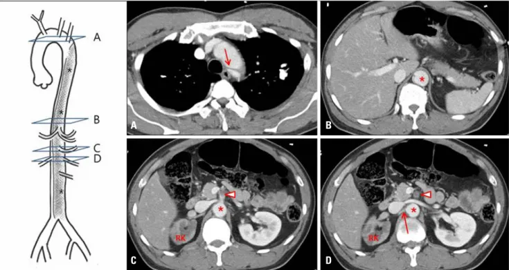
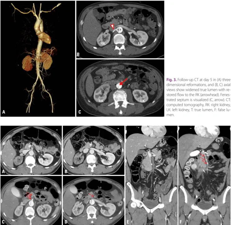
Acute complicated type B aortic dissection (TBAD) is a potentially catastrophic, life-threatening condition. Several risk factors have been associated with acute