Antioxidant Properties of Various Microorganisms Isolated from Arctic Lichen Stereocaulon spp.
Kim, Mi-Kyeong
1, Hyun Park
2, and Tae-Jin Oh
1*
1
Department of Pharmaceutical Engineering, SunMoon University, Asansi, Chungnam 336-708, Korea
2
Korea Polar Research Institute (KOPRI), Songdo TechnoPark, Yeonsu-gu, Incheon 406-840, Korea
Received : March 13, 2013 / Revised : July 10, 2013 / Accepted : August 14, 2013
Introduction
Lichens are typical land plant of Arctica and they can sur- vive in extreme environmental conditions from desert to polar area through their symbiotic relationship between a fungus, algae, or cyanobacteria [35]. Lichens and their nat- ural products have been used as food, dyes, perfumes and decoration [36]. Also, they have used as natural medicines because they show antiviral, anti-proliferative, anti-inflam- matory, anti-tumor, anti-mycobacterial and analgesic activi- ties [22, 26, 27, 32, 34] but most of these are come from lichen or fungal symbionts of lichen [29, 48, 49]. Recent report about bacterial symbioses in lichen suggested that some bacterial symbionts have antibacterial and antioxi- dant activity [17, 38, 46].
Stereocaulon is genus of lichen and commonly found on
the rocks or on the ground in humid regions or warmer regions [11]. Although Stereocaulon species have a num- ber of subspecies, they have not been extensively investi- gated except for their habitat and simple characteristics [11, 21]. There are few studies that metabolite of Stereocaulon species show antioxidant, antibacterial, antimitotic and pro- tein tyrosine phosphatase 1B inhibitory activities [8, 9, 33, 45]. In addition, diversity of bacterial community and their biological activity is not known in Stereocaulon.
Antioxidants are important in the prevention of human disease [19]. Living organisms have a natural defense mechanism of antioxidant, but sometime low level of antiox- idant molecules or inhibition of these antioxidant enzymes causes oxidative stress and may cause damage or kill cells [15]. Several strong synthetic antioxidants have already been reported such as butylated hydroxyanisole (BHA), butylated hydroxytoluene (BHT) and tertiary butylhydro- quinone (TBHQ) which have proven to be highly carcino- genic compound [18, 50]. For this reason, finding new antioxidants from natural sources is highly desirable.
Lichens are symbiotic organisms composed of fungi, algae, or cyanobacteria which are able to survive in extreme environmen- tal conditions ranging from deserts to polar areas. Some lichen symbionts produce a wide range of secondary metabolites that have many biological activities such as antibacterial, antifungal, antiviral, antitumor, antioxidant and anti-inflammatory etc.
Among the symbionts of lichens, of the bacterial communities of lichen symbionts little is known. In this study, we isolated 4 microbial species from the Arctic lichen Stereocaulon spp. and evaluated their antioxidant properties using 1,1-diphenyl-2-pic- ryl-hydrazyl assay as well as 2,2'-azino-bis(3-ethyl benzothiazoline-6-sulphonic acid) assay. Total phenolic contents and total flavonoid contents were also measured. A potent radical scavenging activity was detected in a number of the lichen extracts.
Among the 4 species tested in this study, the ethyl acetate extract of Bosea vestrisii 36546(T) exhibited the strongest free radi- cal scavenging activity, with an inhibition rate of 86.8% in DPPH and 75.2% in ABTS assays. Overall, these results suggest that lichen-bacteria could be a potential source of natural antioxidants.
Keywords: ABTS, antioxidant property, Arctic lichen, DPPH, Stereocaulon spp, TPC
*Corresponding author
Tel: +82-41-530-2677, Fax: +82-41-530-2279 E-mail: [email protected]
© 2013, The Korean Society for Microbiology and Biotechnology
In our previous study, 4 bacterial species were isolated from the Arctic lichens including Stereocaulon spp. There are very few reports about microorganism isolated from lichens in Arctic region and their biological properties have not been fully explored yet. We expected that Arctic region have potential to provide valuable and variety of natural resources. On this background, the aim of this study is to evaluate antioxidant activities of cell extracts from four novel bacterial species isolated from the Arctic lichens Ste- reocaulon spp. for explore the properties as natural antioxi- dants.
Materials and Methods
Lichen species
Lichen Stereocaulon spp. was collected by Korean Polar Research Institute (KOPRI, Incheon, Korea) in 2010. Stere- ocaulon was located around the Dasan, Korean Arctic Sta- tion at Ny-Alesund, Svalbard, Norway (S78
o, E11
o). All microorganisms were identified by KOPRI (Table 1) and the numbers were also assigned by KOPRI.
Screening of polar microorganisms associated with lichens
For isolation of bacteria from Arctic lichen Stereocaulon spp., we carried out screening experiment according to Yamamoto method with some modifications [51]. A seg- ment of lichen thallus was separated by scissors and then added the 1 ml of sterilized 0.85% saline solution. Vortex in ten minute and then discard the solution and repeat above steps about two more times. Break the tissue with mortar added 1 ml of sterilized 0.85% saline solution and then spread on selective media. Cultures were incubated at 28
oC for 15-21 days. Selective media is Humic acid vitamin agar (Humic acid 10.0 g, Na
2HPO
40.5 g, KCl 1.71 g, MgSO
4·7H
2O 0.05 g, FeSO
4·7H
2O 0.01 g, CaCO
30.02 g, vitamin 1.0 ml, distilled water 1.0 L, agar 16.0 g), Bennett’s
vitamin agar (D-glucose 10.0 g, yeast extract 1.0 g, pep- tone 2.0 g, beef extract 1.0 g, vitamin 1.0 ml, distilled water 1.0 L, agar 16.0 g), ISP4 (Difco soluble starch 10.0 g, K
2HPO
4anhydrous 1.0 g, MgSO
4·7H
2O 1.0 g, NaCl 1.0 g, (NH
4)
2SO
42.0 g, CaCO
32.0 g, ISP trace salt solution 1.0 ml, distilled water 1.0 L, agar 16.0 g) and water agar (dis- tilled water 1.0 L, agar 16.0 g). Agar was purchased from Difco (USA) and all other reagents were purchased from Daejung (Korea). Obtained single colony was taken from the Bennett's vitamin agar media and ISP4 agar media.
They have kept on fresh media that mentioned above, respectively. To identify these isolates, we carried out col- ony PCR. Each fresh single colony were picked in PCR tube with 20 ul of distilled water and used as PCR template.
16S rRNA gene sequences were amplified by PCR using universal primer 27F (5'-AGA GTT TGA TCM TGG CTC AG-3') and 1492R (5'-TAC GGY TAC CTT GTT ACG ACT T-3'). The sequences were submitted to the Basic Local Alignment Search Tool (BLAST) search program (http://blast.
ncbi.nlm.nih.gov/Blast.cgi) to identify the closely related organisms.
Preparation of the lichen associated with bacterial cell culture extract
We used solvent extraction system which is the com- monly used technique to recover bioactive compound from bacteria cell culture according to Coleman’s method (2011) with some modifications [14]. For the extraction, 50 ml of total 4 microorganisms were cultured on Bennett’s vitamin liquid media and ISP4 liquid at 15
oC for 10-15 days. Culture broth centrifuged and the supernatant were taken and added to double volume of various solvents including aque- ous, acetone, methanol, ethyl acetate, chloroform, ethanol, diethyl ether and petroleum ether (Daejung, Korea) at room temperature. Subsequently solvent was evaporated in vac- uum at 40°C. The residuals were then dissolved with sol- vent and stored at −20
oC until further study.
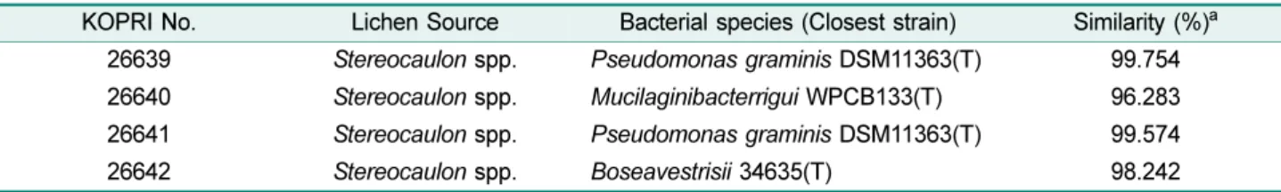
Table 1. Bacterial species isolated from the Arctic lichen.
KOPRI No. Lichen Source Bacterial species (Closest strain) Similarity (%)
a26639 Stereocaulon spp. Pseudomonas graminis DSM11363(T) 99.754 26640 Stereocaulon spp. Mucilaginibacterrigui WPCB133(T) 96.283 26641 Stereocaulon spp. Pseudomonas graminis DSM11363(T) 99.574
26642 Stereocaulon spp. Boseavestrisii 34635(T) 98.242
a
The value are expresses as sequence similarity with the closely related organisms and generated by Basic Local Alignment
Search Tool (BLAST) search program (http://blast.ncbi.nlm.nig.gov/Blast).
Evaluation of total phenolic contents (TPC) and total fla- vonoid contents (TFC)
Total quantity of total phenolic content of the lichen-bacte- rial cell extract was determined with the Folin-Ciocalteu reagent according to the method of Slinkard and Singleton with some modifications [47]. All of chemical reagents were purchased from Sigma-Aldrich (USA). 30 µl of test extract was thoroughly mixed with 30 µl of 1 N Folin-Ciocalteu reagent and incubated for 3 min at room temperature. Sub- sequently, 600 µl of 2% Na
2CO
3was added to the reaction mixture and the mixture was incubated for 30 min at room temperature. Finally the absorbance was measured at 760 nm. Gallic acid was used as a positive control, whereas reaction mixture without the extraction sample was used as negative control. The concentration of TPC was expressed in micrograms of gallic acid equivalent per milligram of lichen-bacterial cell extract. A single extract were measured three times. Total flavonoid content was determined by col- orimetric method described previously [53]. Dried solvent extracts (0.2 g) were dissolved in 20 ml of 80% methanol, extracted for 2 hr at room temperature and centrifuged at 3,000 rpm for 15 min. The volume of the extract was made up to 100 mL with 80% methanol. A portion of 0.5 ml was taken and 0.5 ml of 2% AlCl
3ethanol solution was added to it. After 1 hr at room temperature, the absorbance was measured at 420 nm. Total flavonoid contents were calcu- lated as catechin from a calibration curve.
Evaluation of free radical scavenging activity using DPPH and ABTS
The free radical scavenging activity of the lichen-bacterial cell extract was determined by DPPH and ABTS methods.
The DPPH free-radical scavenging activity of the 4 lichen- bacterial cell extract was determined by the method of Blois with some modifications [10]. All of chemical reagents were purchased from Sigma-Aldrich (USA). 0.1 mM of 1, 1- diphenyl-2-picryl-hydazil (DPPH) was prepared in metha- nol. Then, 950 µl of DPPH solution was mixed with 50 µl of lichens extraction samples with various solvents. The mix- ture was incubated for 30 min at room temperature and the absorbance was measured at 517 nm using a UV-Visible spectrophotometer (Biochrome, USA). For ABTS assay, the procedure followed the method of Arnao with some modifications [3]. 7.4 mM of 2, 2’-azino-bis (3-ethyl benzthi- azoline-6-sulfolic acid, ABTS) was prepared in methanol.
ABTS was kept in the dark for 12 h to generate free radicals
from the ABTS salt and then 950 µl of ABTS solution was mixed with 50 µl of lichens extraction samples with various solvents. The mixture was incubated for 30 min at room temperature and the absorbance was measured at 734 nm using a UV-Visible spectrophotometer (Biochrome, USA).
1 mM of ascorbic acid was used as positive control and a reaction mixture without the test sample was taken as a negative control in both assays. Free radical scavenging activity described as the inhibitory percentage of DPPH and ABTS was calculated according to the following equation. A single extract were measured three times.
Scavenging activity (%) = [ 1 − (Abs sample/Abs control)]
x 100
Ferric reducing antioxidant power (FRAP) assay The FRAP assay was done according to the modified Benzie and Strain method with some modifications [6]. All of chemical reagents were purchased from Sigma-Aldrich (USA). Briefly, 900 µl of FRAP reagent, freshly prepared and warmed at 37
oC, were mixed with 90 µl distilled water and either 30 µl of samples in different concentrations or standard or appropriate blank reagent. The FRAP reagent contained 2.5 ml of a 10 mM 2,4,6-tripyridyl-striazine (TPTZ) solution in 40 mM HCl, plus 2.5 ml of 20 mM FeCl
3·6H
2O and 25 ml of 0.3 mM acetate buffer pH 3.6. Readings at the absorption maximum (593 nm) were taken every 15 sec, using a spectrophotometer (Epoch, Biotech Instruments).
Temperature was maintained at 37
oC. The readings at 30 min were selected for calculation of FRAP values.
Statistical analysis
Data were expresses as Mean ± SD. Statistical analysis was done using Microsoft Office Excel 2007 and a one-way analysis of variance (ANOVA). Differences were consid- ered significant at p < 0.05.
Results
Screening and identification of polar microorganisms from the Arctic lichen Stereocaulon spp
A total of 4 microorganisms were isolated from the Arctic
lichen Stereocaulon spp. They grew well on the Bennett’s
vitamin agar media at 28
oC and obtained colonies have
pigments and their shapes were either circlar or irregular
(data now shown). Their 16S rRNA sequence analysis
showed the identity of the each microorganism (Table 1).
Based on these results, we constructed phylogenetic tree (Fig. 1).
Total phenolic and flavonoid contents of lichen-associ- ated with bacterial cell extracts
Generally, antioxidant activities are dependent on their phenolic constituents [16]. Thus, we evaluated the total TPC of lichen-bacterial cell culture extracts using various
solvent such as aqueous, acetone, methanol, ethyl acetate, chloroform, ethanol, diethyl ether and petroleum ether. As shown in Table 2, phenolic compound of some lichen-bac- terial species showed high-level of TPC. Especially, extract of Bosea vestrisii 34635(T) had the highest TPC in compar- ison to other bacterial species. In case of solvents, ethyl acetate and chloroform extracts showed higher TPC than others. Finally, these high levels of TPC indicate that lichen- bacteria have properties as useful natural antioxidant. Fla- Fig. 1. Phylogenetic dendrogram of polar microorganisms isolated from the Arctic lichen Stereocaulon spp.
The tree was constructed by the neighbor-joining method. Bar 0.02 changes per nucleotide. KOPRI26639, Pseudomonas graminis DSM11363
T; 26640, Mucilaginibacter rigui WPCB133
T; 26641, Pseudomonas graminis DSM11363
T; 26642, Bosea vestrisii 34635
T.
Table 2. Total phenolic/flavonoid contents of lichen-bacterial cell culture extracts.
Sample No.
Solvents Aqueous Acetone Chloroform Diethyl
ether Ethanol Ethyl
acetate Methanol Petroleum ether TPC
a26639 05.22 ± 0.91 12.01 ± 0.21 13.11 ± 1.20 15.14 ± 2.32 19.12 ± 0.37 65.37 ± 1.90 33.12 ± 0.82 014.2 ± 2.33
26640 09.92 ± 2.02 27.33 ± 1.30 28.12 ± 0.32 50.32 ± 0.47 44.39 ± 0.22 88.32 ± 2.16 44.00 ± 0.12 24.18 ± 0.36 26641 05.84 ± 1.42 08.20 ± 1.32 32.02 ± 0.30 44.33 ± 0.58 39.99 ± 0.61 83.05 ± 0.33 30.21 ± 1.02 10.20 ± 1.38 26642 17.01 ± 3.62 36.42 ± 3.41 81.03 ± 0.52 76.42 ± 0.42 53.21 ± 0.32 99.84 ± 0.22 55.02 ± 0.23 34.15 ± 0.32 TFC
b26639 00.31 ± 0.31 05.34 ± 1.02 08.02 ± 0.41 07.98 ± 0.98 08.84 ± 1.15 19.84 ± 1.98 13.59 ± 0.97 06.31 ± 0.21 26640 01.32 ± 1.02 10.39 ± 2.03 11.07 ± 1.01 11.74 ± 0.99 15.34 ± 1.34 25.32 ± 0.88 16.76 ± 0.78 08.07 ± 0.31 26641 01.22 ± 0.11 03.07 ± 0.59 19.09 ± 1.12 13.65 ± 0.78 17.33 ± 1.98 30.26 ± 1.51 11.09 ± 0.52 03.14 ± 0.09 26642 03.31 ± 0.54 04.31 ± 0.51 32.31 ± 1.07 28.47 ± 0.57 20.29 ± 2.01 35.64 ± 1.05 18.01 ± 0.37 10.98 ± 0.04
a
Total phenolic contents are expresses as gallic acid equivalents (mg GAE/g extract).
b
Total flavonoids contents are expresses catechin equivalents (mg CE/g extract).
Each value is expresses as mean ± SD (n = 3). Data with different superscript letters are significantly different (p ≤ 0.05).
vonoids are one of the most powerful antioxidants found in plants [20, 39]. Typically, they possess one or more of the following structural elements that are considered important to their antioxidant activities [42-44]. Thus, total flavonoid content was measured with TFC assay. As shown in the bottom of Table 2, total flavonoid content in all samples was lower than quantity of total phenolic contents. Pattern of quantity is similar with TPC though in order of 26642 >
26641 > 26640 > 26639. Both TPC and TFC level indi- cated that these strains have antioxidant activities.
DPPH and ABTS free radical scavenging activities of lichen-bacterial cell extracts
The 4 lichen-bacterial species were extracted to evaluate the antioxidant activities. Free radical scavenging activity was described as the percentage of DPPH and ABTS as
summarized in Table 3. The DPPH free radical scavenging activity of each lichen-bacterial cell extract was compared with that of the natural antioxidant, ascorbic acid (vitamin C). All the tested extracts and the control (ascorbic acid) exhibited DPPH and ABTS free radical scavenging activity was depend on their concentration. Also, the rate of scav- enging activity was variable for each extract and each sol- vent. Among 4 bacterial species, Bosea vestrisii 34635(T) showed a stronger activity than ascorbic acid about 1.7 fold and 2.9 fold in DPPH and ABTS assay, respectively. In almost all cases, ethyl acetate extracts of bacterial species that have high level amount of TPC showed high antioxi- dant activity.
Ferric reducing antioxidant power of microorganisms The FRAP assay measures the reduction of ferric iron
Table 3. Evaluation of antioxidant properties using various solvents.
Sample No.
Solvents Aqueous Acetone Chloroform Diethyl
ether Ethanol Ethyl
acetate Methanol Petroleum ether
DPPH
a26639 5.9 ± 1.21 23.6 ± 1.52 39.4 ± 0.98 13.2 ± 2.10 9.1 ± 1.22 70.1 ± 1.03 13.9 ± 1.12 10.1 ± 0.23 26640 8.4 ± 0.71 26.3 ± 3.20 20.4 ± 1.17 34.9 ± 1.23 40.9 ± 0.93 82.1 ± 0.91 7.2 ± 0.82 29.1 ± 0.87 26641 8.9 ± 0.91 22.3 ± 0.92 31.2 ± 2.14 32.3 ± 0.82 39.2 ± 0.44 71.2 ± 0.82 12.6 ± 0.71 31.4 ± 1.40 26642 11.3 ± 1.31 44.1 ± 0.52 60.8 ± 1.51 49.1 ± 0.77 41.9 ± 1.12 86.8 ± 1.02 41.7 ± 2.13 46.9 ± 2.33
Control Ascorbic acid (1 mM) : 51.3 ± 0.32
ABTS
b26639 3.3 ± 0.42 13.7 ± 1.11 23.1 ± 0.30 13.2 ± 1.10 9.8 ± 0.91 44.1 ± 1.10 14.5 ± 1.33 10.4 ± 0.73 26640 5.3 ± 0.79 22.1 ± 0.32 28.0 ± 0.29 13.2 ± 3.23 24.3 ± 0.30 62.0 ± 0.39 22.5 ± 2.39 9.3 ± 0.62 26641 5.7 ± 0.92 8.1 ± 0.36 19.2 ± 1.19 12.3 ± 0.52 19.4 ± 0.33 48.2 ± 2.22 13.9 ± 1.14 12.9 ± 1.66 26642 10.2 ± 1.26 24.8 ± 0.45 28.3 ± 0.51 24.9 ± 0.67 8.6 ± 1.54 75.2 ± 0.94 19.7 ± 0.82 23.8 ± 1.39
Control Ascorbic acid (1 mM) : 25.8 ± 0.41
a
DPPH : 1, 1-diphenyl-2-picryl-hydazil.
b
ABTS : 2’-azino-bis (3-ethyl benzthiazoline-6-sulfolic acid).
Each value is expressed as mean ± SD (n = 3). Data with different superscript letters are significantly different (p ≤ 0.05).
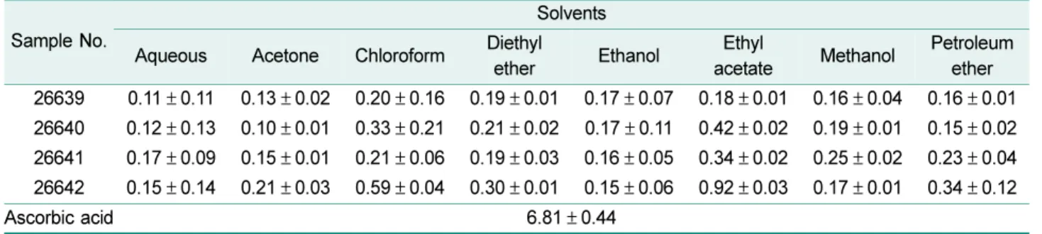
Table 4. Ferric reducing antioxidant power activity of extracts.
FRAP activities are expressed as mM Fe(II)/mg extract and ascorbic acid was used as control.
Sample No.
Solvents Aqueous Acetone Chloroform Diethyl
ether Ethanol Ethyl
acetate Methanol Petroleum ether 26639 0.11 ± 0.11 0.13 ± 0.02 0.20 ± 0.16 0.19 ± 0.01 0.17 ± 0.07 0.18 ± 0.01 0.16 ± 0.04 0.16 ± 0.01 26640 0.12 ± 0.13 0.10 ± 0.01 0.33 ± 0.21 0.21 ± 0.02 0.17 ± 0.11 0.42 ± 0.02 0.19 ± 0.01 0.15 ± 0.02 26641 0.17 ± 0.09 0.15 ± 0.01 0.21 ± 0.06 0.19 ± 0.03 0.16 ± 0.05 0.34 ± 0.02 0.25 ± 0.02 0.23 ± 0.04 26642 0.15 ± 0.14 0.21 ± 0.03 0.59 ± 0.04 0.30 ± 0.01 0.15 ± 0.06 0.92 ± 0.03 0.17 ± 0.01 0.34 ± 0.12
Ascorbic acid 6.81 ± 0.44
Each value is expressed as mean ± SD (n = 3). Data with different superscript letters are significantly different (p ≤ 0.05).
(Fe
3+) to ferrous iron (Fe
2+) in the presence of antioxidants which are reducer with half-reaction reduction potentials above Fe
3+/Fe
2+[6, 7]. This assay is also commonly used for the routine analysis of single antioxidants and total anti- oxidant activity [40]. The FRAP values in various solvent extracts of polar microorganisms are summarized in Table 4. To evaluate the reducing (antioxidant) potential of tomato fractions, the reduction of Fe
3+-TPTZ complex to Fe
2+in the presence of antioxidants were calculated. The assay is based on the total amount of antioxidant to the reducing capacity of the sample. The results showed the ferric reduc- ing antioxidant power were in the order of 26642 > 26640 >
26641 > 26639, which is similar with DPPH and ABTS results.
Discussion
Although many lichen investigations have been reported [23, 30, 31, 37, 41, 49], most of these are based on lichens or their fungal symbionts. Even though bacterial symbionts of lichen are previously demonstrated structurally and eco- logically [5, 12, 13], detail investigation has not been con- ducted yet. Based on this background, we isolated the total 4 microorganisms from the Arctic lichen Stereocaulon spp.
and evaluated their antioxidant activities. In their 16S rRNA sequence analysis of 4 microorganisms, KOPRI 26639 and KOPRI 26641 looks like same strain (Table 1). Neverthe- less, they have not only different color of colony and shape (data not shown), but variation of antioxidant activities (Table 2-4). In the further studies, analysis of morphological traits (e.g., pigmentation, cell wall characteristics) and met- abolic characteristics are needs to identify their major differ- ences. Little is known about these microorganisms such as their source, general morphological and biochemical char- acteristics etc [4, 25], except Pseudomonas graminis that is known as control the foodborne pathogen on fresh-cut fruit [1, 2]. Actually, finding microorganisms from the lichen have been investigating continuously [12, 13] but important thing is that these microorganisms have some biological activity like our results. As we mentioned above, most of active compound come from fungal symbionts in lichen [29, 47, 48] but our results showed the microorganisms associated with lichen have antioxidant activities. We can guess these microorganisms have specific and unique bio-mechanisms because they were taken from lichen which lives in polar area. Therefore, these kinds of works are very important to
understand their unique mechanisms.
Based on our results, among the various solvents used for extraction such as aqueous, acetone, methanol, ethyl acetate, chloroform, ethanol, diethyl ether and petroleum ether, ethyl acetate extracts showed highest activity in all antioxidant activity tests. In particular, ethyl acetate extracts of Bosea vestrisii 34635(T) exhibits highest antioxidant activities in all antioxidant assays including total TPC/TFC assays. In TPC assay, interestingly, chloroform extract of Pseudomonas graminis(T) (26639) showed high antioxi- dant activities than Mucilaginibacter rigui(T) (26640) albeit total phenol contents and total flavonoid contents of 26639 is lower than ones in 26640. This means that all phenolics do not have same antioxidant activity (i.e. even if amounts of phenolic are high, its antioxidant activity can low). It may be due to the synergistic of phenolic compounds or antago- nistic interactions with other phenolics, or differing types of components such as carbohydrates and proteins [30, 42].
In bacterial communities, some lactic acid bacteria (LAB) such as Lactobacillus acidophilus, Bifidobacterium longum and Lactobacillus ferementum possess antioxidant activity and they can decrease the risk of accumulation of ROS [24, 28]. Recently, Zhang et al. (2011) reported the antioxidative activity of lactic acid bacteria in yogurt [52]. Although exper- iment condition from each study have some difference such as culture condition, type of solvents, and evaluation meth- ods etc, our results showed 4 microorganisms isolated from Stereocaulon spp. exhibited higher antioxidant activity than lactic acid bacteria in DPPH assay. These results open that microorganisms isolated from lichen possess antioxidant activity and are able to use as natural antioxidant.
This study is the first time to investigate the polar microor- ganisms isolated from the Arctic lichen Stereocaulon spp.
As mentioned above, several widely known strong syn- thetic antioxidants are often carcinogenic compound [18, 50]. Therefore, trials to find potential antioxidants are impor- tant and meaningful investigation, especially in undevel- oped environment like polar area. In the future study, we will optimize the growth of our polar microorganisms and then investigate other biological activities of these strains and will carry out screening experiment using nature source of polar area.
Acknowledgments
This study was supported by the functional genomics on polar
organisms (PE12020) grants funded by the Korea Polar Research Institute (KOPRI).
References
1. Alegre I, Viñas I, Usall J, Anguera M, Altisent R, Abadias M.
2013. Antagonistic effect of Pseudomonas graminis CPA-7 against foodborne pathogens in fresh-cut apples under simu- lated commercial conditions. Food Microbiol. 33: 139-148.
2. Alegre I, Viñas I, Usall J, Teixidó N, Figge MJ, Abadias M.
2013. Control of foodborne pathogens on fresh-cut fruit by a novel strain of Pseudomonas graminis. Food Microbiol. 34:
390-399.
3. Arnao MB, Cano A, Acosta M. 2010. The hydrophilic and lipo- philic contribution to total antioxidant activity. Food Chem. 73:
239-244.
4. Baik KS, Park SC, Kim EM, Lim CH, Seong CN. 2010. Muci- laginibacter rigui sp. nov., isolated from wetland freshwater, and emended description of the genus Mucilaginibacter. Int. J.
Syst. Evol. Microbiol. 60: 134-139.
5. Bates ST, Cropsey GWG, Caporaso G, Knight R, Fierer N.
2011. Bacterial communities associated with the lichen symbi- osis. Appl. Environ. Microbiol. 77: 1309-1314.
6. Benzie IFF, Strain JJ. 1996. The ferric reducing antioxidant ability of plasma (FRAP) as a measure of “antioxidant power” : the FRAP assay. Anal. Biochem. 239: 70-76.
7. Benzie IFF, Strain JJ. 1999. Ferric reducing/antioxidant power assay: direct measure of total antioxidant activity of biological fluids and modified version for simultaneous measurement of total antioxidant power and ascorbic acid concentration.
Method. Enzymol. 299: 15-27.
8. Bhattarai HD, Kim T, Oh H, Yim JH. 2008. Stereocalpin A, a bioactive cyclic depsipeptide from the Antarctic lichen Stereo- caulon alpinum. Tetrahedron Lett. 49: 29-31.
9. Bhattarai HD, Kim T, Oh H, Yim JH. 2013. A new pseudodepsidone from the Antarctic lichen Stereocaulon alpi- num and its antioxidant, antibacterial activity. J. Antibiot.
(Tokyo). [Epub ahead of print]
10. Blois MS. 1958. Antioxidant determinations by the use of a stable free radical. Nature 26: 1199-1200.
11. Brodo IM, Sharnoff SD, Sharnoff S. 2001. Stereocaulon (pp.
663-670) In, Lichens of North America. Yale University Press, New Haven.
12. Cardinale M, Puglia AM, Grube M. 2006. Molecular analysis of lichen-associated bacterial communities. FEMS Microbiol.
Ecol. 57: 484-495.
13. Cardinale M, Jr Castro JVD, Müller H, Berg G, Grube M. 2008.
In situanalysis of the bacterial community associated with the reindeer lichen Cladonia arbuscula reveals predominance of Alphaproteobacteria. FEMS Microbiol. Ecol. 66: 63-71.
14. Coleman JJ, Ghosh S, Okoli I, Mylonakis E. 2011. Antifungal activity of microbial secondary metabolites. PLoS One. 6:
e25321.
15. Devasagayam TPA, Tilak JC, Boloor KK, Sane KS, Ghaskadbi SS, Lele RD. 2004. Free radicals and antioxidants in human health: current status and future prospects. J. Assoc. Physic.
India. 52: 794-804.
16. Gardner PT, White TAC, McPhail DB, Duthie GG. 2000. The relative contributions of vitamin C, carotenoids and phenolics to the antioxidant potential of fruit juices. Food Chem. 68: 471- 474.
17. Gonzalez I, Ayuso-Sacido A, Anderson A, Genilloud O. 2005.
Actinomycetes isolated from lichens: Evaluation of their diver- sity and detection of biosynthetic gene sequences. FEMS Microbiol. Ecol. 54: 401-415.
18. Grice HC. 1986. Safety evaluation of butylatedhydroxytoluene (BHT) in the liver, lung and gastrointestinal tract. Food Chem.
Toxicol. 24: 1127-1130.
19. Halliwell B. 1997. Antioxidant and human disease: a general introduction. Nutr. Rev. 55: 44-49.
20. Halvorsen BL, Holte K, Myhrstad MCW, Barikmo I, Hvattum E, Remberg SF, et al. 2002. A systematic screening of total anti- oxidants in dietary plants. J. Nutr. 132: 461-471.
21. Hawksworth DL, Kirk PM, Sutton BC, Pegler DN. 1995.
Ainsworth & Bisby’s dictionary of the fungi. 8
thedition. CAB international, Wallingford.
22. Ingolfsdottir K, Chung GAC, Skulason VG, Gissurarson SR, Vilhelmsdottir M. 1998. Antimycobacterial activity of lichens metabolites in vitro. Eur. J. Pharm. Sci. 6: 141-144.
23. Kosani MM, Rankovi BR, Stanojkovi TP. 2012. Antioxi- dant, antimicrobial and anticancer activities of three Parmelia species. J. Sci. Food Agric. 9: 1909-1916.
24. Kullisaar T, Zilmer M, Mikelsaar M, Vihalemm T, Annuk H, Kairane C, et al. 2002. Two antioxidative lactobacilli strains as promising probiotics. Int. J. Food Microbiol. 70: 388-391.
25. La Scola B, Mallet MN, Grimont PA, Raoult D. 2003. Bosea eneae sp. nov, Bosea massiliensis sp. nov. and Bosea ves- trisii sp. nov, isolated from hospital water supplies, and emen- dation of the genus Bosea (Das et al. 1996) Int. J. Syst. Evol.
Microbiol. 53: 15-20.
26. Lauterwein M, Oethinger M, Belsner K, Peters T, Marre R.
1995. In vitro activities of the lichen secondary metabolites vulpinic acid,(+)-usnic acid against aerobic and anaerobic microorganisms. Antimicrob. Agents Chemother. 39: 2541- 2543.
27. Lawrey JD. 1989. Lichen secondary compounds: evidence for a correspondence between antiherbivore and antimicrobial function. J. Bryol. 92: 326-328.
28. Lin MY, Chang FY. 2000. Antioxidative effect of intestinal bac- teria Bifidobacterium longum ATCC 15708 and Lactobacillus acidophilus ATCC 4356. Digest. Dis. Sci. 45: 1617-1622.
29. Luo H, Yamamoto Y, Jeon HS, Liu YP, Jung JS, Koh YJ, et al.
2011. Production of anti-Helicobacter pylori metabolite by the lichen-forming fungus Nephromopsis pallescens. J. Microbiol.
49: 66-70.
30. Luo H, Yamamoto Y, Liu Y, Jung JS, Kahng HY, Koh YJ, et al.
c é é c c é
2010. The in vitro antioxidant properties of Chinese highland lichens. J. Microbiol. Biotechnol. 20: 1524-1528.
31. Manojlovi N, Rankovi B, Kosani M, Vasiljevi P, Stanojk- ovi T. 2012. Chemical composition of three Parmelia lichens and antioxidant, antimicrobial and cytotoxic activities of some their major metabolites. Phytomedicine. 19: 1166-1172.
32. Molnár K, Farkas E. 2010. Current results on biological activi- ties of lichen secondary metabolites: a review. Z Naturforsch.
C. 65: 157-173.
33. Morita H, Tsuchiya T, Kishibe K, Noya S, Shiro M, Hirasawa Y.
2009. Antimitotic activity of lobaric acid and a new benzofu- ran, sakisacaulon A from Stereocaulonsasakii. Bioorg. Med.
Chem. 19: 3679-3681.
34. Muller K. 2001. Pharmaceutically relevant metabolites from lichens. Appl. Microbiol. Biotechnol. 56: 9-16.
35. Nash III TH. 1996. Introduction. In: Nash TH III (ed) Lichen biology. Cambridge University Press, Cambridge, pp 1-7.
36. Oksanen I. 2006. Ecological and biotechnological aspects of lichens. J. Microbial. Biotechnol. 73: 723-734.
37. Paudel B, Bhattarai HD, Prasad Pandey D, Hur JS, Hong SG, Kim IC, et al. 2012. Antioxidant, antibacterial activity and brine shrimp toxicity test of some mountainous lichens from Nepal.
Biol. Res. 45: 387-391.
38. Paudel B, Bhattarai HD, Lee JS, Hong SG, Shin HW, Yim JH.
2008. Antibacterial potential of Antarctic lichens against human pathogenic Gram-positive bacteria. Phytother. Res.
22: 1269-1271.
39. Pietta PG. 2000. Flavonoids as Antioxidants. J. Nat. Prod. 63:
1035-1042.
40. Prior RL, Wu X, Schaich K. 2005. Standardized methods for the determination of antioxidant capacity and phenolics in food and dietary supplements. J. Agric. Food Chem. 53:
4290-4302.
41. Rankovi B, Rankovi D, Mari D. 2010. Antioxidant and anti- microbial activity of some lichen species. Mikrobiologiia 79:
812-818.
42. Rice-Evans CA, Miller NJ, Paganga G. 1997. Antioxidant
properties of phenolic compounds. Trends Plant Sci. 2: 152- 159.
43. Rice-Evans CA, Miller NJ, Bolwell PG, Bramley PM, Pridham JB. 1995. The relative activities of plant-derived polyphenolic flavonoids. Free Radic. Res. 22: 375-383.
44. Rice-Evans CA, Nicholas J, Miller J, Paganga G. 1996. Struc- ture-antioxidant activity relationships of flavonoids and phe- nolic acids. Free Radic. Biol. Med. 20: 933-956.
45. Seo C, Sohn JH, Park SM, Yim JH, Lee HK, Oh H. 2008.
Usimines A-C, bioactive usnic acid derivatives from the Ant- arctic lichen Stereocaulon alpinum. J. Nat. Prod. 71: 710-712.
46. Silva NMV, Pereira TM, Filho SA, Matsuura T. 2011. Taxo- nomic characterization and antimicrobial activity of actino- mycetes associated with foliose lichens from the Amazonian ecosystem. Aust. J. Basic. Appl. Sci. 5: 910-918.
47. Slinkard K, Singleton VL. 1977. Total phenol analysis: Auto- mation and comparison with manual methods. Am. J. Enol.
Vitic. 28: 49-55.
48. Stocker-Wörgötter E. 2008. Metabolic diversity of lichen-form- ing ascomycetous fungi: culturing, polyketide and shikimate metabolite production, and PKS genes. Nat. Prod. Rep. 25:
188-200.
49. Thadhani VM, Choudhary MI, Ali S, Omar I, Siddique H, Karunaratne V. 2011. Antioxidant activity of some lichen metabolites. Nat. Prod. Res. 25: 1827-1837.
50. Wichi HP. 1988. Enhanced tumor development by butylat- edhydroxyanisole (BHA) from the prospective of effect on forestomach and oesophageal squamous epithelium. Food Chem. Toxicol. 26: 717-723.
51. Yamamoto Y. 2002. Discharge and germination of lichen ascospores in the laboratory. Lichenol. 1: 11-22.
52. Zhang S, Liu L, Su Y, Li H, Sun Q, Liang X, et al. 2011. Antiox- idative activity of lactic acid bacteria in yogurt. Afr. J. Microbial.
Res. 5: 5149-5201.
53. Zhishen JT, Mengcheng WJ. 1999. The determination of fla- vonoid contents in mulberry and their scavenging effects on super oxide radicals. Food Chem. 64: 555-559.
c é c é c é c é
é c
é c é c é c
국문초록
북극 지의류 Stereocaulon spp 로부터 분리한 여러 미생물의 항산화 성질 . 김미경
1, 박현
2, 오태진
1*.
1선문대학교 제약공학과 ,
2