감초추출물의 지방세포와 조골세포에 대한 분화효과
서초롱1․변종선2․안재진3․이재환1․홍정우4․장상호2․박계원1†
1성균관대학교 식품생명공학과, 2(주)애드바이오텍
3(주)바이오셀트란, 4경희대학교 동서의학대학원
Effects of Glycyrrhiza inflata Batal Extracts on Adipocyte and Osteoblast Differentiation
Cho-Rong Seo1, Jong Seon Byun2, Jae Jin An3, JaeHwan Lee1, Joung-Woo Hong4, Sang Ho Jang2, and Kye Won Park1†
1Dept. of Food Science and Biotechnology, Sungkyunkwan University, Gyeonggi 440-746, Korea
2Adbiotech Co., Ltd., Gwangwon 200-200, Korea
3Bioceltran Co., Ltd., Gwangwon 200-702, Korea
4Graduate School of East-West Medical Science, Kyung Hee University, Gyeonggi 440-701, Korea
ABSTRACT Glycyrrhiza inflata Batal, an important species of licorice, is one of the most widely used medicinal plants for over 4000 years. Glycyrrhiza plant species has been well known for its various therapeutic activities such as anti-inflammatory, anti-allergic, and anti-ulcer. The purpose of this study was to determine the effects of Glycyrrhiza inflata Batal ethanol extracts (GBE) on adipocyte and osteoblast differentiation. Mesenchymal C3H10T1/2 cells were treated with sub-cytotoxic doses of GBE, and its effects on adipocyte differentiation were assessed. We found that GBE dose-dependently increased lipid accumulation and also induced the expression of adipocyte markers, such as PPARγ and its target genes, aP2, and adiponectin, in C3H10T1/2 cells. Consistently, similar effects of GBE on lipid accumulation were also observed in preadipocyte 3T3-L1 cells that further supports the pro-adipogenic activities of GBE. We also investigated the effects of GBE on osteoblast differentiation of mesenchymal C3H10T1/2 cells. As a results, we found that GBE increased the activity of alkaline phosphatase in a dose-dependent manner and also promoted the expression of osteoblast markers, such as ALP and RUNX2, during osteoblast differentiation of C3H10T1/2 cells. Similar pro-osteogenic effects of GBE were also observed in preosteoblast MC3T3-E1 cells. Finally, our data show that a major bioactive compound found in Glycyrrhiza inflata Batal, licochalcone A (LA) but not glycyrrhizic acid (GA), can mediate the pro-adipogenic and pro-osteogenic effects of GBE. Taken together, this study provides data to show the possibility of GBE and its bioactive component LA as putative strategies for type 2 diabetes and bone diseases.
Key words: Glycyrrhiza inflata Batal, adipocyte, osteoblast, differentiation
Received 11 March 2013; Accepted 3 April 2013
†Corresponding author.
E-mail: [email protected], Phone: 82-31-290-7804
서 론
현대사회에서 서구화된 식습관과 생활습관으로 인한 대 사증후군이 대두되고 있다. 대사질환 중 당뇨병은 여러 가지 합병증을 유발함으로써 현대인의 건강을 위협하고 있는 질 병으로 인슐린을 생산하지 못하는 제1형 당뇨와 인슐린이 상대적으로 부족한 제2형 당뇨로 구분된다(1-3). 연령이 증 가함에 따라 발병률이 증가하는 제2형 당뇨는 고칼로리식 단, 운동부족, 스트레스 등이 원인이 되며 인슐린 저항성이 문제가 되어 발생하는 질병이다. 대표적인 치료법으로는 혈 당강하제로 쓰이는 바이구아나이드(biguanide) 계열 약물
인 티아졸리딘디온(TZD, thiazolidinedione)이 있다(4,5).
TZD의 작용기전을 살펴보면 지방세포의 핵심적인 전사조 절인자인 peroxisome proliferator-activated receptors γ(PPARγ)에 결합하여 인슐린 저항성과 관련된 단백질 합성 을 촉진하고 또한 세포내의 지방산을 적절한 조직으로 배치 시키고 원활한 순환을 도우며 지방세포분화를 촉진함으로 써 결과적으로 인슐린 저항성을 낮추는 역할을 한다(6-8).
그러나 TZD는 간독성, 심혈관계 질환, 부종 등 심각한 부작 용을 동반하고(9,10) 최근에는 TZD를 복용하는 환자의 골 절 위험률이 높다는 연구결과가 많이 보고되고 있다(11- 14). 이에 따라 이를 대체할 수 있는 천연물 소재를 탐색하는 연구가 집중되고 있다.
감초는 콩과에 속하는 다년초로 중국감초(Glycyrrhiza uralensis Fisch), 유럽감초(Glycyrrhiza glabra Linne) 또
는 창과감초(Glycyrrhiza inflata Batal)의 뿌리 및 뿌리줄 기가 생약으로 주로 이용된다(15). 이 중 창과감초는 신강감 초라고도 하며 표면이 거칠고 크기가 큰 것이 특징이다. 감 초는 독성이 있는 약재성분을 완화시키고 여러 한약재가 조 화롭게 어우러지도록 도와주는 약용식물로 조선시대 의서 동의보감에서 감초는 5장6부의 한열과 사기를 다스리며 눈, 코, 입, 귀와 대소변의 생리를 정상으로 되게 하고 모든 혈맥 을 소통시키며 근육과 뼈를 튼튼하게 하고 영양 상태를 좋게 한다고 기록되어 있다. 대표적인 약리작용으로는 해독, 이 담, 항궤양, 항염증, 진경, 진해, 간보호 등이 있어 한방에서 널리 이용되고 있다(16). 따라서 본 연구에서는 지방세포와 조골세포에서 감초의 생리활성을 확인하고자 다분화능 세 포 C3H10T1/2와 지방전구세포 3T3-L1, 조골전구세포 MC3T3-E1에서 창과감초를 이용한 감초추출물(GBE, Glycyrrhiza inflata Batal extracts)의 분화촉진여부를 조 사하였다.
재료 및 방법
재료 및 시약
감초추출물은 중국 신강지역에서 채취한 창과감초(Gly- cyrrhiza inflata Batal)를 이용한 95% 에탄올추출물로서 (주)애드바이오텍(강원도)에서 제공받아 실험에 사용하였 고 glycyrrhizic acid(glycyrrhizic acid ammonium salt, GA)는 Sigma-Aldrich(St. Louis, MO, USA)에서, lico- chalcone A(LA)는 Calbiochem(Darmstadt, Germany)에 서 구매하여 사용하였다.
MTT 시험법
GBE의 세포독성여부를 알아보기 위해 MTT(3-(4,5- dimethylthiazol-2-yl)-2,5-diphenyltetrazolium bromide) (Sigma-Aldrich)를 이용하여 간접적으로 생존세포수를 확 인하였다. 24 well plate에서 세포가 confluent한 상태가 되었을 때 GBE를 농도별로 처리한 후 24, 48, 72시간째에 각각 5 mg/mL MTT 시약을 첨가하였다. 4시간 동안 배양 한 후 배지를 제거하고 DMSO에 녹여서 540 nm에서 흡광 도를 측정하였다.
세포배양과 분화
지방전구세포 3T3-L1(American Type Culture Col- lection, Rockville, MD, USA)의 유지 시에는 Dulbecco's modified Eagle's medium(DMEM)(Hyclone, Logan, UT, USA)과 calf serum(CS)(Hyclone), 1% penicillin-strep- tomycin(Hyclone)을 혼합하여 사용하였고 그 외에 3T3- L1의 분화와 C3H10T1/2(American Type Culture Col- lection)의 세포배양과 분화를 위해 DMEM과 10% fetal bovine serum(FBS)(Hyclone), 1% penicillin-strepto- mycin을 혼합하여 사용하였다. 분화유도를 위해서는 1 μM
dexamethasone(DMS)(Sigma-Aldrich), 0.5 mM isobutyl- 1-methylxanthine(IBMX)(Sigma-Aldrich), 5 μg/mL in- sulin(Sigma-Aldrich), GW7845(kindly provided by Tontonoz Lab., Los Angeles, CA, USA)를 혼합하여 사용 하였다. 37°C, 5% CO2 incubator에서 배양하여 지방세포 가 confluent 상태가 되면 지방세포분화를 유도하여 2일 후 에 배양액을 갈아주고 이후에는 3일 간격으로 배양액을 갈 아주었다. 배양액을 갈아줄 때에는 DMS와 IBMX는 넣지 않았다. 보통 지방세포 분화 차이가 최대가 되는 4~7일 동 안 분화시켰다.
조골세포에 있어서 C3H10T1/2 세포의 배양과 분화를 위해 사용한 배지는 DMEM과 5%의 fetal bovine serum (FBS), 1% penicillin-streptomycin을 혼합하여 사용하였 고 MC3T3-E1(American Type Culture Collection) 세포 의 배양과 분화를 위해 사용한 배지는 MEM alpha mod- ification(Hyclone)과 10% FBS, 1% penicillin-strepto- mycin을 혼합하여 사용하였다. 조골세포는 10 mM β-gly- cerophosphate(Sigma-Aldrich)와 50 μg/mL ascorbic acid(Sigma-Aldrich)를 첨가하여 분화를 유도하였다. 37
°C, 5% CO2 incubator에서 배양하여 조골세포가 confluent 상태가 되면 상기 배양액으로 조골세포분화를 유도한 후 3 일마다 배양액을 갈아주며 3~10일 동안 분화시켰다.
Oil Red O 염색
배양중인 지방세포의 분화정도를 시각화하기 위해 분화 가 완료된 세포의 지방구에 특이적으로 반응하는 Oil Red O(Sigma-Aldrich)를 사용하여 염색하였다. 분화가 완료된 세포들은 phosphate buffer saline(PBS)으로 1회 세척한 후, 4% formaldehyde solution으로 1시간 동안 고정시켰 다. 남아있는 고정액을 제거하고 0.2% Oil Red O solution 에서 1시간 동안 세포를 염색하였다. 염색시약의 여분을 제 거하고 증류수로 세척을 반복하여 염색액이 plate에 남아있 지 않도록 하고 건조시켰다.
Alkaline phosphatase(ALP) 염색
분화가 완료된 조골세포의 plate의 배양액을 제거한 후 2 mM MgCl2를 넣고 바로 제거한 후 AP buffer(100 mM Tris-HCl pH 9.5, 100 mM NaCl, 10 mM MgCl2)로 ALP 효소의 pH를 맞춰주었다. 15분 후 AP buffer를 제거하고 효소와 반응할 기질을 0.4 mg/mL nitroblue tetrazolium (NBT)(Sigma-Aldrich)과 0.2 mg/mL 5-bromo-4-chloro- 3-indolyl phosphate(BCIP)(Sigma-Aldrich)로 만들어 넣 어준 후 반응시켰다. 분화 차이가 최대로 보일 때에 5 mM EDTA를 넣고 반응을 중지시켰다. 10~15분 후에 제거해준 후 fix 용액(4% formaldehyde solution)을 넣어서 그 상태 를 고정시켰다. 마지막으로 1차 증류수로 2번 세척해준 후 건조시켰다.
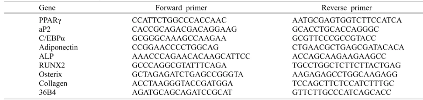
Table 1. Oligonucleotide sequence of mouse primer
Gene Forward primer Reverse primer PPARγ
aP2 C/EBPα Adiponectin ALP RUNX2 Osterix Collagen 36B4
CCATTCTGGCCCACCAAC CACCGCAGACGACAGGAAG GCGGGCAAAGCCAAGAA CCGGAACCCCTGGCAG
AAACCCAGAACACAAGCATTCC GCCCAGGCGTATTTCAGA GCTAGAGATCTGAGCCGGGTA ACCTAAGGGTACCGATGGA AGATGCAGCAGATCCGCAT
AATGCGAGTGGTCTTCCATCA GCACCTGCACCAGGGC GCGTTCCCGCCGTACC CTGAACGCTGAGCGATACACA ACCAGCAAGAAGAAGCC TGCCTGGCTCTTCTTACTGAG AAGAGAGCCTGGCAAGAGG TCCAGCTTCTCCATCTTTGC GTTCTTGCCCATCAGCACC
Fig. 1. Cell viability of C3H10T1/2 pluripotent stem cells with treatment of Glycyrrhiza inflata Batal extracts (GBE) at various concentrations. (A)~(C) Cell viability in C3H10T1/2 cells were measured with various GBE concentrations (1, 10, 20, and 30 μg/mL) treated for 24, 48, and 72 hours after confluent state.
유전자 발현 분석법
TRIzol 시약(Invitrogen, Carlsbad, CA, USA)을 이용하 여 C3H10T1/2 세포로부터 총 RNA를 추출 및 분리하였다.
0.5 μg RNA를 AMV Reverse Transcription System kit (Promega Co., Madison, WI, USA)와 random primer를 이용하여 complementary DNA(cDNA)를 합성하였다.
Thermal Cycler Dice(Takara, Shiga, Japan)를 이용하여 Power SYBR Green PCR Master mix(Applied Biosys- tems, Foster City, CA, USA)와 primer가 포함된 증폭 혼 합물 25 μL와 cDNA를 polymerase chain reation(PCR) 증폭회로를 40회 돌렸다. 발현량은 36B4에 의해 정규화 되 었고 모든 real-time PCR은 최소 2회 수행하였다. PCR에 사용된 oligonucleotide primer(Integrated DNA Tech- nologies, San Diego, CA, USA) 서열은 Table 1에 명시하 였다. ΔCT값은 각 샘플의 CT값과 control(36B4) 간의 차 이에 대하여 계산하였다. ΔCT=CT(target)-CT(control).
상대적인 발현수준은 2-ΔCT으로 계산하였다.
GBE의 GA와 LA 함량 분석
GBE 내의 GA와 LA의 성분 분석은 Kim 등(17)의 방법에 따라 분석하였다. 에탄올로 추출된 감초분말 0.5 g을 메탄올 500 mL로 용해시키고 5분간 초음파 추출을 한 후 상온에서 30분간 냉각시킨 후 여과하였다. 이 용액을 희석하여 검액 으로 사용하였다. GA와 LA 각각의 표준물질 10 mg을 메탄 올 10 mL로 용해시킨 뒤 10,000 rpm에서 10분 동안 원심 분리한 후 여과하여 표준액으로 사용하였다. 여과 시에는 상층액 일부를 취하여 0.2 μm PTFE syringe filter(Cat No.
25JP020AN, Advantec, Tokyo, Japan)로 여과하여 HPLC 시료로 이용하였다. 분석조건은 Agilent Technology의 HPLC(1200 series, Palo Alto, CA, USA)를 이용하였으며 칼럼은 Kromasil C18(100-5-C18 4.6×250 nm, Eka Shemicals, Bohus, Sweden), 이동상으로는 물 : 아세토나 이트릴 : 초산(61:31:2)을 사용하였고 UV detector의 측정 파장은 GA는 254 nm, LA는 370 nm에서 측정하였으며 sample injection volume은 10 μL, flow rate는 0.7 mL/
min이었다.
통계처리
실험결과는 평균±표준오차(mean±SEM) 또는 평균±표 준편차(mean±SD)로 나타내었고 집단 간 평균치 차이는 two-tailed unpaired Student's t-test를 이용하여 P<0.05 수준에서 검증하였다.
결과 및 고찰
세포독성 시험
MTT 시험법을 이용하여 C3H10T1/2 세포에서 GBE의 독성여부를 확인하였다(Fig. 1). DMSO를 처리한 대조군을 포함하여 1, 10, 20, 30 μg/mL의 농도에서 세포독성여부를 조사해본 결과 GBE는 24, 48, 72시간에서 모두 세포의 생 장에 영향을 주지 않았다(Fig. 1A~1C). 따라서 GBE는 비교 적 독성이 낮은 것으로 사료되며 이후 모든 실험에서는 1~30 μg/mL의 범위 내에서 실험을 진행하였다.
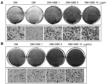
Fig. 2. GBE promotes adipocyte differentiation in C3H10T1/2 and 3T3-L1 cells. (A) (top) C3H10T1/2 cells were treated with various GBE concentrations (1, 5, and 10 μg/mL) and were induced to differentiate for 5 days followed by Oil Red O staining. DM, dif- ferentiation media; GM, growth media containing 10% FBS. (bottom) Microscopic view at ×200 mag- nification. (B) (top) 3T3-L1 cells were treated with various GBE concentrations (1, 5, and 10 μg/mL) and adipocytes were induced to differentiate for 7 days followed by Oil Red O staining. (bottom) Microscopic view at ×200 magnification.
Fig. 3. GBE promotes osteoblast differentiation in C3H10T1/2 and MC3T3-E1 cells. (A) (top) C3H10T1/
2 cells were treated with various GBE concen- trations (1, 5, and 10 μg/mL) and osteoblasts were induced to differentiate for 3 days followed by ALP staining. DM, differentiation media; GM, growth media containing 5% FBS. (bottom) Microscopic view at ×200 magnification. (B) (top) MC3T3-E1 cells were treated with various GBE concentrations (1, 5, and 10 μg/mL) and osteoblasts were induced to differentiate for 5 days followed by ALP staining.
(bottom) Microscopic view at ×200 magnification.
GBE의 지방세포분화 촉진
감초가 지방세포분화에 미치는 영향을 알아보기 위해 다 분화능 세포인 C3H10T1/2에서 1, 5, 10 μg/mL의 농도로 GBE를 처리하여 지방세포로 분화시켰다(Fig. 2A). Oil Red O 염색을 통해 지방세포분화 정도를 확인한 결과 대조군 DM에 비해 지방세포 분화에 필요한 DMS, IBMX, insulin, GW7845를 첨가하지 않은 GM은 분화가 거의 되지 않았다 는 것을 통해 실험이 올바르게 진행되고 있음을 확인하였다.
GBE를 처리한 실험군에서는 Oil Red O 염색이 진해짐에 따라 농도 의존적으로 지방세포분화가 촉진되는 것을 볼 수 있었다. 이 외에 지방전구세포인 3T3-L1에서도 위와 동일 하게 1, 5, 10 μg/mL의 농도로 GBE를 처리하여 지방세포로 분화시킨 후 지방세포분화 정도를 확인한 결과 농도 의존적
으로 지방세포분화를 촉진하는 것을 확인하였다(Fig. 2B).
따라서 GBE는 다분화능 세포 C3H10T1/2와 지방전구세포 3T3-L1에서 모두 지방세포분화를 촉진하는 물질이라 사료 된다.
GBE의 골세포분화 촉진
감초가 뼈와 근육을 튼튼하게 한다는 동의보감 기록을 참 고하여 GBE의 골세포 분화효과를 확인해보았다. 먼저 다분 화능 세포인 C3H10T1/2에서 1, 5, 10 μg/mL의 농도로 GBE 를 처리하여 골세포로 분화시킨 후 ALP 염색을 통해 시각적 으로 골세포분화 정도를 조사하였다(Fig. 3A). 대조군인 DM에 비해 골세포 분화에 필요한 β-glycerophosphate와 ascorbic acid가 첨가되지 않은 GM은 분화가 거의 되지 않
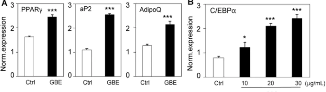
Fig. 4. GBE increases the expression of adipocyte markers in C3H10T1/2 cells. (A) C3H10T1/2 cells were differentiated into adipo- cytes and treated with 10 μg/mL of GBE for 4 days and mRNA expression of PPARγ, aP2, and AdipoQ (adiponectin) was measured by real time PCR. (B) C3H10T1/2 cells were differentiated into adipocytes and treated with various concentrations of GBE for 4 days and mRNA expression of C/EBPα was measured by real time PCR. Data shown represent the mean±SEM from three in- dependent experiments. Statistical significance was determined relative to a control by the Student's t-test (*P<0.05, ***P<0.0005).
Fig. 5. GBE increases the expression of osteoblast markers in C3H10T1/2. (A) C3H10T1/2 cells were differentiated into osteoblasts and treated with 1 μg/mL of GBE for 5 days and mRNA expression of RUNX2, osterix, and collagen was measured by real time PCR. (B) C3H10T1/2 cells were differentiated into osteoblasts and treated with various concentrations of GBE for 5 days and mRNA expression of ALP was measured by real time PCR. Data shown represent the mean±SEM from three independent experiments.
Statistical significance was determined relative to a control by the Student's t-test (*P<0.05, **P<0.005, ***P<0.0005).
았다는 것을 통해 실험이 올바르게 진행되고 있음을 확인하 였다. GBE를 처리한 실험군에서는 ALP 염색이 진해짐에 따라 농도 의존적으로 골세포분화가 촉진되는 것을 볼 수 있었다. 이 외에 조골세포인 MC3T3-E1에서도 위와 동일 하게 1, 5, 10 μg/mL의 농도로 GBE를 처리하여 골세포로 분화시킨 후 골세포분화 정도를 확인한 결과 농도 의존적으 로 골세포분화를 촉진하는 것을 확인하였다(Fig. 3B). 따라 서 GBE는 다분화능 세포 C3H10T1/2와 조골전구세포 MC3T3-E1에서 모두 골세포분화를 촉진하는 물질이라 사 료된다.
GBE의 유전자 발현
지방세포형성 표지유전자 발현량 차이를 통해 GBE의 지 방세포 분화효과를 재확인하고자 지방세포분화의 핵심 전 사조절자 PPARγ와 그의 표지유전자인 adipocyte Protein 2(aP2), adiponectin(AdipoQ)의 발현량 차이를 측정하였 다(Fig. 4A). PPARγ와 aP2, AdipoQ 모두 대조군에 비해 높은 발현량을 나타내며 유의적인 차이를 보였다. 또 다른 표지유전자 CCAAT-enhancer-binding protein α(C/EBP α)는 10, 20, 30 μg/mL의 농도에서 유전자 발현량이 농도 의존적으로 증가하였다(Fig. 4B). Fig. 2의 Oil Red O 염색 을 통해 GBE가 지방세포분화를 촉진한다는 것을 확인한 바
있는데 이와 동일하게 지방세포 관련 표지유전자 발현량 증 가를 통해 GBE의 지방세포분화 촉진효과를 재확인하였다.
골형성 표지유전자 발현량 차이를 통해 GBE의 골세포 분화효과를 재확인하기 위해 조골세포분화의 핵심 전사인 자인 runt-related transcription factor 2(RUNX2)를 포함 하여 골형성 표지유전자인 osterix와 collagen의 발현량 차 이를 측정하였다(Fig. 5A). RUNX2와 osterix는 대조군에 비해 GBE를 처리한 실험군에서 발현량이 높은 것을 볼 수 있었다. Collagen은 유의적인 차이를 보이진 않았지만 GBE 를 처리했을 때 발현량이 소폭 증가하였다. 보편적인 골형성 표지인자 ALP는 10, 20, 30 μg/mL의 농도에서 유전자 발 현량을 측정한 결과 농도 의존적으로 ALP 발현량이 증가하 였다(Fig. 5B). Fig. 3에서 ALP 염색 결과와 동일하게 골 관련 표지유전자 발현량 증가를 통해 GBE의 조골세포분화 촉진효과를 재확인하였다.
GA와 LA의 세포분화 측정
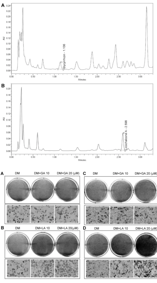
GBE의 어떤 성분이 지방세포와 조골세포의 분화촉진효 과를 매개하는지 확인하기 위해 HPLC를 통해 GBE의 구성 성분을 조사하였다. 그 결과 건물 중 2.6%를 차지하며 감초 의 주요 활성성분으로 알려져 있는 트리테르페노이드(tri- terpenoid)계 사포닌(saponin)인 GA(Fig. 6A)와 0.6%를
Fig. 6. HPLC chromatogram of GBE.
(A) Glycyrrhizic acid (GA). (B) Lico- chalcone A (LA).
Fig. 7. The effects of glycyrrhizic acid (GA) and licochalcone A (LA) in differentiation of C3H10T1/2 cells.
C3H10T1/2 cells were treated with GA (upper A) and LA (upper B), and adipocytes were induced to differ- entiate for 6 days followed by Oil Red O staining. (bottom A and B) Micro- scopic view at ×200 magnification.
C3H10T1/2 cells were treated with GA (upper C) and LA (upper D), and osteoblasts were induced to differ- entiate for 3days followed by ALP staining. (bottom C and D) Micro- scopic view at ×100 magnification.
차지하는 플라보노이드(flavonoid)인 LA(Fig. 6B)의 세포 분화여부를 조사하였다. 다분화능 세포 C3H10T1/2에서 동 일한 농도로 처리하여 지방세포와 조골세포로 분화시켜 보
았을 때, 먼저 지방세포 분화조건에서 GA는 10, 20 μM 두 농도에서 모두 대조군과 비슷한 분화결과를 가져왔고(Fig.
7A) 반면에 LA는 농도 의존적으로 지방세포분화를 촉진한
다는 것을 알 수 있었다(Fig. 7B). 마찬가지로 조골세포 분 화조건에서도 GA는 두 농도에서 조골세포 분화효과가 미미 한 것을 알 수 있었고(Fig. 7C) 반면에 LA는 20 μM에서 조골세포분화를 촉진한다는 것을 알 수 있었다(Fig. 7D).
이렇게 GBE의 지방세포와 조골세포에 대한 분화효과를 매개하는 물질을 규명하기 위해 창과감초(Glycyrrhiza in- flata Batal)의 구성성분 중에서 GA와 LA의 분화효과를 조 사해본 결과, GA는 세포분화에 영향을 주지 않았으나 LA가 GBE와 유사한 지방세포와 조골세포의 분화촉진 기능을 나 타내었다. 그러나 감초 중 LA가 차지하는 0.6%의 함량으로 는 본 연구에서 보여준 감초의 모든 기능을 설명할 수는 없 다. 대신에 GBE에 함유된 다른 화합물 혹은 LA와 이들과의 상승효과로 설명이 가능할 수 있다. 따라서 지방세포와 조골 세포에 대하여 GBE에서 분리되는 다양한 화합물의 동정 및 생리활성 효과, LA와의 상승효과 등의 추후 연구가 기대된다.
요 약
창과감초(Glycyrrhiza inflata Batal)는 한약재의 조화를 돕 고 해독, 항염증, 항궤양 등의 약리작용으로 한방에서 널리 이용되는 약용식물이다. 본 연구에서는 지방세포와 조골세 포에서 감초의 생리활성을 확인하고자 감초에탄올추출물 (GBE)을 이용하여 세포분화 촉진여부를 조사하였다. GBE 의 세포독성여부를 통해 안전하다고 확인된 1~30 μg/mL의 농도 내에서 실험이 진행되었고 지방세포 분화조건에서 다 분화능 세포 C3H10T1/2과 지방전구세포 3T3-L1의 Oil Red O 염색을 통해 GBE의 지방세포분화 촉진효과를 확인 할 수 있었다. 또한 지방세포의 핵심전사조절인자인 PPARγ 와 그의 표지유전자 aP2, AdipoQ, C/EBPα의 발현량 증가 를 통해 GBE의 지방세포분화 촉진효과를 재확인하였다. 이 와 일관된 결과로서 조골세포 분화조건에서 다분화능 세포 C3H10T1/2과 조골전구세포 MC3T3-E1의 ALP 염색을 통해 GBE의 조골세포분화 촉진효과를 확인하였고 조골세 포 표지유전자인 ALP, RUNX2, osterix, collagen의 발현 량 증가를 통해 GBE의 조골세포분화 촉진효과를 재확인하 였다. 이에 따라 본 연구에서는 GBE의 지방세포와 조골세 포 분화효과를 매개하는 감초의 구성성분을 조사하기 위해 GA(glycyrrhizic acid)와 LA(licochalcone A)의 분화촉진 여부를 확인한 결과, GA는 영향을 주지 않으나 LA가 GBE 의 세포분화효과를 매개한다고 사료된다. 따라서 본 연구에 서는 제2형 당뇨와 그에 수반되는 골질환과 골다공증에 대 한 치료 소재로서 GBE와 그의 생리활성을 매개할 수 있는 LA의 가능성을 보았으며 GBE에서 분리되는 다양한 화합물 의 동정 및 생리활성 효과, LA와의 상승효과 등의 추후 연구 가 기대된다.
감사의 글
본 연구는 농림수산식품부 고부가 식품기술개발사업과 농 촌진흥청 공동연구사업(PJ008447)의 지원을 받아 수행되 었으며 이에 감사드립니다.
REFERENCES
1. Bird SR, Hawley JA. 2012. Exercise and type 2 diabetes:
new prescription for an old problem. Maturitas 72: 311- 316.
2. Norris JM, Rich SS. 2012. Genetics of glucose homeostasis:
implications for insulin resistance and metabolic syndrome.
Arterioscler Thromb Vasc Biol 32: 2091-2096.
3. Forbes JM, Cooper ME. 2013. Mechanisms of diabetic com- plications. Physiol Rev 93: 137-188.
4. Yamanouchi T. 2010. Concomitant therapy with pioglita- zone and insulin for the treatment of type 2 diabetes. Vasc Health Risk Manag 6: 189-197.
5. Leahy JL. 2009. Thiazolidinediones in prediabetes and early type 2 diabetes: what can be learned about that disease's pathogenesis. Curr Diab Rep 3: 215-220.
6. Lehrke M, Lazar MA. 2005. The many faces of PPARγ.
Cell 123: 993-999.
7. Bhatia V, Viswanathan P. 2006. Insulin resistance and PPAR insulin sensitizers. Curr Opin Investig Drugs 7: 891-897.
8. Terauchi Y. 2007. PPARγ and metabolic syndrome. Rinsho Byori 55: 447-451.
9. Giles TD, Sander GE. 2007. Effects of thiazolidinediones on blood pressure. Curr Hypertens Rep 9: 332-337.
10. Granberry MC, Hawkins JB, Franks AM. 2007. Thiazolidi- nediones in patients with type 2 diabetes mellitus and heart failure. Am J Health Syst Pharm 64: 931-936.
11. Silva AG, Lazaretti-Castro M. 2010. Diabetes mellitus, thia- zolidinediones and fractures: an unfinished story. Arq Bras Endocrinol Metabol 54: 345-351.
12. Montagnani A, Gonnelli S, Alessandri M, Nuti R. 2011.
Osteoporosis and risk of fracture in patients with diabetes:
an update. Aging Clin Exp Res 2: 84-90.
13. Lecka-Czernik B. 2010. Bone loss in diabetes: use of anti- diabetic thiazolidinediones and secondary osteoporosis. Curr Osteoporos Rep 8: 178-184.
14. Schwartz AV, Sellmeyer DE. 2008. Effect of thiazolidine- diones on skeletal health in women with Type 2 diabetes.
Expert Opin Drug Saf 7: 69-78.
15. Kim NJ, Jin YH, Hong ND. 1995. Studies on the processing of crude drugs (Ⅳ).-Physico-chemical transformation of Glycyrrhizin in Glycyrrhizae Radix by processing. Kor J Pharmacogn 26: 31-39.
16. Kim SH, Jung KK, Kang SY, Kim TG, Kim CO, Moon A, Ryu K, Lee SD, Ryeu HM. 1997. The effects of Jackyak- gamcho-tang on follicular maturation and estrogen production in the immature rat. Kor J Pharmacogn 28: 104-111.
17. Kim SE, Lee SW, Yeum DM, Lee MJ. 2012. Quality charac- teristics of tofu with added alfalfa (Medicago sativa L.) extracts. J Korean Soc Food Sci Nutr 41: 123-128.