1. Introduction
Eosinophils are multifunctional inflammatory
cells that play an essential role in allergic responses and parasitic infections. Although eosinophils are circulating leukocytes, they primarily reside in the mucosal and submucosal areas of the gastrointestinal(GI), respiratory and genitourinary tracts[1]. The GI tract is a principal target for migration of eosinophils, which are part of the normal component of the lamina propria in the
論文
대한민국 공군 장병을 대상으로 한 위점막 조직 호산구 증가증과 유문나선균 간의 관계 연구
김현수*, 이상화*, 이 석**, 최원호**, 김지호**
Relationship between gastric mucosal eosinophilia and the presence of Helicobacter pylori in Republic of Korea Air Force soldiers
Hyun-Soo Kim*, Sang Hwa Lee*, Seok Lee**, Won-Ho Choi** and Ji Ho Kim**
ABSTRACT
호산구는 염증 반응에 의해 활성화되며, 주로 기생충 감염이나 알러지 질환 등에 대한 면역 작용을 담당한다고 알려져 있다. 호산구 증가증은 약물 반응, 알러지, 국소적인 기생충 감염 등 에 의한 경우가 많지만, 자가면역성 질환이나 종양에 의한 경우도 있다. 최근 연구를 통해 위염 의 대표적인 원인균 중 하나인 유문나선균 역시 위점막에서 나타나는 조직 호산구 증가증의 원 인이 될 수 있다고 밝혀지고 있으나, 유문나선균에 의한 호산구 증가증 발생 기전이나 빈도는 아직까지 확립되지 않고 있다. 위점막 내 호산구 침윤과 동반되는 위염은 복통, 오심, 구토, 설 사, 장폐색 등을 일으킬 뿐만 아니라, 아토피성 피부염, 천식, 위식도 역류, 염증성 장질환 등의 발생과 관련이 있다고 보고되고 있다. 위염 및 다양한 관련 질환에 의한 증상은 공중 근무자들 의 임무 수행 능력을 저하시켜 항공기 사고를 발생시킬 수 있는 가능성을 가지고 있다는 점에 서 항공의학적으로 매우 중요하며, 실제로 호산구성 위염이나 유문나선균 감염의 치료 여부가 공중 근무자에게 일시적 또는 영구적 비행임무정지를 부과할 수 있는 기준이 되기도 한다. 본 연구에서는 대한민국 공군 장병 환자를 대상으로 내시경을 통해 얻은 위점막 조직 내 호산구 수를 측정하고, 이를 위점막 표면의 유문나선균 존재 유무와 관련지어 보았다. 111명 중 20명의 환자에서 한 고배율 시야 당 30개 이상의 호산구가 관찰되었고, 63명의 환자의 위점막 표면에서 유문나선균을 확인하였다. 또한 위점막 내 호산구의 밀도와 유문나선균의 존재 간의 관계는 통 계학적으로 유의하였다. 본 연구의 결과는 대한민국 공군 장병을 대상으로 하여 조직 호산구 증 가증과 유문나선균의 빈도 및 상호 관계를 최초로 분석했다는 점에서 의의가 있으며, 추후 유문 나선균이 어떤 기전으로 위점막 조직 내 호산구의 증가에 관여하는지에 대한 연구를 진행하기 위한 기초 자료로서 활용될 수 있을 것이다.
Key Words : Stomach(위), Mucosal eosinophilia(점막 호산구 증가증), Helicobacter pylori (유문나선균), Gastritis(위염)
2012년 월 일 접수 ~ 2012년 월 일 심사완료
* 공군 항공우주의료원 항공우주의학연구센터
** 공군 항공우주의료원 진료부 내과 연락저자, E-mail : [email protected] 충청북도 청원군 남일면 쌍수리 사서함 335-21호
stomach, small intestine and colon[2]. The number of intramucosal eosinophils is believed to vary widely among normal individuals, depending on age, exposure to food allergens, geography and exposure to infectious agents.
Furthermore, eosinophil counts in the same individual vary within different portions of an organ, for example, the cecum versus the sigmoid colon. Thus far, what constitutes the normal eosinophilic infiltrates in each segment of GI tract have not been agreed upon.
Although most observers consider the presence of a few eosinophils in the lamina propria of normal stomach, the Updated Sydney System acknowledged that intraepithelial eosinophils in the gastric mucosa are always viewed as abnormal[3]. Histologic evidence of increased number of eosinophils in human tissues affected by inflammatory diseases has led to the speculation that eosinophils play a significant role in disease pathogenesis, but their specific functions is still unknown. Particularly, although rare cases have been reported sporadically through the literature[4,5], starting from the 1980s Helicobacter pylori virtually monopolized the efforts of gastric researchers, so the few cases or description of eosinophilia in the gastric mucosa were believed to be related to either current or treated H. pylori infection[6]. The aim of the present study is to investigate the level of eosinophils in the gastric mucosa and to determine whether the density of gastric mucosal eosinophils is correlated with the presence of H. pylori-like organisms(HLO).
2. Materials and methods
2.1 Subjects
We reviewed all histopathology slides of gastric biopsy tissue obtained from 111 Republic of Korea Air Force soldiers(median age, 48 years). Between January 1, 2011 and December 31, 2011, they underwent endoscopy in the Department of Internal Medicine at the
Aerospace Medical Center because of dyspeptic complaints. All the subjects had no known history of prior eradication therapy for H.
pylori, anti-ulcer drug use within the past 1 month, GI malignancies, inflammatory and infectious diseases and prior gastric surgery.
2.2 Endoscopic procedure
Endoscopic examination was performed following overnight fasting using a conventional, high-resolution, single-channel GI endoscope(GIF-H260; Olympus Optical, Tokyo, Japan). In each patient, multiple gastric biopsies were taken for histopathological examination.
2.3 Histopathological assessment
The biopsy samples were immediately fixed in 10% buffered formalin solution. After 48 to 72 hours of formalin fixation, tissues were embedded in paraffin and processed for routine hematoxylin and eosin staining. For each section, five high power fields(HPFs;
original magnification, ×400) were randomly selected. The number of eosinophils was individually counted by the same pathologist and the mean score of five fields was obtained from each patient. The presence of HLO in the gastric mucosa was also assessed by histology.
2.4 Statistical analysis
The Fisher’s exact test was performed to determine whether the density of mucosal eosinophils was correlated with the presence of HLO. Statistical analyses were performed using SPSS(version 15.0, SPSS Incorporated, Chicago, IL, USA). Statistical significance was defined as a p value of less than 0.05.
3. Results
Endoscopic pictures showed various abnormal lesions, including mucosal atrophy,
hyperemia, erosion, ulcer, nodule and polyp.
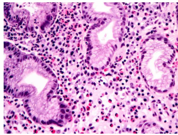
Histologically, the biopsied tissue demonstrated a wide range of eosinophilic infiltration within the lamina propria, as well as the glands and superficial foveolar epithelium(Figure 1).
Microscopically, in 20(17.1%) cases, 30 or more eosinophils/HPF were detected(range, 30 to 120 eosinophils/HPF). The majority of gastric mucosal tissue obtained from these patients revealed erosive superficial mucosa and frequently distorted foveolar glands with neutrophilic infiltrate, but in a few cases only minimal to mild lymphoplasmacytic infiltrate with well-preserved glandular architecture was associated. In addition, in 63(53.8%) cases, a
moderate number of HLO were identified in the epithelial surface. The Fisher’s exact test indicated that there was a statistically
significant relationship between the density of gastric mucosal eosinophils and the presence of HLO(Table 1). However, in 23(20.7%) cases, mean eosinophil count was merely 4 or less/HPF despite the presence of HLO.
Conversely, in 2(0.2%) cases without HLO, 30 or more eosinophils/HPF were observed, suggesting that an increase in eosinophil count is not specific finding to the presence of HLO in the gastric mucosal tissue or H. pylori infection.
4. Discussion
Eosinophilic gastritis is eosinophilic infiltration of the stomach with signs and symptoms related to the layers(mucosa, submucosa, muscularis propria and/or subserosa) and extent of gastric wall involved.
In mucosal disease, typical symptoms are abdominal discomfort, nausea, vomiting and diarrhea with endoscopic biopsy frequently confirming eosinophilic infiltration. In muscle disease, symptoms of gastric outlet and intestinal obstruction are common due to a thickened and rigid gut from the eosinophilic infiltration. In particular, symptoms relevant to aviation include chest pain, dysphagia, food impaction, nausea, vomiting and abdominal pain. These symptoms are of concern primarily due to the potential threat to aviation safety and mission completion. It has been shown that eosinophils increase in the stomach in H.
pylori infection. Similarly, previous studies have suggested that the presense of H. pylori contribute to inflammatory changes seen in eosinophilic gastritis. In this regard, it is important for military and civilian aircrew to diagnose and treat both eosinophilic gastritis and H. pylori infection early and effectively.
We herein demonstrated a statistically significant relationship between the density of gastric mucosal eosinophils and the presence of HLO in Republic of Korea Air Force soldiers.
Association of the presence of H. pylori with the severity of eosinophilic infiltration was Mean eosinophil
count/HPF HLO(+) HLO(-) p value 30 or more
5 to 29 4 or less
18 (90.0) 22 (61.1) 23 (41.8)
2 (10.0) 14 (38.9) 32 (58.2)
0.0002*
HPF indicates high power field; HLO, Helicobacter pylori-like organisms.
* Statistically significant.
Table 1. Association between the density of gastric mucosal eosinophils and the
presence of Helicobacter pylori-like organisms
Figure 1. Representative photomicrograph showing eosinophilic infiltration in the gastric mucosa (Hematoxylin-eosin stain.
Original magnification, ×400)
reported once previously in Korea. Consistent with our result, Lee et al. showed that 159 of 210(75.7%) patients with severe eosinophilic infiltration demonstrated moderate gastric H.
pylori infection[7]. Furthermore, the severity of eosinophilic infiltration was increased in proportion to the number of H. pylori. The slightly higher prevalence of H. pylori infection in Republic of Korea Air Force soldiers than in general population may at least partly be attributable to several factors related to institutional clustering of H. pylori infection, such as inadequate nutritional status, food prepared under inadequate conditions or exposed to contaminated water or soil(inadequate sanitation practices) and crowded or high-density living conditions.
However, there is currently no definite cause of H. pylori infection specifically applicable to the military group. Further investigations are necessary to elucidate the possible causes of the difference in infection prevalence between the soldiers and general population.
In addition to the current or treated H.
pylori infection, increased numbers of GI eosinophils in the lamina propria have been documented in infection with Anisakis species or Strongyloides stercoralis, drugs, inflammatory bowel disease, pyloric obstruction, malignancies, connective tissue diseases, hematopoietic disorders, food allergy and in patients with rare eosinophil-associated GI disorders. Our finding of numerous eosinophils in the gastric mucosal tissue without HLO indicates that a number of disease conditions and factors account for resident GI eosinophils, including environmental temperature, local food products, commensal infectious populations and allergen exposures. Therefore, pathological reports used to make therapeutic decisions should be interpreted in the context of the phenotypic and environmental of each patient.
The pathogenetic mechanisms that might explain a causal role of H. pylori infection in intense mucosal eosinophilic infiltration are still unclear, but it is possible to speculate about them. The chronic immunological stimulation
of the gastric mucosa sustained by H. pylori infection could be among the determinants of this finding. In particular, the long-lasting presence of H. pylori in the gastric mucosa results in a progressive recruitment of inflammatory cells, including neutrophils, eosinophils and lymphocytes, and in a prolonged activation of the immune system, which produces local inflammatory mediators, including cytokines and chemokines[2,8,9].
In conclusion, we demonstrated a significant relationship between gastric mucosal eosinophilia and the presence of HLO in Republic of Korea Air Force soldiers. Further studies are necessary to clarify the precise mechanism by which the presence of H. pylori in the gastric mucosa result in a recruitment of eosinophils and the reason why there is a difference in infection prevalence between the soldiers and general population.
Acknowledgement
The views and opinions expressed in this article are those of the authors and do not reflect the official policy or position of the Republic of Korea Air Force.
References
[1] Weller P. F., “The immunology of eosinophils”, New England Journal of Medicine, Vol. 324, 1991, pp. 1110~1118.
[2] Powell N, Walker M. M., Talley N. J.,
“Gastrointestinal eosinophils in health, disease and functional disorders”, Nature Reviews Gastroenterology and Hepatology, Vol. 7, 2010, pp. 146~156.
[3] Dixon M. F., Genta R. M., Yardley J. H., Correa P., “Classification and grading of gastritis. The updated Sydney System”, American Journal of Surgical Pathology, Vol.
20, 1996, pp. 1161~1181.
[4] Cromwell T. A., Campbell D. A.,
“Eosinophilic gastritis: a case report and
etiological investigation”, Surgery, Vol. 69, 1971, pp. 300~305.
[5] Doniach I., McKeown K. C., “A case of eosinophilic gastritis”, British Journal of Surgery, Vol. 39, 1951, pp. 247~250.
[6] Genta R. M., Lew G. M., Graham D. Y.,
“Changes in the gastric mucosa following eradication of Helicobacter pylori”, Modern Pathology, Vol. 6, 1993, pp. 281~289.
[7] Lee, H., Yu, E., Lee, I., “Histopathologic analysis of Helicobacter pylori associated gastritis”, Korean Journal of Pathology, Vol. 30, 1996, pp. 764~774.
[8] Crabtree J. E., “Immune and inflammatory responses to H. pylori infection”, Scandinavian Journal of Gastroenterology, Vol.
31, 1996, pp. 3~10.
[9] Yoshida N., Granger D. N., Evans D. J., Graham D. Y, Anderson D. C., “Mechanism involved in H. pylori-induced inflammation”, Gastroenterology, Vol. 105, 1993, pp. 1431~1440.