- 35 - 대한두경부종양학회지, 제35권 제2호, 2019. pp.35-38
Korean Journal of Head & Neck Oncology, Vol.35, No.2
https://doi.org/10.21593/kjhno/2019.35.2.35 ISSN 1229-5183(Print) / ISSN 2586-2553(Online)
악하선에 발생한 호산성 지방선종 1례
이범상1⋅이종규1⋅장수경1⋅서강현1⋅김진환1⋅방희진2⋅이동진1+
한림대학교 강남성심병원 이비인후과학교실1, 병리학교실2
A Case of Oncocytic Lipoadenoma Arising in the Submandibular Gland
Bum Sang Lee, MD1, Jong Kyu Lee, MD1, Soo Kyung Jang, MD1, Kang Hyun Seo, MD1, Jin Hwan Kim, MD, PhD1, Heejin Bang, MD2, Dong Jin Lee, MD, PhD1+
Department of Otolaryngology-Head and Neck surgery1, Department of Pathology2 Kangnam Sacred Heart Hospital, Hallym University College of Medicine, Seoul, Korea
= Abstract =
Oncocytic lipoadenoma is a rare salivary gland tumor composed of adipose tissue and oncocytic epithelial cells in varied proportions. We report a case of an oncocytic lipoadenoma of the submandibular gland, which presented as a submandibular gland mass. The patient was a 65-year-old woman with a right submandibular mass measuring 2 x 2 x 1.6 cm. As a sonographic evaluation and computed tomograph scan gave us the impression of benign submandibular gland tumor such as pleomorphic adenoma, we resected the right side submandibular gland. Grossly, the tumor was well circumscribed with yellow to brown cut surface. Microscopically, the tumor was surrounded by a thin, fibrous capsule and composed of oncocytic epithelial cells admixed with mature adipose tissue. Final diagnosis was an oncocytic lipoadenoma. We discussed here radiologic and pathologic finding of this rare salivary gland tumor.
Key Words : Oncocytic lipoadenoma⋅Submandibular gland
Received R e v i s e d Accepted
: July 10, 2019 : August 8, 2019 : August 27, 2019
+Corresponding author: Dong Jin Lee, MD, PhD
Present/Permanent address 1 Singil-ro Yeongdeungpo-gu, Seoul 150-950, Republic of Korea
Tel: +82-2-829-5217, Fax: +82-2-842-5217 E-mail: [email protected]
Introduction
Mesenchymal neoplasm of salivary glands are very un- commonly diagnosed and reported tumors (1.9%-4.7%).1) Majority of these tumors present as painless masses which grow slowly over many years.1) Most common location of these tumors is the parotid gland (85%) and the second is the submandibular gland. On the histological view, they can be subclassified into two subgroups according to their compo-
sition : monophasic (lipoma component only) and “biphasic”
(lipoma component+epithelial component). Oncocytic lip- oadenoma is a biphasic mesenchymal tumor, which was first described by Hirokawa et al. in 1998.2) It consists of adipo- cytes and serous acini with focal oncocytic component.3) We herein present a case of a submandibular gland onco- cytic lipoadenoma. We present the radiologic and histo- pathological findings of this tumor in detail to increase the understainding of this very rare tumor.
Case report
A 65-year-old female patient, without any trauma or med- ical history, came to the Department of Otolaryngology Head and Neck surgery with complaints of slowly growing right neck mass for four months. Physical examination re-
- 36 -
Fig. 1. Preoperative clinical photographs showed a right sub- mandibular area mass (white arrow)
A
B
Fig. 2. Neck ultrasound revealed 1.6cm sized low echoic nodule in right submandibular gland (A) (white arrow) and color doppler image (B) showed the increase of vascularity along the margin of mass (black arrow).
A
B
Fig. 3. Axial CT scan (A) (white arrow) and coronal CT scan (B) (black arrow) showed about 2 x 2 x 1.6 cm sized well-defined enhancing mass containing fat component in right submandibular gland.
vealed a 2cm sized soft and rubbery mass in right sub- mandibular area without redness or tenderness (Fig. 1). To
distinguish the swelling of right submandibular gland from tumorous condition, we performed neck ultrasound. Neck ultrasound revealed 1.6cm sized low echoic nodule in right submandibular gland (Fig. 2A) (white arrow) and color dop- pler image (Fig. 2B) showed the increase of vascularity along the margin of mass (black arrow). As initial im- pression of neck ultrasound is benign submandibular gland mass such as pleomorphic adenoma, we planned surgical excision of the submandibular gland mass. In preoperative axial CT scan (Fig. 3A) (white arrow) and coronal CT scan (Fig. 3B) (black arrow) showed about 2 x 2 x 1.6 cm sized well-defined enhancing mass containing fat component in right submandibular gland. Surgical excision was performed using usual cervical incision for submandibular gland re- section without any complication. Excised surgical speci- men showed about 2 (width) x 2 (thickness) x 1.6 (length) cm sized well-marginated yellowish mass abutting with sub-
- 37 -
Fig. 4. Excised surgical specimen showed about 2 (width) x 2 (thickness) x 1.6 (length) cm sized well-marginated yellowish mass abutting with submandibular gland (white arrow)
A
B
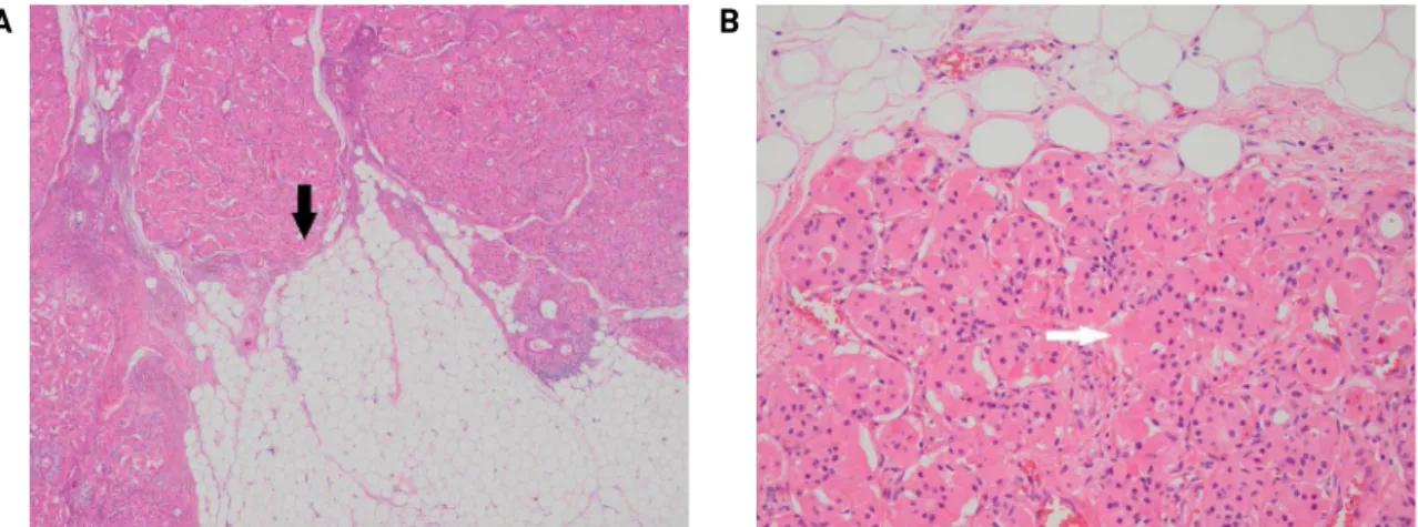
Fig. 5. Microscopically, the tumor was composed of mixed population of oncocytes and adipocytes (black arrow, hematoxylin
& eosin (H&E) x 40) (A). The oncocytes had abundant granular eosinophilic cytoplasm and small prominent nucleoli (white arrow, H&E x 200) (B).
mandibular gland (Fig. 4)(white arrow). The final patho- logic report was oncocytic lipoadenoma. Microscopic ex- amination revealed a well-circumscribed tumor that is sur- rounded by a thin, fibrous capsule. The tumor was com- posed of mixed population of oncocytes and adipocytes (Fig. 5A, H&E x40)(black arrow). The oncocytes were ar- ranged in compact nests and tubules with abundant granular eosinophilic cytoplasm and small variably prominent nucle- oli (Fig. 5B, H&E x 200)(white arrow). The tumor showed somewhat discrete nodules of predominant oncocytic com- ponent within lipomatous area. Cellular pleomorphism, mi- toses or necrosis were not identified. Hemovac drain was removed on third day after surgical excision and the patient discharged without any complication.
Discussion
Lipomatous tumors of salivary gland can be divided into two subgroups: monophasic and biphasic subtypes. Monophasic tumors are more common, which are well circumscribed tumors with a lipomatous component only. Biphasic tumors have epithelial and a lipomatous component. Epithelial component can further be subclassified as "oncocytic" and
"non-oncocytic" component. Oncocytic lipoadenoma con- sists of adipocytes and serous acini with focal oncocytic component.3) The difficulty in the subclassification of these tumors may be due to the reasons that a lipomatous component may be present in pleomorphic adenoma or myoepithelioma.4) The amount of oncocytic component may vary from tumor to tumor and even within the different areas of a tumor.
The differential diagnosis should include all the benign
- 38 - lipomatous salivary gland lesions. A computed tomography (CT) scan can give hints regarding the presence of a lipoma, since a density of 50-150 Hounsfield units is typical for such a tumor.5) A magnetic resonance image may also sug- gest diagnosis and allows for assessment of fatty and non-fatty components.5) Microscopic differential diagnosis of oncocytic lipoadenoma should also include lipomatosis, myoepithelioma and other salivary gland lesions with ex- tensive lipometaplasia.6) Lipomatosis is characterized by in- tensive shrinkage of acinar cells and it is a variant of inter- stitial lipomatosis.7) Oncocytic lipoadenoma has a distinct enlarged main mass, which helps its separation from lipomatosis. Fibrolipoma, the second most common oral lip- oma also contains mature adipose tissue.8) Spindle cell lip- oma is another lesion that needs to be distinguished from sialolipoma. It may affect the parotid region and present typically as a well-circumscribed fatty tumor with bland spindle cells, ropey collagen, and adipocytes.8) Myoepitheliomas can consist of a single type of cells or contain a mixture of cell types lacking ductal differentiation, including spin- dle, plasmacytoid, epithelioid, clear, or oncocytic cell types.9) Moreover, variable amount of myxoid, hyalinized, or clear extracellular matrix produced by myoepitheliomas, contrib- ute to their diverse morphology.9)
The cytologic findings of oncocytic lipoadenoma de- scribed by Chahwala et al.10) included cellular smears in- cluding oncocytic epithelial cells along with occasional fi- brovascular and adipose tissue fragments in prominent lip- oid background. Oncocytes may be seen in aspirates from varying conditions ranging from normal glands of elderly individuals to neoplastic lesions such as oncocytoma, onco- cytic carcinoma, pleomorphic adenoma, Warthin tumor, and mucoepidermoid carcinoma.10) For this reason, accurate di- agnosis by fine-needle aspiration cytology is very difficult.
Furthermore, obtaining sufficient material for accurate diag- nosis is not always possible because of the bulky adipose component. Core needle biopsy can be used as a safe and effective diagnostic tool especially for lipomatous tumor in
salivary gland.11)
In conclusion, we have described an uncommon case of oncocytic lipoadenoma that occurred in the submandibular gland of a 65-year-old woman. Submandibular gland lip- oadenomas are extremely rare benign tumors and may be difficult to diagnose preoperatively. Physicians, surgeons, and pathologists should consider this rare disease entity es- pecially when making a differential diagnosis of lipomatous tumor arising in salivary gland.
References
1) Thompson L. World Health Organization classification of tu- mours: pathology and genetics of head and neck tumours. Ear Nose Throat J. 2006;85:74.
2) Hirokawa M, Shimizu M, Manabe T, Ito J, Ogawa S. Oncocytic lipoadenoma of the submandibular gland. Hum Pathol. 1998;
29:410-412.
3) Agaimy A. Fat-containing salivary gland tumors: A review.
Head Neck Pathol. 2013;7 Suppl 1:S90-96.
4) Jin YT, Lian JD, Yan JJ, Hwang TZ, Tsai ST. Pleomorphic ad- enoma with extensive adipose content. Histopathology. 1996;
28:87-89.
5) Dogan S, Can IH, Unlu I, Sungu N, Gonultas MA, Samim EE.
Sialolipoma of the parotid gland. J Craniofac Surg. 2009;
20:847-848.
6) Ramer N, Lumerman HS, Ramer Y. Sialolipoma: report of two cases and review of the literature. Oral Surg Oral Med Oral Pathol Oral Radiol Endod. 2007;104:809-813.
7) Jang YW, Kim SG, Pai H, Park JW, Lee YC, Rotaru H.
Sialolipoma: case report and review of 27 cases. Oral Maxillofac Surg. 2009;13:109-113.
8) Furlong MA, Fanburg-Smith JC, Childers EL. Lipoma of the oral and maxillofacial region: Site and subclassification of 125 cases.
Oral Surg Oral Med Oral Pathol Oral Radiol Endod. 2004;
98:441-450.
9) Kwon MJ, Kim HJ, Park B, Cho SJ, Shin HS, Park HR, et al. A case report of spindle cell myoepithelioma with extensive lip- omatous metaplasia and thick collagen bundles in the sub- mandibular gland. Diagn Cytopathol. 2016;44:764-769.
10) Chahwala Q, Siddaraju N, Singh N, Goneppanavar M, Basu D.
Fine needle aspiration cytology of oncocytic lipoadenoma of the parotid gland: report of a rare case. Acta Cytol. 2009;53:437-439.
11) Kim HJ, Kim JS. Ultrasound-guided core needle biopsy in sali- vary glands: A meta-analysis. Laryngoscope. 2018;128:118-125.