The Effect of Alkali- and Heat-Treated Titanium Surfaces on Differentiation of Osteoblast
Choong Hee Kang, Mong-Sook Vang, Hong-so Yang, Sang-Won Park, Hyun-Pil Lim
Department of Prosthodontics, Division of Dentistry, School of Dentistry, Chonnam National University
In this study, the biological response of fetal rat calvarial cells on alkali- and heat-treated titanium was assessed. The results were as follows; Cell proliferation on alkali- and heat-treated surfaces showed significantly higher level than on the titanium-6aluminum-4vanadium (weight percentage: 6 % aluminum, 4 % vanadium, Ti-6Al-4V) surface (p<0.01). In ELISA analysis, concentration of IL-1β and IL-6 were raised when the cells were grown to day 7. Pre-treatment with herbimycin, a known tyrosine kinase inhibitor, suppressed the production of IL-6 (p<0.01). In comparison to commercially pure titanium (grade II, cp-Ti) and Ti-6Al-4V alloy, alkali- and heat-treated titanium enhanced alkaline phosphatase activity (p<0.001). In RT-PCR analysis, alkaline phosphatase, bone sialoprotein, receptor activated nuclear factor ligand mRNA expression was increased alkali- and heat-treated titanium. Herbimycin and SB203580, p38 MAPK inhibitor, were repressed of IL-1β-induced IL-6 mRNA expression. These results suggest that alkali- and heat-treated titanium stimulate osteoblasts differentiation and facilitate bone remodeling.
Key words: alkali-treated, differentiation, heat-treated, osteoblast, titanium
(J.K.A.of Stomatognathic Function and Occlusion
2009:25(3):293~306)INTRODUCTION
Titanium and titanium alloys are among the most popular materials for dental and orthopedic implants due to their biocompatibility, excellent corrosion resistance, good mechanical properties
1).There have been efforts to improve the osseointegration capability of the titanium implants by enhancing osteoconduction on their surfaces using surface
Correspondence to : Mong-suk Vang
Department of Prosthodontics, School of Dentistry, Chonnam National University 300 Yongbong-dong, Buk-gu, Gwang-Ju, 500-070, Korea
+82 62 530 5638, E-mail: [email protected]
Received :May 15, 2009, Last Revision :June 21, 2009, Accepted: September 25, 2009 morphology and chemistry
2-4).
Various techniques have been used to produce microrough titanium surfaces for promoting bone ingrowth and fixation between implants and bone.
Besides surface topography, surface chemistry is
another key variable for peri-implant bone
apposition. Kokubo et al
3)introduced an alkaline
and heat treatment on titanium surfaces, which
provided strong bone-bonding ability and high bone
affinity. After alkaline and heat treatments, titanium-based metals form bone-like apatite in simulated body fluid (SBF), which has ion concentrations nearly equal to human body fluid
4). This phenomenon also occurs on the surfaces of bioactive glass and glass ceramics.
Osteoblasts are established as cells who respond to their substrate and rely heavily on signals induced to continue with an osteoblastic phenotype.
If insufficient signals are provided by the substrate then a fibroblast phenotype is induced
5). Other inflammatory mediators also regulate cellular activity, as do other molecules such as osteoprotegerin (OPG) or RANKL
6).
There had been reported that titanium prepared by alkali treatment could form bone-like apatite when it was soaked in SBF in vitro and had strong bone-bonding ability and high bone affinity in vivo
3,7-10)
. Considering these features of alkali- and
heat-treated titanium, alkali- and heat-treated titanium implant is a useful candidate for the preparation of medical device.
Cytokines are a group of communicating molecules, originally identified in the leukocyte population, that are produced by both macrophages and osteoblasts. These protein molecules regulate many aspects of bone biology, including the cellular activity of both osteoblasts and osteoclasts
11). The way in which osteoblasts or osteogenic cells react with alkali- and heat-treated titanium implants has not yet been elucidated. Osteoblasts may attach to the apatite formed on alkali- and heat-treated surfaces, and this may enhance growth and differentiation. As osteoblasts are pivotal in their control of bone remodeling, a biological response would need to be demonstrated to clarify the action of alkali- and heat-treated titanium in vitro. Thus, the purpose of this study was to elucidate cellular events that follow cell adhesion to alkali- and heat-treated
titanium surface by cell proliferation, enzyme-linked immunosorbent assay (ELISA), alkaline phosphatase activity analysis and reverse transcription polymerase chain reaction (RT-PCR) analysis.
MATERIALS AND METHODS 1. Alloy preparation
In this study, three different titanium alloys were prepared as follows; (1) commercially pure titanium (grade II, cp-Ti) (2) titanium-6aluminum-4vanadium (weight percentage: 6 % aluminum, 4 % vanadium, Ti-6Al-4V), (3) alkali- and heat-treated titanium. All Ti and Ti-based alloy disks were formed into disks 12 or 25 mm diameter and 1 mm thickness and kindly provided by the Department of Materials Science and Engineering, Chonnam National University. Cp-Ti disks were wet ground with 240, 400, and 600 grit silicon carbide papers. These surfaces were ultrasonically degreased in acetone and ethanol for 10 minutes each, with deionized water rinsing between applications of each solvent.
Alkali treatment was performed by soaking the samples in a solution of 10 M NaOH at a temperature of 60 ℃ for 8 hours. They were then gently washed with deionized water and dried at 40
℃ for 24 hours. The samples were heated to 500, 600, and 700 ℃ at a rate of 5 ℃
-1, kept at the given temperature for 1 hour and then allowed to cool to room temperature in the furnace.
2. Surface characteristics of disks by atomic force microscopy (AFM).
Cp-Ti, Ti-6Al-4V, and alkali- and heat-treated Ti
disks were examined with atomic force microscopy
to provide qualitative and quantitative information
on the 3 metallic surfaces.
3. Sample preparation
cp-Ti and alkali- and heat-treated titanium disks were placed under aseptic conditions in the bottom of 12- or 6-well culture dishes, then rinsed 3 times in 70 % ethanol, exposed to UV light for 1 hour and air dried in the cell culture hood.
4. Cell culture of fetal rat calvarial cells
Osteoblast-enriched cell preparations were obtained from Sprague-Dawley 21 day fetal calvaria by sequential collagenase digestion (Type II;
Invitrogen, U.S.A). The resultant cells from the third to fifth 15-minute digestions were pooled and cultured in BGJb media (Life Technologies, U.S.A) supplemented with 10 % heat-inactivated fetal bovine serum (FBS), 100 ㎎/㎖ penicillin, and 100
㎎/㎖ streptomycin at 37 ℃ in humidified atmosphere of 5 % CO
2-95 % air.
5. Measurement of cell proliferation
Cells were cultured on cp-Ti, Ti-6Al-4V, and Ti-8Ta-3Nb disks in 12-well plates at a density of 1×10
5cells/㎖ in the BGJb medium. The control was cultured on tissue culture plate (TCP). After 24 hours, the medium was changed and the cells were cultured for additional 3 days. Following incubation, cell proliferation was assessed following the manufacturer's guidelines. In these experiments, the amount of reduced fomazan product is directly proportional to the number of viable cells. Fomazan accumulation was quantitated by absorbency at 490 nm by an enzyme-linked immunosorbent assay (ELISA) plate reader and analyzed. All experiments were carried out in triplicate.
6. Scanning electron microscopy (SEM)
Fetal rat calvarial cells were evaluated for cell attachment and growth using scanning electron microscopy (SEM). Cells were seeded at a density of 1×10
5cells/㎖ in the BGJb medium. After 3-day incubation period, dishes were washed three times with PBS, fixed with 2.5 % glutaraldehyde in 100 mM cacodylate buffer. Samples were dehydrated in increasing concentrations of ethanol (30 %, 60 %, 95 %, and 100 %) at each concentration, immersed in hexamethyldisilazane (Sigma, U.S.A) for 15 mins, air-dried, and immediately mounted on aluminum stubs and coated with carbon. SEM was performed.
7. Alkaline phosphatase (ALP) activity
The assay for ALP activity was carried out
according to Bretaudiere and Spillman
13). For this
purpose cells were seeded onto 12 well dishes at a
density of 1×10
5cell/㎖ in the BGJb medium
containing 10 % FBS, ascorbic acid 40 ㎍/㎖ and
20 ㎍/㎖ β-glycerol phosphate. Determination of
ALP activity was performed as follows; At the day
7, Cells were washed with phosphated buffered
saline (PBS), lysed in Triton 0.1 % (Triton X-100)
in PBS, then frozen at -20 ℃ and thawed. 100 ㎕
of cell lysates was mixed with 200 ㎕ of 10 mM
p-nitrophenyl phosphate and 100 ㎕ of 1.5 M
2-amino-2-methyl-1-propanol buffer. Samples were
then incubated for 1 hour at 37 ℃. ALP activity
was measured for each sample by absorbance
reading at 490 nm with a spectrophotometer
(SmartSpec
TM, BioRAD, U.S.A) and corrected for
cell number determined in parallel. All experiments
were carried out in triplicate.
8. Measurement of cell products
It was examined standardized cell population and determined the changes in cytokine production, specifically the cytokines responsible for osteoclasts activation, interleukin 1β (IL-1β) and IL-6.
Media were removed from the wells and stored at -70 ℃ until required for ELISA measurements.
For analysis, the media were thawed and centrifuged at 5000 rpm for 5 mins. The Quantikine
TMimmunoassay kits (R & D Systems, Oxford) were used for the analysis of cytokines. The assays employ a quantitative "sandwich" emzyme immunoassay technique which was used per the manufacturer's instructions. A color develops, the intensity of which is in proportion to the amount of cytokine present, and it then is measured and compared with known standards prepared from doubling dilutions. The plates were read at 450 nm with a correction at 570 nm using a spectrophotometer (SmartSpec
TM, BioRAD, U.S.A).
9. Reverse transcription polymerase chain reaction (RT-PCR) gene expression analysis
Gene expression of mineralized matrix markers was evaluated by RT-PCR. Cp-Ti, Ti-8Ta-3-Nb, and Ti-6Al-4V disks were placed under aseptic conditions in the 6-well tissue culture dishes. Then 2.0×10
5cells in BGJb medium containing 10 % FBS were seeded into each well and incubated for 1 day. After an initial attachment period, the media was switched to mineralized media containing 10 % FBS, ascorbic acid 40 ㎍/㎖ and 20 ㎍/㎖ β -glycerol phosphate for the duration of experiment and was changed every 3 days.
Total RNA was isolated at day 7 using the methodology described by the manufacturer.
First-strand cDNA synthesis was carried out in an Amplitron II thermocycler using SuperScript II (Invitrigen, U.S.A). PCR was subsequently performed using amplication primer sets (Sigma- Genosys, U.S.A) for glyceraldehyde-3-phosphate dehydrogenase (GAPDH), collagenase type I (COL-I), bone sialoproteins (BSP), interleukin (IL)- 6, osteoporotegerin (OPG) and receptor activation of nuclear factor κB ligand (RANKL) as listed in Figure 1 for 30 cycles.
RT-PCR products were separated and analyzed by gel electrophoresis. Resulting images were captured using a Gel-Doc (BioRad, U.S.A) imaging system equipped with UV light and a gel scanner.
These studies were performed twice.
10. Statistical analysis
An analysis of variance (ANOVA) for replicate measurements and DUNCAN multiple range test was done using SAS program.
RESULTS
1. Surface characteristics and roughness test
Fig. 2 showed SEM and AFM images of three different test surfaces, cp-Ti, Ti-6Al-4V, and alkali- and heat-treated Ti. Roughness measurements of the AFM information yielded root mean square (RMS) values of the surface topographies. For all three tested disks, there was little difference in overall surface roughness (Table I).
2. Scanning electron microscopy
For each specimen, cells were examined by SEM.
The cells spreaded extensively and totally flattened
PCR programs
GAPDH
94 ℃ 94 ℃ 60 ℃ 72 ℃ 72 ℃
1 min 1 min 2 min 1 min 10 min
25 Cycles ALP, BSP,
COL I
94 ℃ 94 ℃ 55 ℃ 72 ℃ 72 ℃
1 min 1 min 2 min 1 min 10 min
30 Cycles IL-6, OPG,
RANKL
94 ℃ 94 ℃ 50 ℃ 72 ℃ 72 ℃
1 min 1 min 2 min 1 min 10 min
30 Cycles PCR primers
Fig. 1. Amplification primer sets and conditions used in polymerase chain reaction. GAPDH, glyceraldehyde-3-phosphate dehydrogenase; ALP, alkaline phosphatase; BSP, bone sialoprotein; COL I, type 1 collagen; IL-6, interleukin-6; OPG, osteoproteogerin; and RANKL
Primer Expected
base pairs Sequence (5'-3')
GAPDH-sense (+) 418 CACCATGGAGAAGGCCGGGG
GAPDH-antisense (-) GACGGACACATTGGGGGTAG
COL I-sense (+) 250 TCTCCACTCTTCTAGGTTCCT
COL I-antisense (-) TTGGGTCATTTCCACATGC
BSP-sense (+) 1068 AACAATCCGTGCCACTCA
BSP-antisense (-) GGAGGGGGCTTCACTGAT
IL-6-sense (+) 638 ATGAAGTTCCTCTCTGCAAGAGACT
IL-6-antisense (-) CACTAGGTTTGCCGAGTAGATCTC
RANKL-sense (+) 499 CAGCACTCACTGCTTTTATAGAATCC
RANKL-antisense (-) AGCTGAAGATAGTCTGTAGGTACGC
OPG-sense (+) 492 TGTAGAGAGGATAAAACGG
OPG-antisense (-) CTAGTTATAAGCAGCTTTAT
ALP-sense (+) 372 CCATGATCACGTCGATATCC
ALP-antisense (-) TCTGACAAACCTTCATGTCC
(A) (B)
(C)
Fig. 2. Surface characteristics of cp- Ti (A), alkali- and heat-treated surfaces (B), and Ti-6Al-4V alloy (C)
Type of Ti-based alloys RMS Roughness (nm)
cp-Ti 3.3
AH-Ti
*19.1
Ti-6Al-4V 7.7
AH-Ti
*: alkali- and heat-treated titanium
Table I. Summary of AFM surface roughness of Ti-based alloys
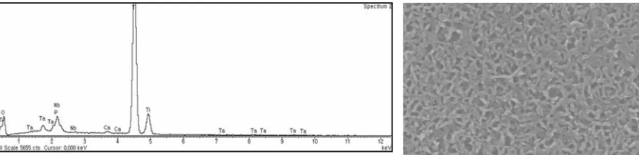
Fig. 3. EDX profile of alkali- and heat-treated titanium-8tantalum-3neobium alloy surface soaked in simulated body fluid for 21 days
on alkali- and heat-treated surfaces. They were polygonal shapes with filopodial extensions indicative of cell spreading. They did not have regular orientation and looked scattered in all directions.
On cp-Ti and alkali- and heat-treated surfaces,
cells spreaded polygonally and cell projections
connecting cells were visible (Fig. 4).
(A) (B)
Fig. 4. Photomicrographs of SEM findings at day 3. cp-Ti (A) and alkali- and heat-treated surfaces (B) show that cells grown on both Ti and alkali- and heat-treated surfaces , presented a flat, elongated spindle-like morphology (x 500)
3. Cell proliferation
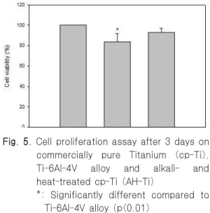
Cell proliferation was measured by the MTT assay. After 3 days, there was a significant difference among glass, cp-Ti, Ti-6Al-4V, and alkali- and heat-treated titanium (p<0.01). Cell proliferation on alkali- and heat-treated surfaces showed significantly higher level than on the Ti-6Al-4V surface (p<0.01, Fig. 5).
4. Alkaline phosphatase (ALP) activity
Alkaline phosphatase activity on alkali- and heat-treated surfaces was the highest of all 3 test materials. Alkaline phosphatase activity on Ti-6Al-4V
Fig. 5. Cell proliferation assay after 3 days on commercially pure Titanium (cp-Ti), Ti-6Al-4V alloy and alkali- and heat-treated cp-Ti (AH-Ti)
*: Significantly different compared to
Ti-6Al-4V alloy (p<0.01)
Fig. 6. Alkaline phosphatase activity of primary rat calvarial cells on commercially pure titanium (cp-Ti), Ti-6Al-4V alloy and alkali- and heat-treated cp-Ti (AH-Ti) at day 7 (U/ 100,000 cells)
*: A significantly higher value of alkali- and heat-treated Ti was observed.
(p<0.001)
alloy was lower than cp-Ti and on tissue culture plate (TCP) controls (p<0.001, Fig. 6)
5. ELISA results
Cells without metallic disks served as control.
The concentration of IL-1β were raised from day 3 to day 7 in all samples. However, there was statistical difference between control and Ti-based metal samples at day 7 (p<0.001, Table II). The concentration of IL-6 was negligible at day 3 (16
~31 pg/㎖) however, was raised at day 7 in alkali- and heat-treated Ti. There was statistical difference between control and alkali- and heat-treated Ti.
Culture on alkali- and heat-treated Ti with herbimycin produced media containing negligible quantities of IL-6 as compared to TCP controls (p<0.001, Table III).
day
Group 3 days 7 days
TCP 42 ± 19 * 132 ± 48
cp-Ti 75 ± 13 237 ± 60
AH-Ti 68 ± 22 308 ± 35
+TCP:tissue culture plate, AH-Ti: alkali- and heat-treated titanium
* : Mean ± standard deviation (pg/ml)
+
: Significantly different compared to tissue culture plate (p<0.01)
Table II. Mean IL-1β levels in TCP, cp-Ti, and alkali- and heat- treated Ti
day
Group 3 days 7 days
TCP 16 ± 10* 68 ± 30
cp-Ti 20 ± 17 89 ± 21
AH-Ti 31 ± 10 156 ± 78
+AH-Ti+HB 31 ± 8 60 ± 20
AH-Ti: alkali- and heat-treated titanium, HB;herbimycin
*: Mean ± standard deviation (pg/ml)
+
: A significantly higher value of alkali- and heat-treated Ti was observed (p<0.01)
Table III. Mean IL-6 levels in TCP, cp-Ti, and alkali- and heat-treated Ti(AH-Ti)
6. RT-PCR gene expression analysis
After 7 days culture, the expression of COL-I,
BSP, ALP, OPG, and RANKL mRNA from cells
was evaluated by RT-PCR. Total RNA isolated
from cells on Ti-6Al-4V alloy did not yield enough
products for use with RT-PCR analysis. In contrast,
enough total RNA was obtained on alkali- and
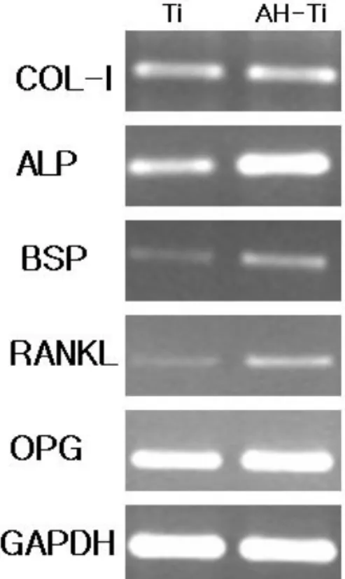
Fig. 7. Reverse transcription-polymerase chain reaction (RT-PCR) analysis of collagen type I (COL-1), alkaline phosphatase (ALP), bone sialoprotein (BSP), receptor activated nuclear factor ligand (RANKL), osteoproteogerin (OPG), and g l y c e r a l d e h y d e - 3 - p h o s p h a t e dehydrogenase (GAPDH) m RNA expression in rat calvarial cells (lane 1;
cp-Ti, lane 2; alkali- and heat-treated titanium)
heat-treated Ti and cp-Ti. For the type I collagen gene(COL-I), the expression level of COL-I mRNA was not different on cp-Ti, alkali- and heat-treated surfaces. Alkali- and heat-treated Ti was shown that increased of alkaline phosphatase mRNA expression (2.5 folds), bone sialoprotein mRNA expression (2.3
Fig. 8. Reverse transcription-polymerase chain reaction (RT-PCR) analysis of interleukin -6 (IL-6) and glyceraldehyde-3- phosphate dehydrogenase (GAPDH) m RNA expression in rat calvarial cells (lane 1: control, lane 2; IL-1b (1 ng/ml), lane 3;SB203580+IL-1b, lane 4;PD + IL-1b lane 5; Herbimycin + IL-1b )
folds), and RANKL mRNA expression (2.8 folds) than those of cp-Ti-induced genes(Fig. 7).
Herbimycin, a known tyrosine kinase inhibitor was repressed of IL-1β-induced IL-6 mRNA expression(Fig. 8).
DISCUSSION
For surface chemistry modification, various
calcium phosphate (Ca-P) coatings on titanium
implants have been proposed to improve
osseointegration. Whether primary cell or
immortalized cell lines would be used when testing
biomaterial is controversial. Primary osteoblasts
have a diploid chromosome pattern, are
characterized by growing slowly and have a finite
lifespan. Established cell lines have been used in
previous studies investing cell morphology and
cytotoxicity in the presence of various titanium
alloys
12,13). Primary cell strains derived from living
tissues are necessary and have been recommended
by the ISO for specific testing to simulate the in
vivo situation
14).
In this study, primary osteoblasts were obtained from fetal rat calvaria. This is an excellent source of osteoblasts because cells from young animals proliferate rapidly. Cells from the third, fourth and fifth digests were collected because these later digests provide a more pure culture, containing most cells that express an osteoblast-like phenotype
15).
Surface roughness can greatly affect the proliferation and protein synthesis of osteoblast cells that are cultured on a metal substrate characteristics in the healing of bone
16,17). Many studies have demonstrated that roughness has a great influence on cell responses. According to this respects, the test samples used in this study was controlled that they had a similar roughness after they were polished.
Alkali- and heat-treated Ti implants have a thin reactive layer (Ca-P layer) which can form apatite in simulated body fluid. In this study, as the same as other reports
3,9,10,18,19), a layer of Ca-P was formed and confirmed by EDX profile however, the elements of calcium and phosphorus were not found on cp-Ti and Ti-6Al-4V alloy when soaked in culture media for 21 days(Fig. 3).
The osteoblastic cells undergo a temporal sequence of phase during the development of their completely differentiated phenotype: proliferation, differentiation, and mineralization
20). Briefly, cells initially increase their number and produce extracellular matrix. The phase of differentiation follows, which is characterized by the production of high levels of alkaline phosphatase and modifications of the matrix that lead to the deposition of hydroxyapatite crystals. This phase of mineralization is also characterized by the synthesis of collagen, bone sialoprotein, and osteocalcin (OCN) that play a fundamental role in bone remodeling and represents the specific markers of
final differentiation of osteoblasts.
In general, cell synthesis activity is sensitive to the type of material
16). Cells grown on alkali- and heat-treated Ti showed alkaline phosphatase levels showed significantly higher than on cp-Ti and Ti-6Al-4V alloy (p<0.001). Alkaline phosphatase activity of cp-Ti and TCP exhibited the similar activity, but showed significantly higher than on Ti-6Al-4V alloy. This might be in case of Ti-6Al-4V alloy, due to the release of aluminum and/or vanadium in culture medium. In particular, vanadium is known to cytotoxic
21), this might affect cells to protein synthesis. This result means that Ti chemical composition interfered with alkaline phosphatase activity.
IL-1β and IL-6 are cytokines that involved indirectly or directly in osteoclastogenesis
22). In RT-PCR analysis, mRNA expression was analyzed at 7 days. Type I collagen expression is an essential component of the extracellular matrix that is required before mineralized matrix formation. In this study, mRNA level of Type I collagen on cp-Ti and alkali- and heat-treated Ti was consistent. This means that cp-Ti and alkali- and heat-treated Ti did not inhibit mineralized matrix expression of fetal rat calvarial cells.
Alkaline phosphatase (ALP) is an enzyme belonging to a group of membrane-bound glycoproteins. Although its physiological function still remains unclear, ALP may play a key role in the formation and calcification of hard tissues
23), and its expression and enzyme activity are frequently used as markers of osteoblastic cells.
Bone sialoprotein (BSP) is a 34-kDa protein that is
highly sulfated, phosphorylated and glycosylated,
and is expressed almost exclusively in mineralizing
connective tissues
24). Studies on the developmental
expression of BSP have shown that BSP mRNA is
expressed at high levels by osteoblasts at the onset
of bone formation
25), and under steady-state conditions in vitro BSP nucleates hydroxyapaptite crystal formation
26)indicating a role for this protein in the initial mineralization of bone. In this study, alkali- and heat-treated surface increased of alkaline phosphatase (ALP) mRNA and bone sialoprotein mRNA expression. This result is in accordance with Chosa et al
27)and could be explained that alkali- and heat-treated surface increased in osteoblastogenesis and bone formation.
Interestingly, in this study, the expression of RANKL mRNA increased in alkali- and heat-treated Ti surface. In previous studies, a number of stimulators of bone resorption such as parathyroid hormone (PTH) and IL-6 were shown to increase RANKL expression in osteoblasts
28,29). In contrast, inhibitors of bone resorption and stimulators of bone formation such as bone morphogenic proteins inhibited RANKL. Li et al
30)indicated the involvement of RANKL in bone resorption by showing osteoclastic bone resorption can be inhibited by inhibitors that interfere with RANKL production. Although the precise role of RANKL in bone resorption, it has been proposed that RANKL differentiate osteoclasts by binding RANKL receptors
31). Thus, it is possible that alkali- and heat-treated Ti stimulate the secretion of RANKL that cause the differentiation of osteoclasts at bone remodeling sites. Taken together, these results indicated that alkali- and heat-treated Ti surface facilitated bone turn over rate and eventually stimulate bone remodeling.
In this study, alkali- and heat-treated Ti had better cell response than cp-Ti for bone remodeling.
This property could make alkali- and heat-treated Ti a useful candidate for the preparation of medical and dental device. However, more animal and human studies will be needed before using this alloy in medical and dental use.
CONCLUSION
Osseointegration of dental implant depends on the cellular response around implant. Changes in osteoblastic proliferation, differentiation, and maturation are important events in bone remodeling.
Alkali- and heat-treated titanium formed an apatite surface layer which was reported to facilitate osteogenic differentiation. As osteoblasts are pivotal in their control of bone remodeling, a biological response would need to be demonstrated to clarify the action of alkali- and heat-treated titanium in vitro.
In this study, the biological response of fetal rat calvarial cells on alkali- and heat-treated titanium was assessed by cell proliferation, ELISA, alkaline phosphatase activity, and RT-PCR method.
The results are as follows;
1. Cell proliferation on alkali- and heat-treated surfaces showed significantly higher level than on the Ti-6Al-4V surface (p<0.01).
2. In ELISA analysis, concentration of IL-1β and IL-6 was raised when the cells were grown to day 7. Pre-treatment with herbimycin, a known tyrosine kinase inhibitor, suppressed the production of IL-6.
3. In comparison to cp-Ti and Ti-6Al-4V alloy, alkali- and heat-treated titanium enhanced alkaline phosphatase activity (p<0.001).
4. In RT-PCR analysis, ALP, BSP, and RANKL mRNA expression increased alkali- and heat-treated titanium. Herbimycin and SB203580, p38 MAPK inhibitor, were repressed of IL-1β-induced IL-6 mRNA expression.
These results suggest that alkali- and heat-treated
titanium stimulate osteoblasts differentiation and
facilitate bone remodeling.
REFERENCES
1. Park JB, Lakes RS. Biomaterials:an introduction, 2nd ed, New York: Plenum Press 1992;89-92.
2. Li D, Ferguson SJ, Beutler T, Cochran DL, Sittig C, Hirt HP. Biochemical comparison of the sand blasted and acid etched and the machined and acid-etched titanium surface for dental implants. J Biomed Mater Res 2002;60:325-332.
3. Kokubo T, Miyaji F, Kim HM, Nakamura T.
Spontaneous apatite formation on chemically surface treated Ti. J Am Ceram Soc 1996;79:1127-1129.
4. Shi S, Kirk M, Kahn AJ. The role of type I collagen in the regulation of osteoblast phenotype. J Bone Mineral Metabolism 1996;11:1139-1145.
5. Sisk MA, Lohmann CH, Cochran DL, Sylvia VI, Simpson JP, Dean DD, Boyan BD, Schwarts Z.
Inhibition of cyclooxygenase by indomethacin modulates osteoblast response to titanium surface roughness in a time-dependent manner. Clin Oral Implants Res 2001;12:52-61.
6. Groot K, Wolke JGC, Jansen JA. Calcium phosphate coatings for medical implants. Proc Instn Mech Engrs 1998:212:134-142.
7. Nishiguchi S, Nakamura T, Kobayashi M, Kim HM, Miyaji F, Kokubo T. The effect of heat treatment on bone-bonding ability of alkali-treated titanium.
Biomaterials 1999;20:491-500.
8. Nishiguchi S, Kato H, Fujita H, Oka M, Kim HM, Kokubo T, Nakamura T. Alkali- and heat-treated porous titanium for orthopedic implants. Biomaterials 2001;22:2525-2533.
9. Nishiguchi S, Kobayashi M, Kim HM, Kokubo T, Nakamura T. Biology of alkali- and heat-treated titanium implants. J Biomed Mater Res 2003;67A:
26-35.
10. Green J, Schotland S, Stauber DJ, Kleeman CR, Clemens TL. Cell matrix interaction in bone: Type I collagen modulates signal transduction in osteoblasts.
Am J Physiol 1995;268:C1090-C1103.
11. McCann TJ, Meikle MC, Northrop AJ, Mason WT, Mcdonald F. A collagen peptide motif activates calcium signalling in SaOS-2 cells. Biochem Soc
Trans 1995 23:404S. 13. Lazarus MD, Cuckler JM, Schumacher HR, Ducheyne P, Baker DG.
Comparison of the inflammatory response to particulate polymethylmethacrylate debris with and without barium sulfate. J Orthop Res 1994;12:
532-541.
12. Lang H and Mertens T. The use of cultures of human osteoblast-like cells as an in vitro test system for dental materials. J Oral Maxillofac Surg 1990;48:
606-611.
13. Naganawa T, IshiharaY, Iwata T, Koide M, Ohguchi M, Ohguchi Y, Murase Y, KameiH, Sato N, Mizuno M, Noguchi T. In vitro biocompatibility of a new titanium-29niobium-13tantalum-4.6zirconium alloy with osteoblast-like MG63 cells. J Periodontol 2004;
75:1701-1707.
14. Schmalz G. Use of cell cultures for toxicity testing of dental materials-advantages and limitations. J Dent 1994;22:PG-S-6-11(suppl 2).
15. Luben RA Wong GL Cohn DV. Biochemical characterization with parathyroid hormone and calcitonin of isolated bone cells: provisional identification of osteoclasts and osteoblasts.
Endocrinol 1976;99:526-534.
16. Lincks J, Boyan BD, Blachard CR. Response of MG63 osteoblast-like cells to titanium and titanium alloy is dependent on surface roughness and composition. Biomaterials 1998;19:2219-2232.
17. Kawahara H, Soeda Y, Niwa K, Takahashi M, Kawahara D, Araki N. In vitro study on bone formation and surface topography from the standpoint of biomechanics. J Mat Sci 2004;15:1297-1307.
18. Sultana R, Kon M, Hirakata LM, Fujihara E, Asaoka K, Ichikawa T. Surface modification of titanium with hydrothermal treatment at high pressure. Dent Mater J 2006;25:470-479.
19. Jonasova L, Muller FA, Helebrant A, Strnad J, Greil P. Biomimetic apatite formation on chemically treated titanium. Biomaterials 2004;25:1187-94.
20. Lian JB, Stein GS. The developmental stages of osteoblast growth and differentiation exhibit selective responses of genes to growth factors and hormones.
J Oral Implantol 1993;19:95-105.
21. Hallab NJ, Anderson SA, Caicedo M, Brasher A, Mikecz K, Jacobs JJ. Effects of soluble metals on human peri-implant cells. J Biomed Mater Res 2005;74A: 124-140.
22. Manolagas SC. The role of IL-6 type cytokines and their receptors in bone. Ann NY Acad Sci 1998;840:194-204.
23. Genge BR, Sauer GR, Wu LNY, Mclean FM, Wuthier RE. Correlation between loss of alkaline phosphatase activity and accumulation of calcium during matrix vesicle mediated mineralization. J Biol Chem 1988;263:18513-18519.
24. Ogata Y, Yamauchi M, Kim RH, Li JJ, Fredman LP, Sodek J. Glucocorticoid regulation of bone sialoprotein (BSP) gene expression: Identification of a glucocorticoid response element in the bone sialoprotein gene promoter. Eur J Biochem 1995;230:
183-192.
25. Ganss B, Kim RH, Sodek J. Bone sialoprotein. Crit Rev Oral Biol Med 1999;10:78-98.
26. Hunter GK, Goldburg HA. Nucleation of hydroxyapatite by bone sialoprotein. Proc Natl Acad Sci 1993;90:8562-8565.
27. Chosa N, Taira M, Saitoh S, Sato N, Araki Y.
Characterization of apatite formed on Alkaline-heat- treated Ti. J Dent Res 2004;83;465- 469.
28. Fu Q, Manolagas SC, O'Brien CA. Parathyroid hormone controls receptor activator of NF-kappaB ligand gene expression via a distant transcriptional enhancer. Mol Cell Biol 2006;26:6453-68.
29. Gorny G, Shaw A, Oursler MJ. IL-6, LIF, and TNF-alpha regulation of GM-CSF inhibition of osteoclastogenesis in vitro. Exp Cell Res 2004;294:
149-58.
30. Li F, Rogers J, Rossa C, Kim YJ, Kriegel J, Giannobile W, Protter A, Kirkwood K. p38 MAP inhibitors block LPS-induced alveolar bone loss in vivo. AADR abstract No. 0932, pp 106, 2006.
31. Khosla S. Mini-review: The OPG/RANKL/RANK
system. Endocrinol, 2001;142:5050-5055.
티타니움 표면의 알칼리-열처리가 골모세포의 분화에 미치는 영향
전남대학교 치의학전문대학원 보철학교실
강충희․방몽숙․양홍서․박상원․임현필
본 연구에서는 알카리 열처리 티타늄 임플란트상에서 백서 태자 두개관 세포의 생물학적 반응을 알아보고자 하 여 다음과 같은 결과를 얻었다. 알카리 열처리 티타니움 표면에서 Ti-6Al-4V 합금 표면에서보다 세포증식도가 유의 하게 높았다 (p<0.01). 면역효소 분석에서 interleukin (IL)-1β와 IL-6의 합성은 배양 시간이 길수록 증가하였다.
Herbimycin으로 전처치한 경우에는 세포 배양 7일 째에 IL-6의 합성이 유의하게 억제되었다(p<0.01). 알카리인산 분 해 효소 활성은 알카리 열처리 임플란트에서 타이타늄, Ti-6Al-4V 합금 표면에서 보다 유의하게 더 높았다 (p<0.001). 유전자 분석에서 알카리 열처리 임플란트에서 배양된 세포들의 alkaline phosphatase, bone sialoprotein, receptor activated nuclear factor ligand 유전자의 발현이 증가하였다. Herbimycin과 SB203580으로 전처치시 IL-1β -induced IL-6 유전자의 발현이 감소하였다. 이상의 결과는 알카리 열처리된 티타늄 임플란트가 조골세포의 분화를 자극하고 골개조를 촉진시킴을 시사한다.
주요어: 분화, 알카리처리, 열처리, 조골세포, 티타늄
교신저자 :
방몽숙
전남대학교 치의학전문대학원 보철학교실, 광주 광역시 북구 용봉동 300번지, 500-070, 팩스:+82-62-530-5638. E-mail: [email protected]
원고접수일 :2009년 05월 15일, 원고수정일 :2009년 06월 21일, 원고채택일 :2009년 09월 25일