ISSN 0378-6471 (Print)⋅ISSN 2092-9374 (Online)
https://doi.org/10.3341/jkos.2017.58.6.731
Case Report
양안 동공잔류막 제거술과 수정체유화술 및 인공수정체 삽입술을 동시에 시행한 1예
One Step Operation of the Persistent Pupillary Membrane Removal and Cataract Operation: A Case Report
송원석⋅박성표⋅윤삼영
Won Seok Song, MD, Sung Pyo Park, MD, Sam Young Yoon, MD
한림대학교 의과대학 강동성심병원 안과학교실
Department of Ophthalmology, Kangdong Sacred Heart Hospital, Hallym University College of Medicine, Seoul, Korea
Purpose: We report a case of one-step operation of persistent pupillary membrane removal, phacoemulsification, and posterior chamber lens implantation that was performed in a patient with persistent pupillary membrane and brunescent cataracts in both eyes.
Case summary: A 64-year-old male with no baseline disease visited our clinic with bilateral decreased visual acuity. His best cor- rected visual acuity at first visit was 0.1 in the right eye and 0.2 in the left eye. On anterior segment examination, both bilateral persistent pupillary membranes and brunescent cataracts were observed. First, we surgically removed the left pupillary mem- brane and performed phacoemulsification using posterior chamber lens implantation via one-stage operation. After one week, the same operation was performed for the right eye using the same method. At 6-months postoperative, his best corrected visual acuity was 0.2 in the right eye and 0.8 in the left eye. No complications such as anterior segment inflammation, uveitis, or intra- ocular pressure elevation were observed during the follow-up period.
Conclusions: We report a case of one-step operation of persistent pupillary membrane removal and cataract operation, which can improve visual acuity without any complications.
J Korean Ophthalmol Soc 2017;58(6):731-735
Keywords: Cataract, One-step operation, Pupillary membrane
■Received: 2017. 2. 9. ■ Revised: 2017. 4. 22.
■Accepted: 2017. 5. 19.
■Address reprint requests to Sam Young Yoon, MD
Department of Ophthalmology, Hallym University Kangdong Sacred Heart Hospital, #150 Seongan-ro, Gangdong-gu, Seoul 05355, Korea
Tel: 82-2-2224-2274, Fax: 82-2-470-2088 E-mail: [email protected]
ⓒ2017 The Korean Ophthalmological Society
This is an Open Access article distributed under the terms of the Creative Commons Attribution Non-Commercial License (http://creativecommons.org/licenses/by-nc/3.0/) which permits unrestricted non-commercial use, distribution, and reproduction in any medium, provided the original work is properly cited.
동공막은 홍채의 윤상혈관에서 발아하는 태생기의 중배 엽성 조직이며 동공 중심 쪽으로 자라서 앞 수정체 혈관막 을 형성한다.1 동공막은 정상적인 경우 임신 8개월까지 존 재하다가 퇴화하는 것으로 알려져 있지만 동공막의 퇴화와
위축이 불완전하게 일어나면 동공막이 잔존하게 된다.1 경 도의 동공잔류막 조직은 시력저하를 일으키지 않으나 중증 도 이상의 동공잔류막 조직은 시축을 침범하여 시력 저하 를 유발하고 심한 경우 약시까지 초래할 수 있다.1,2 따라서 동공잔류막 조직의 범위와 크기, 시축 침범 여부에 따라 치 료방향을 결정하게 된다.1 작은 동공잔류막의 경우 산동제, 굴절이상 교정, 약시 치료 등을 통하여 보존적으로 치료하 여 좋은 효과를 보였다는 보고가 있다.1 그러나 동공잔류막 의 크기가 크거나 시축을 침범할 경우 수술적 치료로 교정 해야 한다.1 특히 동공잔류막 환자에서 백내장이 동반될 경 우 심한 시력저하가 발생할 수 있어 동공잔류막을 제거하
Figure 1. Slit lamp photographs of preoperative persistent pupillary membrane of left eye. (A) Before mydriasis. (B) After
mydriasis.고 혼탁해진 수정체를 제거하는 것이 환자의 시력 개선에 도움이 될 수 있다.1,2 이전 연구에서 동공잔류막 제거와 백 내장수술을 시행한 예가 보고된 바 있으며 동공잔류막 제 거 후 2차 수술로 백내장수술을 시행하는 두 단계의 수술 법으로 환자의 시력 호전을 보인 증례가 보고된 적이 있 다.3,4 저자들은 동공잔류막과 양안 갈색 백내장이 동반된 환자에서 동공잔류막 제거술과 수정체유화술 및 인공수정 체 삽입술을 동시에 시행한 1예를 경험하여 이를 보고하고 자 한다.
증례보고
양안 시력저하를 주소로 64세 남자 환자가 외래 내원하 였다. 기저 질환은 없었으며 환자 진술상 양안, 특히 우안 시력이 어렸을 때부터 좋지 않았다고 진술하여 약시가 의 심되었다. 초진 시 최대교정시력 우안 0.1, 좌안 0.2였으며 안압은 우안 15 mmHg, 좌안 19 mmHg로 측정되었다. 전 안부 검사에서 양안 시축을 가리는 심한 동공잔류막이 관 찰되었고, 우안에서 더 심한 양상을 보였다(Fig. 1). 수정체 는 양안 심한 핵경화가 동반된 갈색백내장 소견을 보였다.
안저는 동공잔류막과 수정체 혼탁으로 관찰할 수 없었으나 B-scan 초음파검사에서 유리체 혼탁이나 망막박리 소견은 없었다. 수술 전 안축장 길이는 우안 21.75 mm, 좌안 22.17 mm로 측정되었다. Yttrium-aluminum-garnet (YAG) 레이 저를 시행하기에는 홍채가닥(iris strand)이 넓고 잔존조직 량이 많을 것으로 예상되어 수술적 제거를 선택하였다. 먼 저 좌안 동공잔류막 제거술과 수정체유화술 및 인공수정체 삽입술을 동시에 시행하였고 1주 후 우안에 동일한 수술을 시행하였다. 수술 과정은 다음과 같았다(Fig. 2). 각막의 10
시 부위에 2.2 mm ClearCut slit Knife (Alcon Laboratories, Fort Worth, TX, USA)를 사용하여 주 절개창을 만들고 I knifeTM II (Alcon Laboratories, Fort Worth, TX, USA)를 이용하여 1시, 4시, 7시 방향에 보조 기구 삽입을 위한 1.0 mm 보조 절개창을 만들었다. 절개창을 통해 Viscoat (Alcon Laboratories, Fort Worth, TX, USA) 점탄물질을 전방으로 주입하였고 특히 동공잔류막과 수정체 사이에 충분히 주입 하여 동공잔류막 제거 시 수정체낭의 손상을 방지하고 동 공잔류막을 최대한 남김없이 제거하고자 하였다. 이후 세 방향의 보조 절개창을 통해 Harris-Sinskey lens hook, an- gled (Katena products Inc., Denville, NJ, USA) 혹은 Nagahara Phaco Chopper (Katena products Inc., Denville, NJ, USA)를 삽입하여 잔존동공막 홍채가닥을 팽팽하게 당 긴 후 주 절개창을 통해 Vannas scissors (Katena products Inc., Denville, NJ, USA)를 이용하여 동공 조임근 손상을 방지하면서 최대한 남김없이 동공잔류막을 절제하였다. 홍 채 출혈은 발생하지 않았고 5 mm 이상으로 산동 상태가 유지되어 홍채 견인기(iris retractor) 사용 없이 수정체유화 술 및 인공수정체 삽입을 진행하였다.
술 후 다음 날 경과 관찰 시 동공잔류막은 깨끗하게 제거 된 상태였고 동공조임근의 기능도 잘 보존되었다. 중등도 의 각막 부종은 관찰되었으나 전방출혈은 없었고, 경도의 전방 염증 소견이 있었다. 인공수정체 모두 수정체낭 속에 잘 위치하였다. 수술 1주 후 좌안 시력 0.1, 우안 시력 0.3이 었고 안압은 우안 12 mmHg, 좌안 19 mmHg로 측정되었으 며 이후에도 안압 상승 소견을 보이지 않았다. 수술 6개월 째 최대교정 시력 우안 0.2, 좌안 0.8로 측정되었다. 수술 후 3개월째 전방 염증 소견 보이지 않고 인공 수정체 위치 도 안정적이었다(Fig. 3). 또한 내피세포 수의 경우 Specular
A B
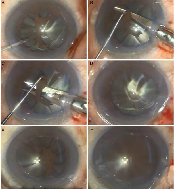
Figure 2. Anterior segment structures as seen under operating microscope showing surgical steps in both eyes. In the left eye, (A)
Dispersive ocular viscoelastic device was injected between persistant pupillary membrane and crystalline lens. (B) After appropriate tension to the iris strand was applied by using Phaco Chopper, the operator cut the persistent pupillary mambrane. (C) The remnant iris strands were cut carefully in the same way. (D) Though the persistent pupillary membrane was removed, mydriasis maintained.In the right eye, (E) preopreative persistent pupillary membrane after mydriasis. (F) Mydriasis was also maintained after removing the pupillary membrane.
microscopy (Konan medical, Nishinomiya, Japan)로 술 전 우안 2,303개/mm3와 좌안 2,332개/mm3, 술 후 내피세포 수 우안 1,970개/mm3와 좌안 1,827개/mm3로 측정되었다.
고 찰
동공잔류막이 두껍거나 동공의 크기가 1.5 mm 미만으로 작아 시력장애를 유발하거나 약시가 생기는 경우 적극적인
치료가 필요하다.1 보존적인 치료로는 산동제를 사용하여 동공을 1.5 mm 이상 유지시킨 상태에서 가림 치료를 하여 약시를 치료 및 예방할 수 있다.1 보존적 치료에 반응하지 않거나 동공막의 크기가 두껍고 커서 시축을 가리는 경우 에는 수술적 치료가 필요하며, 레이저 시술이나 동공잔류 막의 두꺼운 부분을 절제하는 동공잔류막 절제술을 시행할 수 있다.1 Neodymium-doped yttrium aluminium garnet (Nd:YAG) 레이저 시술로 동공잔류막을 성공적으로 제거
A B
C D
E F
Figure 3. Slit lamp photograph 3 months after persistent pupil-
lary membrane removal and phachoemulsification, and poste- rior chamber lens implantation in left eye. There was no evi- dence of anterior chamber inflammation and intraocular lens position was also stable.한 증례가 1987년 Vega, 1994년 Kumar 등에 의해서 보고 된 바 있으며 순차적 Argon YAG 레이저 시술을 통한 성공 적 제거도 2005년 Wang에 의해서 보고되었다.4-7 YAG 레 이저를 통한 동공잔류막 제거는 홍채의 가닥이 동공잔류막 의 상부와 연결되어 있으며 가닥이 좁은 경우에만 가능하 고 소아의 경우 협조가 어려워 시행하기 힘들다.4-7 부작용 으로는 전방출혈을 야기하거나 백내장 생성 혹은 색소 분 산 증후군의 위험성이 존재한다. 동공잔류막의 수술적 제 거에는 forcep과 vitreous scissors를 이용하여 홍채 가닥을 제거하는 방법이 보통 사용된다.1,3 수술 시 전방 출혈, 동공 조임근 손상, 수정체 손상에 의한 백내장 발생 등을 주의해 야 하며 수술 전후 감염과 같은 위험성이 존재한다.8
본 증례에서는 환자 양안 홍채가닥이 넓어 레이저 시술 시 미세 전방 출혈 및 색소 분산 증후군 위험도 높다고 판 단했고 동공잔류막의 크기가 직경 3 mm에 달하여 YAG laser로 홍채가닥을 제거하더라도 많은 양의 잔존 조직이 전방에 남아 염증을 유발할 수 있기 때문에 수술로 제거하 는 방법을 택하였다. 동공잔류막 제거 시 전방이 잘 유지되 지 않거나 얕은 경우 수술기구가 각막에 닿아 각막 내피세 포 손상을 유발할 수 있고, 동공조임근 주변의 동공잔류막 제거 시 수정체낭 혹은 동공조임근의 손상을 유발할 수 있 어 특히 주의해야 하는데 이를 방지하기 위해 분산성의 점 탄물질을 전방과 후방에 각각 충분히 주입하여 홍채가닥을 팽팽하게 하고 동공잔류막과 수정체낭과의 거리를 충분히 확보하였다. 또한 동공잔류막과 연결된 홍채가닥을 Sinskey hook 혹은 Phaco Chopper를 사용하여 팽팽하게 당긴 다음 동공조임근에서 약 1 mm 떨어진 부위에서 홍채가닥 절제
를 시행하였다. 결과적으로 술 중 흔히 발생할 수 있는 미 세 전방 출혈은 발생하지 않았으며 동공조임근 손상 없이 동공잔류막을 제거하였다.
2013년 Ko et al3이 동공잔류막 제거술을 먼저 시행하여 전방염증이 없고 안정된 상태에서 초음파 유화술 및 인공 수정체 삽입술을 시행하는 2단계 수술방법을 시행하여 좋 은 결과를 보고한 적이 있다. 하지만 2단계 수술은 환자의 부담과 불편을 초래하고 비용 부담도 커지는 단점이 있다. 또한 수술 횟수가 많아짐에 따라 각막 내피세포 숫자가 감 소할 수 있고, 감염, 상피내생, 홍채 손상, 녹내장, 황반 부 종과 같은 안내수술 후 발생 수 있는 부작용 가능성이 높아 질 수 있다. 본 증례에서는 술 전 전방 홍채 출혈, 염증 소 견이나 녹내장 소견이 발견되지 않아 동공잔류막 제거술 시행 시 합병증의 위험도가 낮을 것이라 예상되어 동공잔 류막 및 백내장 동시 수술을 계획할 수 있었다. 수술 후 시 력호전을 보였으며 전방염증, 포도막염, 안압 상승 등의 합 병증을 보이지 않았다. 기존 연구에서 동공잔류막 및 백내 장 동시 수술로 인한 안내 감염 및 부작용을 언급하였으나 본 증례에서 수술 후 감염과 같은 부작용이 발견되는 것 없 이 시력 호전을 보였고 오히려 2단계의 수술로 이루어지는 환자의 불편함을 줄이고 만족도를 개선할 수 있었다. 술 후 경도의 염증만 관찰되었지만 술 후 각막 내피세포 밀도가 술 전에 비해 약 15–20% 감소를 보였는데, 이는 갈색백내 장 유화를 위한 고에너지 초음파 사용에 의한 것으로 추정 된다.
동공잔류막 및 백내장 동시 수술 시 상대적으로 긴 수술 시간이 필요하며 동공잔류막 제거 시 출혈, 염증 등의 합병 증이 발생할 수 있고, 홍채를 자극하여 축동이 되면 백내장 수술이 어려울 수 있다. 하지만 본 증례와 같이 술 전 특별 한 위험요인이 없고 술 중 합병증 없이 동공잔류막을 제거 한 경우 백내장 동시 수술을 진행하는 것이 좋을 것이라 사 료된다.
REFERENCES
1) Kraus CL, Lueder GT. Clinical characteristics and surgical ap- proach to visually significant persistent pupillary membranes. J AAPOS 2014;18:596-9.
2) Lambert SR, Buckley EG, Lenhart PD, et al. Congenital fi- brovascular pupillary membranes: clinical and histopathologic findings. Ophthalmology 2012;119:634-41.
3) Ko J, Jung JW, Kim EK. Two-stage operation for the treatment of cataract associated with persistent pupillary membrane. J Cataract Refract Surg 2013;39:1615.
4) Wang JK, Wu CY, Lai PC. Sequential argon-YAG laser mem- branectomy and phacoemulsification for treatment of persistent pupillary membrane and associated cataract. J Cataract Refract
= 국문초록 =
양안 동공잔류막 제거술과 수정체유화술 및 인공수정체 삽입술을 동시에 시행한 1예
목적: 양안 동공잔류막과 갈색 백내장이 동반된 환자에서 동공잔류막 제거술과 수정체유화술 및 인공수정체 삽입술을 동시에 시행한 1예를 보고하고자 한다.
증례요약: 양안 시력저하를 주소로 기저질환이 없는 64세 남자 환자가 외래로 내원하였다. 초진 시 최대교정시력 우안 0.1, 좌안 0.2였 다. 양안 동공잔류막이 관찰되었고 수정체는 양안 심한 핵경화가 동반된 갈색백내장 소견을 보였다. 먼저 좌안 동공잔류막 제거술과 수정체유화술 및 인공수정체 삽입술을 동시에 시행하고 1주 후 우안도 같은 수술을 시행하였다. 수술 후 6개월째 최대교정시력 우안 0.2, 좌안 0.8로 측정되었고 경과 관찰 기간 동안 전방염증, 포도막염, 안압 상승 등의 합병증은 관찰되지 않았다.
결론: 동공잔류막 제거술과 초음파 유화술 및 인공수정체 삽입술을 동시에 시행 후 특별한 합병증 없이 시력호전을 보여 이를 보고하 고자 한다.
<대한안과학회지 2017;58(6):731-735>
Surg 2005;31:1661-3.
5) Vega LF, Sabates R. Neodymium: YAG laser treatment of persis- tent pupillary membrane. Ophthalmic Surg 1987;18:452-4.
6) Kumar H, Sakhuja N, Sachdev MS. Hyperplastic pupillary mem- brane and laser therapy. Ophthalmic Surg 1994;25:189-90.
7) Mansour AM, Hamade I, Antonios RS. Sequential argon-YAG la-
ser membranotomy of extensive persistent pupillary membrane with visual loss. BMJ Case Rep 2015;2015. pii: bcr2015210140.
8) Kesarwani S, Murthy R, Vemuganti GK. Surgical technique for re- moving congenital fibrovascular pupillary membrane, with clin- icopathological correlation. J AAPOS 2009;13:618-20.