Extramedullary myeloma (Plasmacytoma) is a malig- nant tumor composed entirely of plasma cells in the ab- sence of bone involvement. These tumors can occur anywhere in soft tissue, especially in the upper respira- tory tract and oral cavity (1). Plasmacytoma of the breast is a rare condition that may occur as a solitary finding or in association with multiple myeloma (2). We report here on a case of plasmacytoma of the breast that was associated with a plasmacytoma in left eyelid involving the sphenoid sinus. To our knowledge, this report is the first case of plasmacytoma of the breast in Korea.
Case Report
A 50-year-old woman presented with a palpable mass in her left breast for a month. On physical examination,
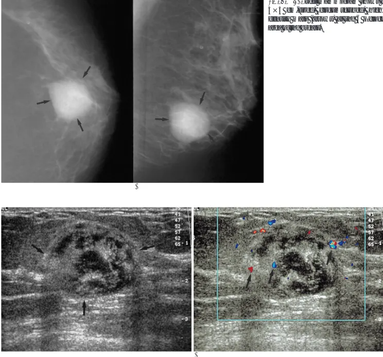
a palpable mass (about 3×3 cm) was identified at the 6 o’clock area of the left breast. The patient had a history of a plasmacytoma of the left eyelid with the involve- ment of the sphenoid sinus 3 years ago, which recurred at the right eyelid and left preauricular area in spite of chemotherapy and radiation therapy after an excision biopsy. The left mammogram revealed a 3×3 cm-sized, circumscribed, high-density mass at the 6 o’clock area (Fig. 1). The patient’s previous mammogram 5 months ago was normal. The high-resolution ultrasound exami- nation using a 12 MHz linear array transducer (HDI 5000, ATL Bothell, WA U.S.A.) demonstrated a relative- ly well-defined, round-shaped, heterogeneously hypoe- choic mass with a mild posterior acoustic enhancement in the left breast (Fig. 2A). Increased vascularity was noted in the solid hyperechoic portion of the tumor on the color Doppler study (Fig. 2B).
Fine needle aspiration biopsy was performed, and specimen was composed of dispersed plasmacytoid cells with abundant, basophilic cytoplasms and eccentric nu- clei, including some in mitosis (Fig. 3). These appear- ances were characteristic of plasmacytoma or multiple myeloma. The bone marrow biopsy was done and mul-
J Korean Radiol Soc 2004;50:385-388
─ 385 ─
Mammographic and Ultrasonographic Appearances of Plasmacytoma of the Breast: Case Report1
Kyoung Ah Kim, M.D., Jeong Mi Park, M.D., Sun Mi Kim, M.D.
Plasmacytoma is a tumor composed of plasma cells in the absence of bone involve- ment. These malignant tumors can occur anywhere in soft tissue, and especially in the upper respiratory tract and oral cavity. Plasmacytoma of the breast is a rare condition that may occur as a solitary finding or in association with multiple myeloma. We re- port the mammographic and ultrasonographic findings of a case of breast plasmacy- toma associated with a recurrent plasmacytoma in left eyelid involving the sphenoid sinus.
Index words :Breast neoplasms Metastases Radiography
1Department of Radiology, Asan Medical Center University of Ulsan College of Medicine
Received July 7, 2003 ; Accepted March 18, 2004
Address reprint requests to : Jeong Mi Park, M.D, Department of Radiology, Asan Medical Center, University of Ulsan College of Medicine, 388-1 Pungnap-dong, Songpa-gu, Seoul 138-736, Korea.
Tel. 82-2-3010-4400 Fax. 82-2-476-0090 E-mail: [email protected]
tiple myeloma was ruled out again. The patient received another round of chemotherapy including cisplatin, etoposide and dexametasone.
Discussion
Plasmacytoma usually arises from the bone marrow, and it is known as myeloma that can be divided into an uncommon solitary and common multiple entity (multi- ple myeloma, myelomatosis). In approximately 70% of patients with multiple myeloma, autopsy findings show
microscopic neoplastic plasma cell infiltrations outside the skeletal system that may occur in almost any organ (3).
However, extamedullary plasmacytomas are rarely detected macroscopically, and so they are seldom no- ticed before death. There is a distinction between a soli- tary form and disseminated form. The latter is indistin- guishable from disseminated multiple myeloma and can be considered as an end-stage, advanced disease.
In the breast, plasmacytoma is very rare. It was first described by Cutler in 1934 (3), and only 12 cases have
Kyoung Ah Kim, et al: Mammographic and Ultrasonographic Appearances of Plasmacytoma of the Breast
─ 386 ─
A B
Fig. 2. A. Transverse ultrasonography of the mass shows a well-defined heterogeneous echoic solid mass (arrows) with a hypoe- choic center in the left breast.
B. Color Doppler study shows an increased vascularity in the mass.
A B
Fig. 1. A, B. Left mammogram shows a 3×3 cm-sized, circumscribed, high- density mass (arrows) at the 6 o’clock area of the breast.
been reported to date. Mammographic findings were mainly a well-circumscribed and dense mass. In 3 cases of the 12, ultrasonography demonstrated a heteroge- neous hypoechoic mass with posterior acoustic shadow- ing in one case, but there was posterior acoustic en- hancement in the other cases. Vascularity was relatively increased in two of the masses (4-6).
Our present study showed a rapidly growing breast mass, as in a case of plasmacytoma. By statistical inci- dence, primary adenocarcinoma of the breast should be considered as the first differential diagnosis.
Mesenchymal sarcomas of the breast stroma also need- ed to be ruled out because of its significant incidence.
Primary and secondary lymphoproliferative processes, such as malignant lymphoma and plasmacytoma are, however, exceedingly rare (4). But considering the growth rate of mass and the presence of an underlying plasmacytoma in the eyelid, plasmacytoma can be in-
cluded into the differential diagnosis. Moreover, the mass showed a well-defined margin mammographical- ly, a lobulated heterogeneous hypoechogenicity sono- graphically, and an increased vascularity on the color Doppler image. These findings were similar to those of plasmacytoma of the breast, as was described in previ- ous cases (5-7).
Recognition of extramedullary plasmacytoma as a dis- tinct variant of myeloma is important, because this con- dition responds to systemic and local treatment much better than multiple myeloma, and its natural history is also different(8).
We report here on the first case of plasmacytoma of the breast in Korea with its mammographic and ultra- sonographic findings.
References
1. De Chiara A, Losito S, Terraciano L, Di Giacomo R, Iaccarino G, Rubolotta MR. Primary plasmacytoma of the breast. Arch Pathol Lab Med 2001;125:1078-1080
2. Brem RF, Revelon G, Willey SC, Gatewood OM, Zeiger MA.
Bilateral plasmacytoma of the breast: a case report. Breast J 2002;8:
393-395
3. van Nieuwkoop C, Giard R, Veen HF, Dees A. Extramedullary plasmacytoma of the breast simulating breast cancer. Netherlands J Med 2001;58:174-176
4. Ross J, King TM, Spector I, Zimbler H, Basile RM. Plasmacytoma of the breast. An unusual case of recurrent myeloma. Arch Intern Med 1987;147:1838-1840
5. Coll D, Spence L, Cardenosa G. Plasmacytoma of the breast: mam- mographic and sonographic findings. AJR Am J Roentgenol 1999;173:1135-6
6. Collins CD, Kedar RP, Cosgrove DO. Case report: myeolma of the breast-appearances on ultrasound and color Doppler. Br J Radiol 1994;67:399-400
7. Bloomberg TJ, Glees JP, Williams JE. Bilateral breast lumps: an unusual feature of extramedullary plasmacytoma. Br J Radiol 1980;53:498-501
8. Momiyama N, Ishikawa T, Doi T, et al. Extramedullary Plasma- cytoma of the brest with serum IgD Monoclonal Protein: a case re- port and reviw of the literature. Breast Cancer 1999;6:217-221 J Korean Radiol Soc 2004;50:385-388
─ 387 ─ Fig. 3. Photomicrograph of fine needle aspiration biopsy speci- men shows dispersed plasmacytoid cells with abundant ba- sophilic cytoplasm and eccentric nuclei, including some mito- sis (arrow) (×400, Wright stain).
Kyoung Ah Kim, et al: Mammographic and Ultrasonographic Appearances of Plasmacytoma of the Breast
─ 388 ─
대한영상의학회지 2004;50:385-388
유방의 형질세포종의 유방촬영술과 초음파 소견: 증례 보고1
1울산대학교 서울아산병원 방사선과 김경아・박정미・김선미
형질세포종은 형질세포로 이루어진 악성종양으로 다발성 골수종이 골수에서 생기는 반면 골수에 병발하지 않는 점 에서 다르다. 비교적 드문 혈구 기원 종양 질환으로 신체 어디서나 발생할 수 있으나 주로 상부기도와 구강에 발생하는 것으로 알려져 있다. 저자들은 좌측안검과 접형동의 형질세포종으로 절제 생검을 받았던 50세 여자 환자의 좌측 유방 에 병발한 형질세포종의 유방촬영술과 초음파 소견을 보고하고자 한다.