A composite glandular/exocrine-endocrine carcinoma of the gastrointestinal tract is characterized by the co-existence of two adjacent, but histologically-distinct tumors in an organ. Composite glandular/exocrine-endocrine carcinomas are a special type of tumor comprised of common adenocarcinomas and neuroendocrine components that account for at least one-third of the entire tumor area. Composite tumors have been reported in a range of organs, but are relatively rare in the stomach. We report a case of a composite neuroendocrine carcinoma with an adenocarcinoma of the stomach (mixed exocrine-endocrine carcinoma), which was misdiagnosed as a giant submu- cosal tumor preoperatively based on esophagogastroduodenoscopy and a contrast-enhanced axial computed tomographic scan.
Key Words: Composite tumor, Stomach, Submucosal tumor
Introduction
Human cancers exhibiting a combination of conventional (glan- dular, squamous, or urothelial) and neuroendocrine features occur in various organs. Such lesions are classified into two subgroups:
composite and collision-type tumors.(1,2) Composite neuroendo- crine carcinomas with adenocarcinomas occur throughout the gas- trointestinal tract, but rarely occur in the stomach.(3) The histogen- esis of composite tumors is unclear, but both types of carcinomas may be derived from the same cell, most likely a pluripotent stem cell. Composite neuroendocrine carcinomas with adenocarcinoma in the stomach can be diagnosed if neuroendocrine marker shows a positive reaction in the immunohistochemical staining of the component of the neuroendocrine carcinoma.(4)
We report a case of a composite neuroendocrine carcinoma with adenocarcinoma of the stomach (mixed exocrine-endocrine
carcinoma), which was misdiagnosed as a giant gastric submucosal tumor preoperatively by esophagogastroduodenoscopy (EGD) and contrast-enhanced axial computed tomographic (CT) scan. By immunohistochemical staining of the neuroendocrine carcinoma component, the tumor was identified as a composite neuroendo- crine carcinoma with adenocarcinoma of the stomach. We report this rare case with a review of the literature.
Case Report
A 62-year-old woman underwent EGD, abdominal CT and abdominal ultrasonography as part of an evaluation for epigastric discomfort. Her past medical and family histories were unremark- able. The physical examination on admission, routine blood tests, and urine analysis were also unremarkable. The levels of tumor markers were within the reference range, as follows: carcinoem- bryonic antigen (CEA), 5.21 ng/ml (normal, ≤8 ng/ml); CA 19- 9, 10.49 U/ml (normal, ≤37 U/ml); and CA 72-4, 0.289 ng/ml (normal, ≤6.9 ng/ml).
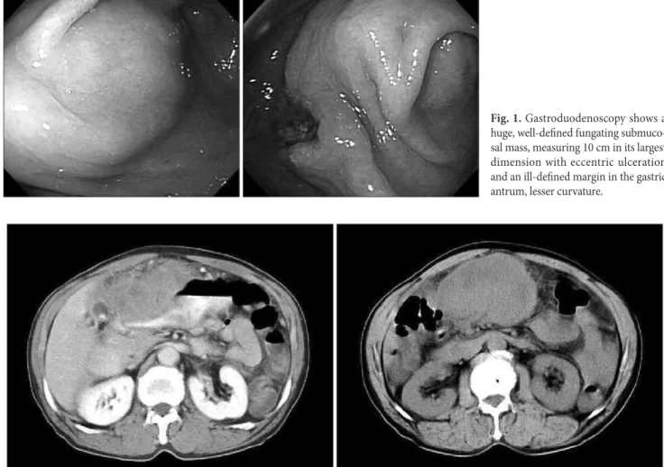
The EGD revealed a huge, well-defined, fungating, submuco- sal mass with eccentric ulceration characterized by an ill-defined margin in lesser curvature of the gastric antrum (Fig. 1). An EGD Correspondence to: Hyun-Dong Chae
Department of Surgery, Catholic University of Daegu School of Me
dicine, 30566, Daemyung 4dong, Namgu, Daegu 705718, Korea Tel: +82536504429, Fax: +82536247185
Email: hdchae@cu.ac.kr Received December 9, 2010 Accepted February 17, 2011
This is an openaccess article distributed under the terms of the Creative Commons Attribution NonCommercial License (http://creativecommons.org/
licenses/bync/3.0) which permits unrestricted noncommercial use, distribution, and reproduction in any medium, provided the original work is properly cited.
Copyrights © 2011 by The Korean Gastric Cancer Association www.jgc-online.org
biopsy was not performed because a biopsy is not generally recom- mended for the diagnosis of gastric submucosal tumors. A contrast- enhanced axial CT scan demonstrated a huge, solid, ovoid, hetero- geneous, exophytic mass measuring approximately 15×10 cm in the pre-pyloric antrum of the stomach with several metastasis into the lymphatic glands around the stomach was observed (Fig. 2). A diagnosis of a giant submucosal tumor of the stomach with lymph node metastasis was made, and a radical subtotal gastrectomy with D2 lymph node dissection was subsequently performed.
Grossly, a diffuse submucosal bulging mass was noted in the surgically-resected stomach. The mass-measure 17×15×6.5 cm, and had an intact mucosa, with the exception of a small polypoid nodule with a central ulcerated umbilication measuring 1.5×1 cm.
On sectioning, the cut surface of the mass showed a diffuse, geo- graphic, hemorrhagic, necrotic area with a cleft-like cystic change and grayish-brown solid area which infiltrated to the surrounding omental fat tissue.
Microscopically, the tumor was comprised of a conventional
adenocarcinoma with anaplastic glandular nests and a neuroen- docrine carcinoma with central necrosis (Fig. 3). The glandular carcinoma was confined to the mucosa and submucosa, the neuro- endocrine carcinoma had invaded into the serosa and the lymphatic glands. Two of 39 lymph nodes had metastases, and lymphatic and perineural invasion were observed. Immunohistochemically, the tumor cells exhibited immunopositivity for chromogranin A and synaptophysin, but negativity for CD56, CD117, CD34, and SMA.
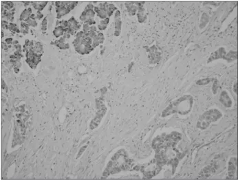
The neuroendocrine carcinoma components showed strong immu- nopositivity for chromogranin A, but were negative for the adeno- carcinoma component (Fig. 4).
The post-operative course was uneventful and the patient was discharged on the 9th postoperative day.
Discussion
Submucosal tumors refer to tumors that exist below the mucosal layer and protrude into the lumen in a hemispherical or spherical Fig. 2. Preoperative contrast-enhanced axial CT scan shows a huge, solid, ovoid, heterogenous and exophytic mass measuring approximately 15×10 cm in the prepyloric antrum of the stomach.
Fig. 1. Gastroduodenoscopy shows a huge, well-defined fungating submuco- sal mass, measuring 10 cm in its largest dimension with eccentric ulceration and an ill-defined margin in the gastric antrum, lesser curvature.
shape because of the surrounding mucosa on the surface.
Submucosal tumors can cause symptoms, such as bleeding, but in most cases, submucosal tumors are detected incidentally dur- ing gastroduodenscopy or an upper GI series. On routine EGD, the rate of diagnosing submucosal tumors is approximately 0.36%.
(5) Submucosal tumors that usually occur in the stomach include gastrointestinal stromal tumors (GISTs), leiomyomas, leiomyo- sarcomas, lipomas, and carcinoids. However, the gastric cancers with a pattern of submucosal tumors are very rare, accounting for 0.1~0.62% of resected gastric cancers.(5,6) The pathologic diagnosis of most gastric cancers is an adenocarcinoma. In our case, a giant protruding lesion covered with gastric mucosa was noted during the pre-operative EGD, and the origin of the tumor was located in the submucosal muscularis propria layer, as demonstrated by a contrast-enhanced axial CT scan. Therefore, we diagnosed the tu- mor clinically as a malignant gastric GIST and performed a radical subtotal gastrectomy.
Composite glandular/exocrine-endocrine carcinomas of the gastrointestinal tract are special tumors comprised of common ad- enocarcinomas with neuroendocrine components, and accounts for at least one-third of the entire tumor area. Glandular-endocrine tumors of the digestive tract have rarely been described in the medical literature because the classification, which restricts the term “mixed” to lesions in which the endocrine cells account for between about one-third and one-half of all cells, and proposed a classification for such neoplasms distinguishing (a) composite (or mixed) glandular-endocrine tumors with both elements in more or less equal proportions, (b) amphicrine tumors with dual differentia-
tion within the same cell, and (c) collision tumors in which the two components are juxtaposed, but not admixed.(6) According to this classification, the case herein was a composite glandular-endocrine carcinoma.
More recently, Fujiyoshi et al.(7) reclassified mixed endo- crine and non-endocrine epithelial tumors by dividing the tu- mors into six categories: 1) neuroendocrine cells interspersed within carcinomas; 2) carcinoids (neuroendocrine tumors [NETs]) with interspersed non-endocrine cells; 3) composite glandular- neuroendocrine cell carcinomas containing areas of carcinoid and conventional carcinomas; 4) collision tumors in which NETs and conventional carcinomas are closely juxtaposed, but not admixed;
5) amphicrine tumors predominantly composed of cells exhibit- ing concurrent neuroendocrine and non-endocrine differentiation;
and 6) combinations of the previous types. According to this clas- sification, our case could be classified as a composite glandular- endocrine carcinoma containing mainly a NET with small areas of a conventional carcinoma.
Composite neuroendocrine carcinomas with adenocarcinomas in the stomach can be diagnosed if at least one of the neuroendo- crine markers, such as chromogranin A, synaptophysin, and NSE has a positive reaction in the immunohistochemical staining of the neuroendocrine carcinoma component.(4) In the case herein, the component of the neuroendocrine carcinoma had a strong positive response to chromogranin A, which is a neuroendocrine marker, but the adenocarcinoma component was negative (Fig. 4).
With the exception of the appendix, only rare instances of composite tumors have been detected in the esophagus, stomach, Fig. 3. The tumor is composed of conventional adenocarcinoma with
anaplastic glandular nests (arrow) and neuroendocrine carcinoma with central necrosis (arrow head) (H&E, ×100).
Fig. 4. Neuroendocrine carcinoma com ponents show strong immu- nopositivity for chromogranin A (left upper), but negative immunos- taining of adenocarcinoma component (right lower) (Immunohisto- chemical stain, ×200).
gallbladder, and the small and large bowel. However, it is possible that before the advent of immunohistochemistry, the true incidence of composite tumors was underestimated.
The histologic origin of composite tumors is unclear. In rats with hypergastrinemia, enterochromaffin-like cells have the ca- pacity to dedifferentiate and become potential precursors of gastric adenocarcinomas.(8,9) Some authors have postulated proliferation of a pluripotential precursor cell,(10-12) and studies describing common genetic alterations in the glandular and neuroendocrine component of mixed tumors support the latter hypothesis.(12)
Because of their rarity and uncertain pathologic criteria, the clinical behavior and histogenesis of composite tumors is still unclear, but Volante et al.(13) reported that the clinical behavior of composite carcinomas depends on the adenocarcinomatous component if the associated endocrine component is well-differ- entiated, and upon the neuroendocrine component if it is poorly- differentiated. In this case, although the glandular carcinoma was confined to the mucosa and submucosa, the neuroendocrine carci- noma had invaded into the serosa and the lymphatic glands. There- fore, the neuroendocrine component of this case was believed to impact heavily on the prognosis and we performed chemotherapy based on the neuroendocrine carcinoma.
Treatment of neuroendocrine cancers that occur in the stomach is based on radical surgical resection. In cases of distant metastasis, such as liver metastasis, a gastrectomy with hepatectomy is effec- tive treatment.(14) Chemotherapeutic regimens, including cisplatin, doxorubicin, and vincristine can be administered to neuroendocrine carcinoma patients. Because combination treatment of cisplatin and etoposide after gastrectomy has been reported to result in reduction of liver metastasis by 96% and lung metastasis by 81%, chemother- apy has recently been recommended to be administered following gastrectomy; however, the efficacy has not been fully verified due to the low incidence.(15)
In summary, we initially misdiagnosed the tumor as a giant gastric submucosal tumor by EGD and contrast-enhanced axial CT scan, and subsequently performed surgical resection. However, in retrospect, metastasis into the lymphatic glands around the stomach was observed in the pre-operative contrast-enhanced axial CT scan, and eccentric ulceration with an ill-defined margin was noted on EGD, which could have been diagnosed as metastasis into the lymphatic glands around the stomach secondary to gastric cancer.
Therefore, if a submucosal tumor is large and eccentric ulceration with ill-defined margins is observed with lymphatic gland metas- tasis around the stomach, an adenocarcinoma, and although ex-
tremely rare, a composite glandular-endocrine carcinoma contain- ing a conventional carcinoma should be taken into consideration in the differential diagnosis.
References
1. Liu SW, Chen GH, Hsieh PP. Collision tumor of the stomach:
a case report of mixed gastrointestinal stromal tumor and ad- enocarcinoma. J Clin Gastroenterol 2002;35:332-334.
2. Fukui H, Takada M, Chiba T, Kashiwagi R, Sakane M, Tabata F, et al. Concurrent occurrence of gastric adenocarcinoma and duodenal neuroendocrine cell carcinoma: a composite tumour or collision tumours? Gut 2001;48:853-856.
3. Chejfec G, Falkmer S, Askensten U, Grimelius L, Gould VE.
Neuroendocrine tumors of the gastrointestinal tract. Pathol Res Pract 1988;183:143-154.
4. Granberg D, Wilander E, Stridsberg M, Granerus G, Skogseid B, Oberg K. Clinical symptoms, hormone profiles, treatment, and prognosis in patients with gastric carcinoids. Gut 1998;43:223- 228.
5. Hedenbro JL, Ekelund M, Wetterberg P. Endoscopic diagnosis of submucosal gastric lesions. The results after routine endos- copy. Surg Endosc 1991;5:20-23.
6. Lewin K. Carcinoid tumors and the mixed (composite) glan- dular-endocrine cell carcinomas. Am J Surg Pathol 1987;11:71- 86.
7. Fujiyoshi Y, Kuhara H, Eimoto T. Composite glandular- endocrine cell carcinoma of the stomach. Report of two cases with goblet cell carcinoid component. Pathol Res Pract 2005;200:823-829.
8. Fossmark R, Zhao CM, Martinsen TC, Kawase S, Chen D, Waldum HL. Dedifferentiation of enterochromaffin-like cells in gastric cancer of hypergastrinemic cotton rats. APMIS 2005;113:436-449.
9. Waldum HL, Rørvik H, Falkmer S, Kawase S. Neuroendocrine (ECL cell) differentiation of spontaneous gastric carcinomas of cotton rats (Sigmodon hispidus). Lab Anim Sci 1999;49:241- 247.
10. Lee EJ, Park SM, Maeng L, Lee A, Kim KM. Composite glan- dular-endocrine cell carcinomas of the stomach: clinicopatho- logic and methylation study. APMIS 2005;113:569-576.
11. Shibuya H, Azumi N, Abe F. Gastric small-cell undifferentiated carcinoma with adeno and squamous cell carcinoma compo- nents. Acta Pathol Jpn 1985;35:473-480.
(neuro)endocrine and non-(neuro)endocrine tumours: a comment on concepts and classification of mixed exocrine- endocrine neoplasms. Virchows Arch 2006;449:499-506.
tric collision tumor (adenocarcinoma and neuroendocrine carcinoma) diagnosed as a advanced gastric cancer. J Korean Surg Soc 2007;73:173-177.