89
책임저자 : 소의영, 경기도 수원시 영통구 원천동 산 5번지
443-721, 아주대학교병원 외과 Tel: 031-219-5200, Fax: 031-219-5755 E-mail: [email protected]
게재승인일:2008년 5월 3일
분화 갑상선암 수술 후 발생한 불현성 갑상선중독증 환자의 골대사 및 골밀도의 특징
아주대학교 의과대학 외과학교실, 1강동성심병원 외과
박승현ㆍ이잔디ㆍ최수윤1ㆍ소의영
The Bone Mineral Density and Bone Metabolism of Patients with Differentiated Thyroid Carcinoma and who are Receiving Long-term TSH Suppre- ssive Therapy
Seung Hyun Park, M.D., Jandee Lee, M.D., Soo Youn Choi, M.D.
1 and Euy-Young Soh, M.D., Ph.D.Purpose: The clinical implications of long-term suppressive
thyroxine (TSH) therapy on the skeletal system are critical, largely because of the favorable prognosis of differentiated thyroid carcinomas (DTC). However, the potentialdeleterious effects of TSH suppressive therapy on the bone metabolism remain controversial. The aim of this prospective study was to assess the effects of chronic L-thyroxine (LT4) treatment with supraphysiologic doses of TSH on the bone mineral density (BMD) and biochemical bone remodeling markers.Methods: This cross-sectional study was designed to com-
pare patients with DTC and who were treated with LT4 for more than 2 years after thyroidectomy with an age-matched and gender-matched healthy control group. A total of 100 female outpatients (mean age: 47.5±13.8; 38 pre and 62 post-menopausal) who were on LT4 for between 2 and 10 yearswere enrolled. One hundred and three age-matched healthy volunteers were recruited as a control group.Laboratory tests were performed to exclude other possible factors for secondary osteoporosis. We measured the BMD by dual energy X-ray absorptiometry (DEXA), and bone turnover was assessed by several biochemical parameters.
Results: Our data showed no significant difference between
the bone markers for the study group and the control group that had a premenopausal status. However, for the patients with a postmenopausal status, the serum levels of bone alkaline phosphatase were significant higher in the studygroup than that in the control group (P=0.038). We also found no significant difference between the study patients and the age- and weight-matched controls for the BMD at any site of measurement.
Conclusion: This preliminary report suggests that bone turn-
over and accelerated bone loss might be related to the long-standing TSH suppression in postmenopausal women.Future prospective studies with an increased number of studied patients and a prolonged time of observation will be necessary in order to better assess the relative risk of osteoporosis in patients who are undergoing TSH sup- pressive treatment. (Korean J Endocrine Surg 2008;8:89-
94)
Key Words: Osteoporosis, TSH suppression, Differentiated
thyroid carcinoma중심 단어: 골다공증 갑상선자극호르몬 억제용법,
분화 갑상선암Department of Surgery, Ajou Universiy School of Medicine,
1Department of Surgery, Kangdong Sacred Heart Hospital, Seoul, Korea
서 론
갑상선호르몬은 골 교체율(bone turnover rate)의 조절에 관여하므로 정상적인 골 성장 및 발육에 주요한 역할을 한 다. 따라서, 갑상선기능항진증이 지속되면 골 교체율의 증 가로 골흡수(bone resorption) 및 골형성(bone formation)의 불 균형이 야기되어 골다공증(osteoporosis) 및 병적 골절(patho- logic fracture)의 발생률이 높아진다고 알려져 있다.(1-5) 골 대사에 직접적인 영향을 미치는 갑상선 호르몬은 활성 형(active)인 T3이며, 각각의 전달과정에서 다양한 성장 인자 및 시토카인(cytokine) 등이 간접적으로 관여한다고 알려져 있었다. 하지만, 최근 갑상선자극호르몬(Thyroid stimulating hormone, TSH)이 혈중 갑상선 호르몬과 관계없이 독립적인 세포내 신호전달체계를 통해 골격 재형성(skeletal remodeling) 에 결정적인 역할을 한다고 보고되어, TSH 억제와 골밀도 변
화에 대한 연관성이 주요한 임상 문제로 부각되고 있다.(3-8) 불현성 갑상선 중독증(subclinical hyperthyroidism)이란 혈 청 free T3, free T4는 정상이지만 TSH가 낮은 수치를 보이 는 경우로 정의하며, 가장 많은 원인은 갑상선암 수술 후 장기간 호르몬 대체 요법으로 발생하는 외인성 요인이다 (2,3-8). 최근 분화 갑상선 암의 급격한 증가로 수술 후 호르 몬 대체 요법을 시행하는 환자에서 발생할 수 있는 불현성 갑상선 중독증에 대한 관심이 증대되고 있다. 불현성 갑상 선 중독증에서 낮은 TSH 수치가 골대사 및 골밀도 변화에 미치는 영향에 대한 여러 임상 보고가 있지만, 아직 이견이 많다. 즉, 분화 갑상선암 수술 후 TSH 억제 정도 및 골밀도 에 대한 선별검사 방법 등에 대한 뚜렷한 지표가 아직 없는 실정이다.(4,5)
이에 저자들은 본원에서 분화 갑상선 수술 후 갑상선 호 르몬 대체요법으로 장기간 불현성 갑상선 중독증 상태인 환자를 대상으로 정상 갑상선 호르몬 수치를 보이는 동일 한 조건의 대조군과 비교하여 차이를 분석하고자 하였다.
대상 및 방법 1) 연구군과 대조군
1997년 11월부터 2005년 10월까지 아주대학교병원에서 유두상 갑상선암으로 갑상선 전절제술 및 추가적인 방사성 요오드 치료를 시행 받은 여성 환자를 조사하였다. 갑상선 호르몬 대체 요법을 시행하면서 혈중 갑상선 기능검사를 적어도 3∼6개월 간격으로 검사한 결과, 활성 갑상선 호르 몬 수치(T3, fT4 등)는 정상범위이지만 TSH 저하(TSH
<0.35 mU/L)를 보이는 불현성 갑상선 중독증이 2년 이상 지속된 환자를 대상으로 하였다. 또한 환자의 과거력 및 투 약 내역을 조사하여 아래의 기준에 1개 이상이라도 해당될 때에는 연구군에서 제외하였다. 제외기준은 첫째, 과거에 골다공증이나 병적 골절의 기왕력이 있는 경우, 둘째, 골 대 사에 영향을 미치는 다른 질환이 동반된 경우(장기간 침상 안정으로 인한 운동 제한, 최근 골절 경험, 당뇨, 염증성 골 관절 질환, 갑상선 질환, 뇌하수체 질환, 성선 기능 저하증, 간 질환, 신장 질환, 알코올중독, 조기 폐경 등), 셋째, 골 대사에 영향을 미치는 약물 복용력(칼슘 제제, Vit. D 제제, 이뇨제, 스테로이드 제제, 비스포스페이트 제제, 경구 피임 제, 에스트로젠 제제, 항경련제, 사이클로스포린제, 리튬제, 헤파린 등)이 있는 경우였다.
본 연구의 기준에 적합한 연구군은 100명의 여성이었으 며, 환자의 문진 및 여포자극호르몬(follicular stimulating hormone, FSH)을 조사하여 폐경 전(premenopausal group) 및 폐경 후(postmenopausal group) 여성군으로 구분하였으며 각 각 38예 및 62예였다. 연구군 각각의 수술전 갑상선 기능 검사상 T4 평균 11.4±0.35 ug/dl, TSH 평균 3.07±0.42 mU/L 등 갑상선 호르몬 수치가 모두 정상 범위에 있음을 확인하
였다. 갑상선 호르몬 제제(sy nthyroid)의 하루 평균 복용량 은 159.4±34.5 ug이었으며, 복용기간은 평균 62±53 (24∼
119)개월이었다.
상기 제외 기준에 해당되지 않으며, TSH를 포함한 갑상 선 호르몬 수치가 모두 정상이며, 연구군과 연령, 신장, 체 중등의 비교가 적절한 대조군은 같은 시기에 검사를 시행 한 건강한 여성 103예를 대상으로 하였다.
2) 연구 방법
대상군과 대조군 비교시 골밀도에 민감한 영향을 미치는 여성호르몬 분비 상태를 고려하여, 폐경전 및 폐경후 상태 를 구분하여 각각을 분석하였다. 즉, 폐경전 여성을 대상군 및 대조군으로, 폐경후 여성을 대상군과 대조군으로 각각 구분하여 갑상선 호르몬 수치, 칼슘, 인, 오스테오칼신, 부 갑상선 호르몬, 알칼리성 인산효소, 데옥시피리디놀린등의 골대사 수치 및 골밀도를 비교 분석하였다.
검사는 아침 공복상태로 이루어졌으며, 혈청 유리 T4, T3 은 방사면역측정법(Radioimmunoassay, IMMUNOTECH a.s.
- Radiova, Czech Republic)으로, TSH는 면역방사계수측정법 (immunoradiometric assay, IMMUNOTECH a.s. - Radiova, Czech Republic)으로, 혈청 PTH (parathyroid hormone)은 면역방사 계수측정법(immunoluminometric assay, ELSA-PTH, CIS bio international, France)으로 측정하였다. 요 데옥시피리디놀린 (Deoxypyridinoline)의 측정은 8∼10시 사이에 채뇨하여 En- zyme-linked immunosorbent assay (Pyrilinks-D kit, Metrasys- tem, USA)를 이용하였으며, 혈청 오스테오칼신(osteocalcin) 은 면역방사계수측정법(immunoradiometric assay, OSTEO- RIACT, CIS bio international, France)으로, 혈청 알카리성 인 산효소(Alkaline phosphatase)는 자동 분석기(Toshiba200FR, Japan)를 이용하여 측정하였다. 콜레스테롤과 중성 지방은 생화학 자동 분석기(Toshiba200FR, Japan)를 이용하여 효소 법으로 측정하였고, 고밀도 지단백 콜레스테롤은 같은 기 종을 이용하여 직접법으로 측정하였다.
골밀도는 이중에너지 방사선 흡수계측법(DEXA on the lumbar spine and the whole body, using LUNAR prodigy, ADVANCE, U.S.A)을 사용하여 측정하였다. 요추부 골밀도 는 요추골 1번과 4번 사이의 골밀도를, 대퇴부 골밀도는 좌 측 대퇴골 경부의 골밀도를 이중 에너지 방사선 흡수 계측 법(dual energy X-ray absorptiometry, DEXA)을 이용하여 측 정하였다. 골다공증(osteoporosis)은 세계보건기구(World Health Organization, WHO)의 표준에 따라 요추골이나 대퇴골의 T 값이 −2.5 이하인 경우로 정의하였으며, 골감소증(osteopenia) 은 요추골이나 대퇴골의 T값이 1 이하인 경우로 정의하였다.
3) 통계학적 분석 방법
통계학적 분석은 SPSS 12.0 (2003 SPSS Inc. Chicago, Illinois, U.S.A)을 이용하였으며, P<0.05인 경우를 유의한
Table 1. Clinical characteristics and biochemical parameters in study patients and controls at the time of bone study
Parameter Study
group
Control group
P value Premenopausal (n=87) n=38 n=49
Age (years) 48.2±11.5 50.1±9.8 0.578 BMI (kg/m2) 20.9±2.8 22.7±3.4 0.607 Total cholesterol (mg/dl) 189.2±4.8 198.7±5.3 0.287 HDL-cholesterol (mg/dl) 53.2±11.9 51.7±16.8 0.654 Triglyceride (mg/dl) 118.3±5.2 120.5±6.1 0.599
T3 (ng/ml) 121.3±21.8 100.5±48.7 0.168
fT4 (ng/ml) 1.14±0.19 0.87±0.56 0.092 TSH (mU/L) 0.18±0.06 1.98±1.54 0.028 Months of therapy
(months)
61±49
Doses of LT4 (ug/day) 142±32
Postmenopause (n=116) n=62 n=54
Age (years) 46.8±10.7 48.5±12.5 0.932 BMI (kg/m2) 23.5±2.8 24.1±2.1 0.389 Total cholesterol (mg/dl) 197.6±5.1 200.1±6.5 0.512 HDL-cholesterol (mg/dl) 54.7±10.5 52.2±14.3 0.452 Triglyceride (mg/dl) 120.2±4.7 116.7±4.6 0.528
T3 (ng/ml) 121.4±11.4 101.5±39.6 0.125
fT4 (ng/ml) 1.17±0.15 0.97±0.24 0.104 TSH (mU/L) 0.11±0.07 1.42±1.89 0.038 Months of therapy
(months)
59±48
Doses of LT4 (ug/day) 157±18
Table 3. Bone mineral measurement of the lumbar spine, femoral neck and total body in study and control group
Parameter Study
group
Control group
P value
Premenopausal (n=87) n=38 n=49
Lumbar spine BMD (g/cm2) 1.07±0.15 1.04±0.52 0.548 z-score 0.06±0.90 0.21±0.70 0.488 t-score 0.005±0.80 0.006±0.80 0.854 Femoral neck BMD (g/cm2) 1.02±0.15 0.97±0.21 0.554 z-score 0.10±0.54 0.95±0.62 0.335 t-score 0.007±0.50 0.006±0.21 0.545 Total body BMD (g/cm2) 1.05±0.15 0.98±0.13 0.657 z-score 0.14±0.78 0.15±0.57 0.655 t-score −0.2±0.92 −0.1±0.82 0.503
Postmenopause (n=116) n=62 n=54
Lumbar spine BMD (g/cm2) 0.79±0.17 0.85±0.50 0.205 z-score −0.1±0.78 −0.09±0.21 0.365 t-score −0.08±1.8 −0.07±0.9 0.526 Femoral neck BMD (g/cm2) 0.75±0.16 0.86±0.19 0.201 z-score −0.12±0.68 −0.09±0.36 0.312 t-score −0.05±0.9 −0.06±0.8 0.257 Total body BMD (g/cm2) 0.72±0.07 0.80±0.09 0.108 z-score −0.11±0.52 −0.159±0.81 0.254 t-score −0.5±0.62 −0.4±0.61 0.541 Table 2. Biochemical markers of bone turnover and bone density in
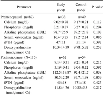
study group and control group
Parameter Study
group
Control
group P value Premenopausal (n=87) n=38 n=49
Calcium (mg/dl) 9.02±0.78 9.17±0.21 0.112 Phosphorus (mg/dl) 3.21±0.87 3.27±0.78 0.204 Alkaline phosphatase (IU/L) 98.7±25.9 89.2±21.8 0.108 Serum osteocalcin (ng/ml) 16.4±3.25 17.2±2.14 0.086
iPTH (pg/ml) 47±11 51±14 0.397
Deoxypyridinoline (nmol/mmol Cr)
10.34±4.39 9.78±5.32 0.257
Postmenopause (N=116) n=62 n=54
Calcium (mg/dl) 9.14±0.31 9.21±0.34 0.215 Phosphorus (mg/dl) 3.19±0.41 3.04±0.12 0.397 Alkaline phosphatase (IU/L) 112.5±19.87 92.4±21.7 0.038 Serum osteocalcin (ng/ml) 30.5±2.29 39.7±1.98 0.059
iPTH (pg/ml) 43±18 471±18 0.425
Deoxypyridinoline (nmol/mmol Cr)
11.8±4.76 10.85±5.3 0.217 것으로 정하였다. 두 군간의 임상적 특징, 골대사 표지자,
골밀도등의 차이를 알아보기 위해 unpaired t-test 방법으로 분석하였고, 통계학적 유의성은 P값이 0.05 이하인 경우로 하였다.
결 과
1) 연구군과 대조군의 임상적 특징 비교
연구군의 평균연령은 47.5±13.8 (17∼77)세였으며, 대조군 의 평균 연령은 49.1±11.6 (23∼75)세였다. 연구군과 대조군 모두 폐경전 여성군(premenopausal group)과 폐경후 여성군 (postmenopausal group)으로 분류하여 각각의 특징을 비교하 였다. 연구군 및 대조군의 연령, 신체용적지수(body mass index), 콜레스테롤 수치 등 골 대사에 영향을 미칠 수 있는 지표를 비교하였을 때 유의한 차이는 없었다. 갑상선 기능 검사 상 T3, fT4는 연구군과 대조군을 비교하였을 때 유의 한 차이가 없었으나, TSH는 연구군에서 저하된 수치를 보 였다(premenopausal group; P=0.028, postmenopausal group;
P=0.038)(Table 1).
2) 연구군과 대조군의 골대사 표지자 및 골밀도 비교 폐경 전 여성군에서 연구군과 대조군을 비교해 보았을 때 칼슘, 인, 오스테오칼신, 부갑상선 호르몬, 및 데옥시피 리디놀린은 양군간에 차이가 없었으며, 알칼리성 인산효소 는 통계학적으로 의미 있는 차이는 없었으나, 연구군에서
약간 증가된 경향을 보였다(P=0.086). 폐경 후 여성군에서 도 칼슘, 인 오스테오칼신, 부갑상선 호르몬 및 데옥시피리 디놀린은 양 군간에 차이가 없었으나, 알칼리성 인산효소 는 연구군에서 의미 있는 증가를 보였다(P=0.038)(Table 2).
골밀도를 비교하였을때, 폐경 전 및 폐경후 여성군 모두에 서 요추골, 대퇴골 및 전신의 Z값 및 T값이 연구군과 대조 군 간에 차이는 없었다(Table 3).
고 찰
갑상선 호르몬은 정상적인 골 대사 및 골 성장에 관여하 므로, 갑상선 호르몬이 장기간 과다 분비되면 골교체율을 증가시켜 골소실로 인한 골다공증 및 병적 골절 등이 발생 할 수 있다.(1-8) 즉, 갑상선 기능 항진 상태가 지속되면 골 형성 및 골흡수가 모두 증가되어, 파골 세포(osteoclast)에 의 한 골 흡수가 조골 세포(osteoblast)에 의한 골 형성을 초과 하여 골밀도(bone density)의 감소를 초래한다.(9-11) 이것은 골형성의 생화학적 지표인 혈청 알카리성 인산효소(alkaline posphatase), 오스테오칼신(osteocalcin)의 증가, 그리고 골흡 수 지표인 히드록시프롤린(hydroxyproline), 데옥시피리디놀 린(deosypyridinoline)의 배설 증가에 의해서 증명된다.(10,11) 갑상선 호르몬이 골격계에 관여하는 생화학적 경로는 활 성형 갑상선 호르몬(active thyroid hormone (T3))이 일련의 세포 내 신호 전달 과정을 통해 골격계의 기본 구조의 상태 변화를 일으킨다고 알려져 있다. T3의 신호전달 과정은 T3 가 갑상선 호르몬 수용체(thyroid receptors: TR α1, TR β1) 와 결합하여 조골 세포(osteoblast)에 직접적인 영향을 줄 수 있지만, 신호 전달의 각 단계에서 interleukin-6, interleukin-8, osteoprotegerin (OPG), receptor activator of nuclear factor-kap- pa B ligand (RANKL), TNF-α, parathyroid hormone, 및 Vitamin D 등의 매개체들이 관여한다고 보고되어 있 다.(6-11) 하지만, T3가 파골 세포에서는 갑상선 수용체에 직접 영향을 미치지 못하고, 조골세포의 신호 전달에 의해 간접적으로 활성화 된다.(12-15)
이와 같이 갑상선 호르몬 과다 분비가 골 대사에 영향을 일으킨다는 이론은 이미 여러 차례 보고되어, 장기간 지속 되는 갑상선 기능 항진증의 경우 이로 인한 골대사 및 골밀 도 변화를 예측할 수 있다. 따라서, 갑상선 기능 항진증 환 자에서 갑상선 기능을 정상으로 유지하여, 골교체율의 감 소로 인한 골다공증으로의 진행을 조기에 예방하고자 하는 노력들이 있었다.(16-19) 하지만, 혈청 free T3, free T4 등 인체에 능동적으로 작용하는 갑상선호르몬의 수치는 정상 이지만 TSH만 낮은 수치를 보이는 경우인 불현성 갑상선중 독증(subclinical hyperthyroidism)이 골대사 및 골밀도에 미 치는 영향에 대해서는 잘 알려지지 않았다.(20) 최근 분화 갑상선암의 급격한 증가로 갑상선암 수술 후 장기간 호르 몬 대체 요법을 시행함에 따라, 외인성으로 불현성 갑상선
중독증 상태가 지속되는 환자가 점점 늘어나게 되었다. 따 라서, 장기간 TSH 억제 상태 유지, 즉 낮은 TSH 수치가 골 대사 및 골밀도의 변화에 얼마나 영향을 미치는가에 대한 근래에 임상 보고가 있지만, 아직 이견이 많은 상태이다.
(21,22)
최근에는 TSH가 활성형 갑상선호르몬 등과 관계 없이 직 접적으로 골격 재형성(skeletal remodeling)에 영향을 미칠 수 있다는 이론이 발표됨에 따라, 불현성 갑상선 중독상태 와 골대사와의 관계에 대해 새로운 관심이 고조되고 있다.
즉, 혈중 T3, T4 등과 상관 없이 TSH가 독립적인 세포 내 신호 전달 체계를 통해 골 상태를 변화 시킬 수 있다는 연 구 결과이며, TSH는 갑상선 외에도 다양한 조직에 분포하 고 있는 TSHR (TSH receptor)와 결합하여 신호 전달 체계를 작동시켜, 조골세포의 과도한 생성 및 파골 세포의 생성 및 활성을 직접적으로 억제하는 역할을 할 수 있다는 이론이 다.(23) 하지만, TSH가 골격계에 미치는 직접적인 신호전달 체계 및 골대사와의 직접적인 연관성에 대해서는 아직 이 견이 많은 상태이며, 임상적인 연구에서는 더욱 결론이 모 호한 상태이다(22,24-26). 저자들은 갑상선암 수술 후 호르 몬 억제 요법을 시행하는 환자에서 TSH 감소가 골대사에 미치는 영향에 대해 조사하였고, 호르몬 억제요법을 시행 하는 갑상선암 환자에서 골다공증의 위험 및 예방에 대한 지침이 마련될 수 있는 기초 자료를 마련하기 위한 선행 과제로 이러한 연구를 계획하였다.
분화 갑상선암 수술 후 갑상선 호르몬 대체요법을 장기 간 시행 받아 불현성 갑상선 중독증 상태가 유지되는 환자 에서 골다공증 및 병적 골절에 대한 빈도, 자연경과, 임상적 문제점 및 치료에 대한 임상연구가 여러 차례 보고 되었으 나, 상이한 결과를 보이고 있어 아직 정립된 결론이 없는 상태이다.(22-27) 즉, 폐경 후 여성의 경우 유의하게 골밀도 가 감소된다는 보고도 있지만, 일부 보고에서는 골밀도의 차이가 없다고 발표되었다.(22-25). 하지만, 폐경 전 여성의 경우 갑상선자극호르몬의 저하가 골밀도에는 영향을 미치 지 않는다는 보고가 대부분이다.(25-27) 이러한 연구 결과 들의 차이는 첫째, 연구군의 선별에 있어서 성별, 나이, 신 체용적지수, 폐경의 여부, 기왕력 등 골대사에 영향을 미칠 수 있는 요소 및 약제 등이 고려되었는지, 둘째, 장기간의 갑상선호르몬 대체요법으로 TSH의 억제가 적절히 이루어 져서 불현성 갑상선중독증이 진단적으로 충족되었는지, 셋 째, 동일조건의 대조군과 골표지자 및 골밀도 등 골격계의 상태를 정확히 비교하였는지, 넷째, 충분한 추적관찰이 이 루어졌는지 등의 요인들과 관계가 있을 것으로 추정되고 있다.(25,27) 따라서 최근 연구들은 골밀도에 변화를 줄 수 있는 다른 요인들을 완전히 배재한 상태에서 갑상선암 수 술후 불현성 갑상선 중독증의 정확한 범위에 해당하는 환 자만을 대상으로 골변화에 대해 정상 대조군과 비교하고자 하였다. 또한, 골변화 정도를 골감소증(osteopenia) 및 골다
공증(osteoporosis)로 구분하고, 골밀도를 대퇴골 및 요추골 등 골성분에 따라 세분하는 등 연구 결과에 정확성을 더하 고자 하였다.(26-28)
본 연구의 결과는 정상 대조군과 비교하여 폐경 후 여성 군의 혈청 알카리성 인산효소가 의미 있게 증가되어 있는 반면, 다른 골표지자 및 골밀도의 차이는 발견되지 않았으 며, 폐경 전 여성에서는 의미있는 차이가 없었다. 이 연구의 결과로 폐경 후 여성에서 지속적인 TSH 억제요법과 골대사 혹은 골소실의 변화를 정확히 결론짓기는 어렵다. 저자들 의 경우 연구 결과에 영향을 줄수 있는 요인들에 대한 충분 한 검토를 통해 연구군과 대조군을 설정하여 대상 선정은 비교적 조건을 만족시켰지만, 아직 연구군의 규모가 부족 하고, 불현성 갑상선중독증 상태의 기간이 충분하지 않으 며, 추적 관찰 기간이 짧은 문제점을 가지고 있다. 따라서 향후 장기간 추적 관찰과, 더 많은 대상군이 수집된다면 보 다 정확한 결과를 기대할 수 있을 것이다. 따라서, 본 연구 는 이런 향후의 대규모 연구에 있어 선행적인 예비 보고인 점에 의의를 두고자 하였다.
향후 계획적인 임상연구가 진행된다면 갑상선 호르몬 억 제요법를 장기간 시행하는 갑상선암 환자에서 주기적인 갑 상선 호르몬 상태의 확인 및 골다공증에 대한 정기적인 선 별 검사에 대한 이해 및 조기 치료로 보다 합병증을 최소화 할 수 있는 방안이 검토될 수 있을 것이다.
결 론
분화 갑상선암 수술 후 장기간 갑상선 호르몬 대체요법 을 시행한 불현성 갑상선 중독증 환자에서 정상 대조군과 비교해 보았을 때 폐경 전 여성의 경우 의미 있는 골표지자 및 골밀도의 차이가 없었지만, 폐경 후 여성의 경우 골표지 자 중 알칼리성 인산효소가 대상군에서 의미 있는 증가를 보였다. 향후 더 많은 대상군의 수집, 보다 오랜 기간의 추 적 관찰, 다기관 연구로의 확대가 이루어진다면, 분화 갑상 선암 환자에서 장기간 갑상선 호르몬 대체요법에 따른 골 다공증의 상대적 위험도를 더 구체적으로 파악할 수 있을 뿐만 아니라, 더 명확한 예방 지침을 기대할 수 있을 것이 다.
REFERENCES
1) Vestergaard P, Mosekilde L. Fractures in patients with hyperthyroidism and hypothyroidism: a nationwide follow-up study in 16,249 patients. Thyroid 2002;12:411-9.
2) Kisakol G, Kaya A, Gonen S, Tunc R. Bone and calcium metabolism in subclinical autoimmune hyperthyroidism and hypothyroidism. Endocr J 2003;50:657-61.
3) Woeber KA. The thyroid. 9th ed. Philadelphia: Lippincott Williams and Wilkins; 2005;864-9.
4) Franklyn JA, Betteridge J, Daykin J, Holder R, Oates GD, Parle JV, et al. Long-term thyroxine treatment and bone mineral density. Lancet 1992;340:9-13.
5) Quan ML, Pasieka JL, Rorstad O. Bone mineral density in well-differentiated thyroid cancer patients treated with suppressive thyroxine: a systematic overview of the literature, Journal of Surgical Oncology 2002;79:62-9.
6) Bauer DC, Ettinger B, Nevitt MC, Stone KL. Risk for fractures in women with low serum levels of thyroid- stimulating hormone. Annals of Internal Medicine 2001;134:
561-8.
7) Mosekilde L, Melsen F, Bagger JP, Myhre-Jensen O, Sch- wartz SN. Bone changes in hyperthyroidism: interrelationships between bone morphometry, thyroid function and calcium- phosphorus metabolism. Acta Endocrinol (Copenh) 1997;85:
515-25.
8) Greenspan SL, Greenspan FS. The effect of thyroid hormone on skeletal integrity. Ann Intern Med 1999;130:750-8.
9) Wejda B, Hintze G, Katschinski B, Olbricht T, Benker G. Hip fractures and the thyroid: a case-control study. J Intern Med 1995:237:241-7.
10) Lee MS, Kim SY, Lee MC, Cho BY, Lee HK, Koh CS, et al. Negative correlation between the change in bone mineral density and serum osteocalcin in patients with hyper- thyroidism. J Clin Endocrinol Metab 1990;70:766-70.
11) Garnero P, Vassy V, Bertholin A, Riou JP, Delmas PD.
Markers of bone turnover in hyperthyroidism and the effects of treatment. J Clin Endocrinol Metab 1994;78:955-9.
12) Basset P, Okada A, Chenard MP, Kannan R, Stoll I, Anglard P, et al. Matrix metalloproteinases as stromal effectors of human carcinoma progression: therapeutic implications. Matrix Biol 1997;15:535-41.
13) Kanatani M, Sugimoto T, Sowa H, Kobayashi T, Kanzawa M, Chihara K. Thyroid hormone stimulates osteoclast differenti- ation by a mechanism independent of RANKLRANK inter- action. J Cell Physiol 2004;201:17-25.
14) Miura M, Tanaka K, Komatsu Y, Suda M, Yasoda A, Sakuma Y, et al. A novel interaction between thyroid hormones and 1,25(OH)(2)D(3) in osteoclast formation. Biochem Biophys Res Commun 2002;291:987-94.
15) Kim HS, Yoon HS, Han SS, Choi HA, Kim SH, Yim CH, et al. Biochemical markers of bone turnover and bone density in patients with subclinical thyrotoxicosis according to menopausal status. Korean J Bone Metab 2003;10:185-92.
16) Abu EO, Bord S, Horner A, Chatterjee VK, Compston JE. The expression of thyroid hormone receptors in human bone. Bone 1997;21:137-42.
17) Allain TJ, Yen PM, Flanagan AM, McGregor AM. The isoform-specific expression of the tri-iodothyronine receptor in osteoblasts and osteoclasts. Eur J Clin Invest 1996;26:418-25.
18) Klaushofer K, Varga F, Glantschnig H, Fratzl-Zelman N, Czerwenka E, Leis HJ, et al. The regulatory role of thyroid
hormones in bone cell growth and differentiation. J Nutr 1995;125:1996S-2003S.
19) Mosekilde L, Eriksen EF, Charles P. Effects of thyroid hormone on bone and mineral metabolism. Clin North Am 1990;19:35-63.
20) Baran DT. Detrimental skeletal effects of thyrotropin suppre- ssive doses of thyroxine: fact or fantasy? J Clin Endocrinol Metab 1994;78:816-7.
21) Engler H, Oettli RE, Riesen WF. Biochemical markers of bone turnover in patients with thyroid dysfunctions and in euthyroid controls: a cross-sectional study. Clin Chim Acta 1999;
289:159-72.
22) Kumeda Y, Inaba M, Tahara H, Kurioka Y, Ishikawa T, Morii H, et al. Persistent increase in bone turnover in Graves' patients with subclinical hyperthyroidism. J Clin Endocrinol Metab 2000;85:4157-61.
23) Abe E, Marians RC, Yu W, Wu X, Ando T, Li Y, et al. TSH
is a negative regulator of skeletal remodeling. Cell 2003;
115:151-62.
24) Woeber KA. Subclinical thyroid dysfunction. Arch Intern Med 1997;157:1065-8.
25) Heemstra KA, Hamdy NA, Romijn JA, Smith JW. The effects of thyrotropin-suppressive therapy on bone metabolism in patients with well-differentiated thyroid carcinoma. Thyroid 2006;16:583-91.
26) Greenspan SL, Greenspan FS. The effect of thyroid hormone on skeletal integrity. Ann Intern Med 1999;130:750-8.
27) Murphy E, Williams GR. The thyroid and the skeleton, Clin Endocrinol (Oxf) 2004;61:285-98.
28) Reverter JL, Holgado S, Alonso N, Salinas I, Granada ML, Sanmarti A. Lack of deleterious effect on bone mineral density of long-term thyroxine suppressive therapy for differentiated thyroid carcinoma. Endocr Relat Cancer 2005;12:973-81.