Copyright © 2017 The Korean Society for Bone and Mineral Research
This is an Open Access article distributed under the terms of the Creative Commons Attribution Non-Commercial Li- cense (http://creativecommons.org/licenses/by-nc/4.0/) which permits unrestricted non-commercial use, distribu- tion, and reproduction in any medium, provided the original work is properly cited.
Factors Affecting Bone Mineral Density
Measurement after Fracture in South Korea
Jin-Woo Kim1, Yong-Chan Ha2, Young-Kyun Lee1
1Department of Orthopaedic Surgery, Seoul National University Bundang Hospital, Seongnam;
2Department of Orthopaedic Surgery, Chung-Ang University College of Medicine, Seoul, Korea
Background: Prior osteoporotic fractures are strongly associated with the subsequent fractures. To prevent this, the diagnosis of osteoporosis following an osteoporotic frac- ture is important. The measurement of bone mineral density (BMD) is the first step for the diagnosis and management of osteoporosis. Therefore, the purposes of this study are 1) to evaluate the rate of BMD measurement after osteoporotic fracture in Korean population; and 2) determine the associated factors with BMD measurement after frac- tures among Korean patients. Methods: From database of Health Insurance Review &
Assessment Service, we identified patients with osteoporotic fractures happened in 2010. The BMD examinations were evaluated by using procedure codes. We evaluated the rate of BMD measurement within 6 months after fracture according to gender, age group (10-year incremental), type of insurance, residency area (rural vs. urban), type of medical institute, department, history of depression, rheumatoid arthritis, medical his- tory suggestive of secondary osteoporosis, osteoporosis-induced drug, and number of family members. Results: During study period, about a half (53.9%) of patients with os- teoporotic fractures had BMD measurement. Men, younger age, urban residency, and depression history were associated with low rate of BMD measurement. However, in- creasing age, use of glucocorticoid use, osteoporosis-inducing comorbid disease includ- ing rheumatologic disease, and osteoporosis-induced drug user were associated with higher likelihood of BMD measurement. Conclusions: Our results showed that about a half of patients with osteoporotic fractures had BMD measurement in South Korea, and provided the basic information to encourage management after fracture by educating not only patient but also physician about post-fracture management.
Key Words: Bone density, Osteoporotic fractures, Risk factors
INTRODUCTION
Considering aging society, osteoporosis and its related fractures have become a growing health problem worldwide.[1,2] Osteoporosis occasionally results in os- teoporotic fracture in hip, spine, humerus, and wrist.[3-5] In Korea, the annual in- cidences of osteoporotic fractures were 1,614 per 100,000 person-years in people aged 50 years or more in 2008.[6,7]
It is obvious that securing an appropriate level of post-fracture management for patients with osteoporosis may significantly reduce the risk of osteoporotic frac- ture. Especially, patients with previous osteoporotic fracture have higher risk of a Corresponding author
Young-Kyun Lee
Department of Orthopaedic Surgery, Seoul National University Bundang Hospital, 82 Gumi-ro 173 beon-gil, Bundang-gu, Seongnam 13620, Korea
Tel: +82-31-787-7204 Fax: +82-31-787-4056 E-mail: ykleemd@gmail.com Received: October 13, 2017 Revised: November 8, 2017 Accepted: November 18, 2017
No potential conflict of interest relevant to this article was reported.
subsequent fracture than those without previous fracture.[8]
Thus, post-fracture management for osteoporosis is high- ly recommended to prevent the occurrence of new fragility fractures.[9-11]
However, even high-risk patients with previous fracture often do not receive preventive management worldwidely.
[8,12-15] Korea is not an exception. Only 52.2% were aware of their diagnosis and 58.4% received pharmacological treatment among those with osteoporotic fractures.[16]
Bone mineral density (BMD) measurement is the first important step to investigate and manage patients with osteoporosis.[17] That is the important opportunity to ini- tiate secondary prevention in patients with previous osteo- porotic fracture.[18] In fact, a few empirical studies have already dealt with this issue in Korea.
There was lack of studies on the rate of BMD measure- ment after osteoporotic fracture in Korea, and what factors are associated with BMD measurement after fractures.
Therefore, our purposes were 1) to evaluate the rate of BMD measurement after osteoporotic fracture in Korean population; and 2) to determine associated factors with BMD measurement after fractures in Korea.
METHODS
We used data from the nationwide claims database of Health Insurance Review & Assessment Service (HIRA). Al- most 97% of the Korean populations have been currently covered with this national insurance system. In other words, the medical claims data include demographic information (age and gender), diagnoses using the International Classi- fication of Diseases, Tenth Revision (ICD-10) codes and pro- cedures for diagnosis and treatment using codes in both of inpatients and outpatients care. Thus, it is firmly certain that all information about health care utilization is avail- able from the HIRA database. Several epidemiologic stud- ies have used this national claim database.[19-21] We ana- lyzed patients aged over 50 years who were diagnosed with osteoporotic fracture by physician at 2010.
We identified patients with hip, spine, humerus and wrist fractures diagnosed in 2010. To identify patients with these fractures, we adopted the diagnostic codes using the ICD- 10 (hip, S720 and S721; spine, M484, M485, S220, S221, and S320; humerus, S422 and S423; wrist fractures, S525 and S526) and the procedure codes according to each anatom-
ic site.[3,7,22,23]
If an individual with fracture had more than one outpa- tient visits or admissions within the time period of six months, the cases were not counted separately, as below.[24,25]
Double recording was avoided by counting only one re- cord in the case that a person had more than one record in the HIRA database. If a patient had both spine and wrist fractures, only the first episode was counted.
The data based on the HIRA came from the patients who had experienced a hip, spine, humerus or wrist fracture and had undergone BMD examinations within 6 months before and after osteoporotic fractures. The procedure codes (HC 341-HC 344) for these examinations included dual x- ray absorptiometry scans (single site, HC 341; multiple sites, HC 342), quantitative computed tomography scans (HC 343), and other methods, including ultrasound (HC 344).
The rates of BMD examinations were estimated within 6 months after osteoporotic fractures.
We evaluated gender, age group (10-year incremental), type of insurance, residency area (rural vs. urban), type of medical institute, department, history of depression, rheu- matoid arthritis, medical history suggestive of secondary osteoporosis, osteoporosis-induced drug, and number of family members as potential associated factors.
The significance of differences was determined with use of a χ2 test. Statistical analyses were performed using SPSS version 16.0 (SPSS Inc., Chicago, IL, USA).
RESULTS
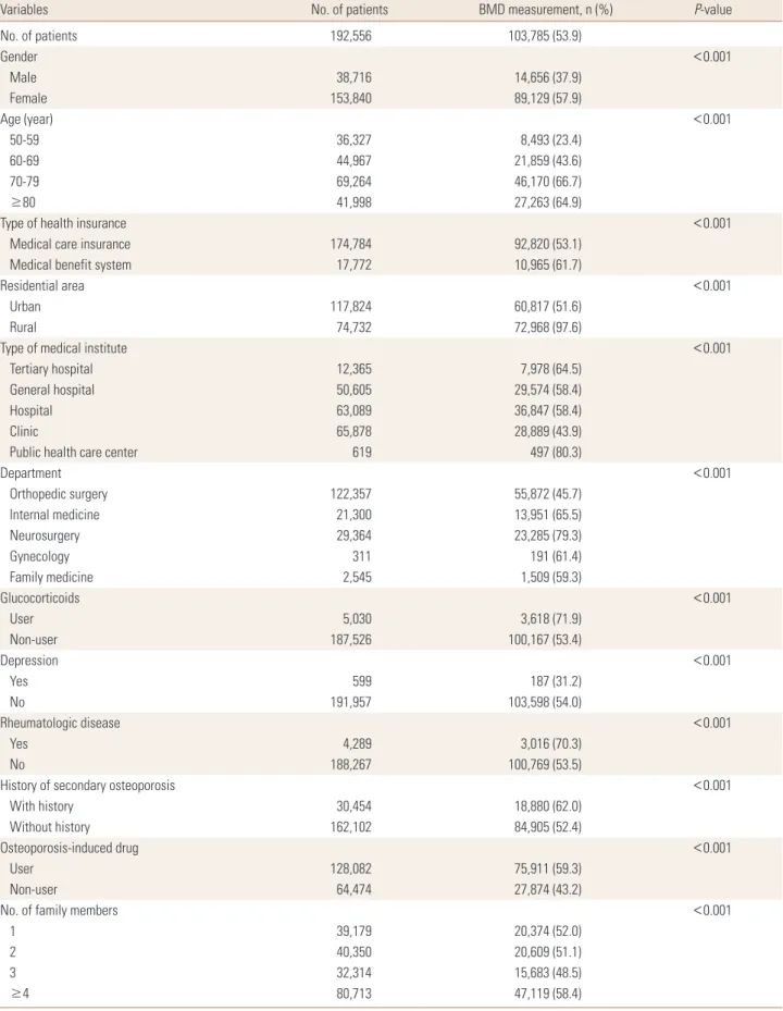
We analyzed a total of 192,556 patients who were diag- nosed of osteoporotic fracture in 2010. Of these patients, only 103,785 (53.9%) had been measured with BMD within 6-months post fracture (Table 1).
Indeed, the number of female showed higher frequency to have BMD measurement as compared with men (57.9%
vs. 37.9%). And, older patients were more likely to have BMD measurement, especially in population aged from 70 to 79 (66.7%). Patients in medical benefit system indicat- ing almost free-ride medical aid program in Korea tended to have BMD measurement more frequently than those with medical care insurance (61.7% vs. 53.1%). Patients liv- ing in urban area were less likely to have BMD measure- ment than those living in rural area (51.6% vs. 97.6%). Pa- tients who utilized public health care center showed high-
Table 1. Baseline characteristics of fracture patients who receive post-fracture therapy
Variables No. of patients BMD measurement, n (%) P-value
No. of patients 192,556 103,785 (53.9)
Gender <0.001
Male 38,716 14,656 (37.9)
Female 153,840 89,129 (57.9)
Age (year) <0.001
50-59 36,327 8,493 (23.4)
60-69 44,967 21,859 (43.6)
70-79 69,264 46,170 (66.7)
≥80 41,998 27,263 (64.9)
Type of health insurance <0.001
Medical care insurance 174,784 92,820 (53.1)
Medical benefit system 17,772 10,965 (61.7)
Residential area <0.001
Urban 117,824 60,817 (51.6)
Rural 74,732 72,968 (97.6)
Type of medical institute <0.001
Tertiary hospital 12,365 7,978 (64.5)
General hospital 50,605 29,574 (58.4)
Hospital 63,089 36,847 (58.4)
Clinic 65,878 28,889 (43.9)
Public health care center 619 497 (80.3)
Department <0.001
Orthopedic surgery 122,357 55,872 (45.7)
Internal medicine 21,300 13,951 (65.5)
Neurosurgery 29,364 23,285 (79.3)
Gynecology 311 191 (61.4)
Family medicine 2,545 1,509 (59.3)
Glucocorticoids <0.001
User 5,030 3,618 (71.9)
Non-user 187,526 100,167 (53.4)
Depression <0.001
Yes 599 187 (31.2)
No 191,957 103,598 (54.0)
Rheumatologic disease <0.001
Yes 4,289 3,016 (70.3)
No 188,267 100,769 (53.5)
History of secondary osteoporosis <0.001
With history 30,454 18,880 (62.0)
Without history 162,102 84,905 (52.4)
Osteoporosis-induced drug <0.001
User 128,082 75,911 (59.3)
Non-user 64,474 27,874 (43.2)
No. of family members <0.001
1 39,179 20,374 (52.0)
2 40,350 20,609 (51.1)
3 32,314 15,683 (48.5)
≥4 80,713 47,119 (58.4)
BMD, bone mineral density.
est likelihood (80.3%) of being taken BMD measurement than those who utilized another type of medical institute.
When patients visited at neurosurgery department after fracture, BMD was measured in the most proportion (79.3%).
As compared with those who had no medications during the baseline period, patients who took glucocorticoids, were taken BMD measurement more frequently (53.4% vs.
71.9%). Patients who had depression history were less like- ly to have BMD measurement than those with no depres- sion history (31.2% vs. 54.0%). Patients who had rheuma- toid arthritis were more likely to have BMD measurement than those with no rheumatoid arthritis (70.3% vs. 53.5%).
Patients who had medical history suggestive of secondary osteoporosis were more likely to have BMD measurement than those with no medical history osteoporosis (62.0% vs.
52.4%). Patients who took osteoporosis-induced drug were more likely to have BMD measurement than those with no medical history osteoporosis (59.3% vs. 43.2%). Patients who had family members of more than four were more likely to have BMD measurement than those with less than four (58.4% vs. 50.7%).
DISCUSSION
This study examined BMD measurement in patients af- ter the first fracture in Korea by using nationwide retro- spective data. We found that only 37.9% of men and 57.9%
of women underwent BMD measurement within 6 months after a fracture.
The low rate of BMD measurement was associated with men, younger age, urban residency, and depression histo- ry, in terms of patients’ factor. Our results show that post- fracture management including BMD measurement is par- ticularly poor among younger men, which was evidently in the line with the previous studies.[26,27]
In addition, physician in orthopedic department made the lowest level of BMD measurement after fracture (45.7%).
Given that the most of fractures have been treated in or- thopedic department, it is important that orthopedic sur- geon have awareness of necessity of BMD measurement.
[4] In our analysis, patients who utilized larger hospital showed less likelihood of being taken BMD measurement than those who utilized another smaller medical institute, which is likely to be related to the possibility of more coor- dinated care between the different departments in small
hospital than in large hospital. Our findings indicate that more coordinated service model is required in Korea to re- duce the care gap for secondary fracture prevention.
According to our results, many of the factors affected the BMD measurement significantly. Increasing age, use of glucocorticoid use, osteoporosis-inducing comorbid dis- ease including rheumatologic disease, and osteoporosis- induced drug user were associated with higher likelihood of BMD measurement, which is in line with prior studies.
[8,28] Patients who utilized public health care center showed highest likelihood (80.3%) of being taken BMD measure- ment than those who utilized another type of medical in- stitute. This means that public health care center play an important role to manage osteoporosis in Korea.
There were several limitations. First, we assumed that all patients with osteoporotic-fracture were eligible for BMD measurement. Second, we could not include patients who had BMD measurement after fracture, when patients with fracture took routine health care examination, because it was not included claim database. Therefore, there is possi- bility for us to under estimate the real gap in the post-frac- ture BMD measurement. Third, we could not evaluate whe- ther the patients with BMD measurement took anti-osteo- porosis treatment after BMD measurement. Finally, we could not perform multivariable analysis, because we could not link age, gender, disease or procedure code, type of insur- ance and so on using joint key of each individual.
CONCLUSION
Given the drawbacks raised above, our findings indicate that lack of BMD measurement after fracture remains a problem in Korea, especially among young men with os- teoporotic fracture. Besides, this study provided the basic information to optimize management after fracture, such as giving education patient and physicians about the im- portance of post-fracture management.
ACKNOWLEDGMENT
This research was supported by grants (HI13C1522, HI15- C1189) of the Korea Health Technology R&D Project through the Korea Health Industry Development Institute (KHIDI) funded by the Ministry of Health & Welfare, Republic of Korea.
REFERENCES
1. Cooper C, Campion G, Melton LJ 3rd. Hip fractures in the elderly: a world-wide projection. Osteoporos Int 1992;2:
285-9.
2. Court-Brown CM, Clement ND, Duckworth AD, et al. The spectrum of fractures in the elderly. Bone Joint J 2014;96B:
366-72.
3. Hodsman AB, Leslie WD, Tsang JF, et al. 10-year probability of recurrent fractures following wrist and other osteopo- rotic fractures in a large clinical cohort: an analysis from the Manitoba Bone Density Program. Arch Intern Med 2008;168:2261-7.
4. Kherad M, Mellstrom D, Rosengren BE, et al. The number and characteristics of prevalent vertebral fractures in el- derly men are associated with low bone mass and osteo- porosis. Bone Joint J 2015;97B:1106-10.
5. Choi WS, Lee HJ, Kim DY, et al. Does osteoporosis have a negative effect on the functional outcome of an osteopo- rotic distal radial fracture treated with a volar locking plate?
Bone Joint J 2015;97B:229-34.
6. Ha YC, Park YG, Nam KW, et al. Trend in hip fracture inci- dence and mortality in Korea: a prospective cohort study from 2002 to 2011. J Korean Med Sci 2015;30:483-8.
7. Park C, Ha YC, Jang S, et al. The incidence and residual life- time risk of osteoporosis-related fractures in Korea. J Bone Miner Metab 2011;29:744-51.
8. Klop C, Gibson-Smith D, Elders PJ, et al. Anti-osteoporosis drug prescribing after hip fracture in the UK: 2000-2010.
Osteoporos Int 2015;26:1919-28.
9. von Friesendorff M, Besjakov J, Åkesson K. Long-term sur- vival and fracture risk after hip fracture: a 22-year follow- up in women. J Bone Miner Res 2008;23:1832-41.
10. Warriner AH, Patkar NM, Yun H, et al. Minor, major, low- trauma, and high-trauma fractures: what are the subse- quent fracture risks and how do they vary? Curr Osteopo- ros Rep 2011;9:122-8.
11. Lee JH, Lee YH, Moon SH, et al. Influence of insurance ben- efit criteria on the administration rate of osteoporosis drugs in postmenopausal females. Clin Orthop Surg 2014;6:56- 61.
12. Elliot-Gibson V, Bogoch ER, Jamal SA, et al. Practice pat- terns in the diagnosis and treatment of osteoporosis after a fragility fracture: a systematic review. Osteoporos Int 2004;15:767-78.
13. Leslie WD, Giangregorio LM, Yogendran M, et al. A popula- tion-based analysis of the post-fracture care gap 1996-2008:
the situation is not improving. Osteoporos Int 2012;23:
1623-9.
14. Panneman MJ, Lips P, Sen SS, et al. Undertreatment with anti-osteoporotic drugs after hospitalization for fracture.
Osteoporos Int 2004;15:120-4.
15. Shepherd AJ, Cass AR, Ray LA, et al. Treatment for older men with fractures. Osteoporos Int 2012;23:1041-51.
16. Lee YK, Yoon BH, Koo KH. Epidemiology of osteoporosis and osteoporotic fractures in South Korea. Endocrinol Metab (Seoul) 2013;28:90-3.
17. Kim TI, Choi JH, Kim SH, et al. The adequacy of diagnosis and treatment for osteoporosis in patients with proximal humeral fractures. Clin Orthop Surg 2016;8:274-9.
18. Mettyas T, Carpenter C. Secondary prevention of osteopo- rosis in non-neck of femur fragility fractures: is it value for money? A retrospective, prospective and cross-sectional cohort study. J Orthop Surg Res 2013;8:44.
19. Yoon HK, Lee YK, Ha YC. Characteristics of patients diag- nosed with osteoporosis in South Korea: results from the national claim registry. J Bone Metab 2017;24:59-63.
20. Park C, Jang S, Lee A, et al. Incidence and mortality after proximal humerus fractures over 50 years of age in South Korea: national claim data from 2008 to 2012. J Bone Metab 2015;22:17-21.
21. Yoon HK, Park C, Jang S, et al. Incidence and mortality fol- lowing hip fracture in Korea. J Korean Med Sci 2011;26:
1087-92.
22. Watts NB, Geusens P, Barton IP, et al. Relationship between changes in BMD and nonvertebral fracture incidence as- sociated with risedronate: reduction in risk of nonverte- bral fracture is not related to change in BMD. J Bone Miner Res 2005;20:2097-104.
23. Yoo JH, Moon SH, Ha YC, et al. Osteoporotic fracture: 2015 position statement of the Korean society for bone and min- eral research. J Bone Metab 2015;22:175-81.
24. Lau E, Ong K, Kurtz S, et al. Mortality following the diag- nosis of a vertebral compression fracture in the medicare population. J Bone Joint Surg Am 2008;90:1479-86.
25. Kang HY, Yang KH, Kim YN, et al. Incidence and mortality of hip fracture among the elderly population in South Ko- rea: a population-based study using the national health insurance claims data. BMC Public Health 2010;10:230.
26. Cadarette SM, Katz JN, Brookhart MA, et al. Trends in drug
prescribing for osteoporosis after hip fracture, 1995-2004.
J Rheumatol 2008;35:319-26.
27. Roerholt C, Eiken P, Abrahamsen B. Initiation of anti-os- teoporotic therapy in patients with recent fractures: a na- tionwide analysis of prescription rates and persistence.
Osteoporos Int 2009;20:299-307.
28. Bessette L, Jean S, Davison KS, et al. Factors influencing the treatment of osteoporosis following fragility fracture.
Osteoporos Int 2009;20:1911-9.