© 2013 Korean Breast Cancer Society. All rights reserved. http://ejbc.kr | pISSN 1738-6756
INTRODUCTION
Hematological malignancies include lymphoid, myeloid, and histiocytic/dendritic neoplasms as defined in the most re- cent World Health Organization classification, updated in 2008 [1]. Lymphoma is the most common hematological ma- lignancy affecting the breast. However, breast lymphoma ac- counts for only approximately 0.04% to 0.7% of all breast can- cer cases [2,3], and this rarity may be related to the fact that the breast contains very little lymphoid tissue [4,5]. In these cases, it is often difficult to determine the relationship between the breast lesion and the hematological malignancy. Although several previous studies have addressed the imaging features of breast lymphoma, these findings are generally nonspecific and are typically presented as a list that was insufficient to guide clinical practice. As a result, clinicians may misinterpret the imaging findings associated with this lesion and conse-
quently miss its significance. However, unlike a breast carci- noma, a mass of lymphoma cells in the breast does not require excision. To avoid unnecessary treatment, clinicians and radi- ologists need to know the characteristic imaging features of breast lymphoma.
The purpose of this pictorial review is to illustrate the clini- cal aspects and multimodal imaging findings of lymphoma af- fecting the breast. We also described the key radiological and clinical findings that allow it to be distinguished from other breast malignancies and inflammations. We identified articles that concerned breast lymphoma, were published in English between 1983 and 2012, and were indexed on PubMed. In ad- dition, we evaluated the radiological, pathological, and clinical findings of 10 patients who had lymphoma affecting the breast and who underwent multimodal breast imaging studies in our institute between January 2003 and March 2012. The multi- modal imaging consisted of mammography, ultrasonography (US), dynamic low-dose computed tomography (CT), and dynamic magnetic resonance imaging (MRI). The protocol for breast low-dose CT differs from that of conventional chest CT, as the former is performed in a prone position to spread the whole breast parenchyma and lower radiation dose set- tings (120 kVP and 50 mA) are used. The American College of Radiology recommends an average glandular dose of ≤3 mGy for standard screening mammography, and thus, the ef-
Lymphoma Affecting the Breast: A Pictorial Review of Multimodal Imaging Findings
Euddeum Shim*, Sung Eun Song*, Bo Kyoung Seo, Young-Sik Kim1, Gil Soo Son2
Departments of Radiology, 1Pathology, and 2General Surgery, Korea University Ansan Hospital, Ansan, Korea REVIEW ARTICLE
Hematological malignancies rarely affect the breast, and the ma- jority of those that do are lymphomas. In this review, we describe the clinical aspects and multimodal imaging findings of breast lymphoma. We also illustrate the key clinical and radiological findings that allow it to be distinguished from various other malig- nant and benign diseases of the breast. Breast lymphoma mani- fests as a breast mass, a change in the subcutaneous tissue or the skin, or enlargement of the associated lymph node on radio- logical examination. Radiological findings associated with other
breast malignancies, such as calcifications, spiculations, or archi- tectural distortions are extremely rare. Skin and subcutaneous changes frequently accompany T-cell lymphoma. Multimodal breast imaging characteristics may aid in the diagnosis of breast lymphoma.
Key Words: Breast, Computed tomography, Lymphoma, Magnetic resonance imaging, Ultrasonography
Correspondence to: Bo Kyoung Seo
Department of Radiology, Korea University Ansan Hospital, 123 Jeokgeum- ro, Danwon-gu, Ansan 425-707, Korea
Tel: +82-31-412-5228, Fax: +82-31-412-5224 E-mail: [email protected]
*These authors contributed equaly to this work.
Received: May 4, 2013 Accepted: September 6, 2013
Cancer
fective radiation dose should be 6 mGy or less for routine, two-view mammography [6]. When measured radiation dos- es administered to a CT phantom at 120 kVp and 50 mA were compared with the recommended dose for mammography, a CTDI100 of ≤6 mGy was obtained (2.01-4.08 mGy) [7]. We have already published both the phantom and patient results of the breast CT scan using this protocol and demonstrated satisfactory image quality and radiation doses [7-9]. The ra- diological findings were evaluated using a modified version of the Breast Imaging Reporting and Data System lexicon [10].
Because this lexicon does not include breast CT, we described CT findings using MRI descriptions, and the density of a breast lesion on a CT scan was compared with that of the pec- toralis muscles. Thus, for example, if a lesion was of identical density to the pectoralis muscle, it was described as “isodense.”
CLINICAL ASPECTS
Epidemiology
Breast lymphoma may occur as either a primary or a sec- ondary lesion. Wiseman and Liao [11] proposed that, for a di- agnosis of primary breast lymphoma, the breast should be the site of the first or major manifestation of the lymphoma and that there should be no evidence of lymphoma elsewhere, ex- cept at the ipsilateral axillary node. Primary breast lymphoma accounts for 0.85% to 2.2% of all extranodal malignant lym- phomas, and secondary breast lymphoma is more common [12]. The age distribution of reported cases is wide (16-93 years at the time of diagnosis) and the reported median age ranges from 55 to 65 years [13,14], which is similar to the me- dian age of extramammary lymphoma patients [15,16]. Of the 10 cases we described, six (60%) were primary breast lympho- mas and the remaining four were secondary lymphomas (Ta- ble 1). The median age of the patients was 49.5 years (range, 21-65 years) and all of the patients were women.
Clinical findings
The most common symptom of breast lymphoma is a pain- less, palpable mass. Nipple retraction or discharge and skin change can also occur, but are rare [12,17]. Most B-cell lym- phomas of the breast are present as palpable masses, whereas skin changes, edema, and local pain are more commonly asso- ciated with T-cell lymphoma [18]. Ipsilateral axillary lymph- adenopathy has been reported in 13% to 50% of cases [19]. In our study, all patients had clinical symptoms. These were pain- less palpable masses in the breast and/or axilla in seven cases (70%), tender subcutaneous nodules in two cases (20%), and breast swelling in one case (10%) (Table 1). Three patients (30%) complained of skin thickening. Both patients (100%)
with subcutaneous nodules and two of the three patients (67%) with skin thickening were found to have T-cell lymphoma on pathological examination. Therefore, skin or subcutaneous changes were more common in T-cell lymphoma, which is consistent with the findings of the previous study [18].
Pathophysiology
Most breast lymphomas are of the B-cell type. In a recent study by Surov et al. [12], 94% of breast lymphomas were found to be of the B-cell type and only 6% were of the T-cell type. However, some forms of T-cell lymphoma are more common in Asia than in Western countries [20], most notably peripheral T-cell, nasal T-cell, and natural killer (NK)/T-cell lymphomas. In the breast, T-cell lymphoma occurs only rarely and is reported on a case-by-case basis [21-24]. Included in these reports are lymphoblastic lymphoma, peripheral T-cell lymphoma, multilobated T-cell lymphoma, NK/T-cell lym- phoma, cutaneous T-cell lymphoma, and anaplastic large cell lymphoma.
Of growing concern is a possible association between ana- plastic large cell lymphoma and breast implants [22,23,25-28].
Previously regarded as chemically inert, silicone has been shown to induce inflammatory T-cell reactions [29]. If im- plants are causally related to lymphoma, the incidence of T- cell lymphoma in the breast may soon increase. In our study, six patients (60%) had diffuse large B-cell lymphomas and the remaining four (40%) had various T-cell lymphomas: two cases of peripheral T-cell lymphoma and a single case each of NK/T-cell lymphoma and precursor T-lymphoblastic lym- phoma (Table 1). We included lymphoma patients who had breast involvement in this study and excluded patients who had only lymph node enlargement, and as a result, T-cell lym- phoma manifesting as breast parenchymal or subcutaneous nodules might have been more common in study.
Treatment and prognosis
The optimal treatment for B-cell lymphoma is a multiagent regimen consisting of anthracycline-based chemotherapy with rituximab, possibly also combined with radiation. In patients with an indolent lymphoma such as lymphoplasmacytic lym- phoma, mucosa-associated lymphoid tissue lymphoma, low- grade follicular lymphoma, or small lymphocytic lymphoma, radiation therapy is potentially curative. Central nervous sys- tem relapse occurs more frequently (3%-27%) in primary breast lymphoma cases than in those involving extramamma- ry lymphoma, and thus, cranial radiation therapy or intrathe- cal chemotherapy may be performed prophylactically [15].
Treatment for T-cell lymphoma varies widely because there are so many different types. Standard lymphoma therapies in-
Table 1. Patients characteristics Case no.AgeSymptomPrimary or secondary lymphomaPathologic typeImaging findings Confirm methodTreatment MammographyUltrasoundCTMRI 1F/61Palpable masses in breastSecondaryDiffuse large B-cell lymphomaBilateral multiple massesBilateral multiple massesBilateral multiple massesNACore needle biopsyExcision and chemotherapy with R-CHOP 2F/43Palpable mass in breastPrimaryDiffuse large B-cell lymphomaBilateral multiple masses with lymphadenopathy Bilateral multiple masses with axillary lymphadenopathy Bilateral multiple masses with axillary lymphadenopathy Bilateral multiple masses with lymphadenopathy
Excisional biopsyExcision and chemotherapy with R-CHOP 3F/46Breast swellingSecondaryDiffuse large B-cell lymphomaNegativeUnilateral multiple massesUnilateral multiple fociNACore needle biopsyChemotherapy with R-CHOP 4F/43Skin thickening and palpable mass in axilla
PrimaryDiffuse large B-cell lymphomaUnilateral trabecular thickening and lymphadenopathy
Unilateral edema and axillary lymphadenopathyUnilateral edema and lymphadenopathyUnilateral edema and lymphadenopathyCore needle biopsyChemotherapy with R-CHOP 5F/65Palpable mass in breast and axillaSecondaryDiffuse large B-cell lymphomaNAUnilateral single mass in subcutis and axillary lymphadenopathy Unilateral single mass in subcutis and lymphadenopathy Unilateral single mass in subcutis and lymphadenopathy
Core needle biopsyChemotherapy with R-CHOP 6F/53Papable mass in breastPrimaryPeripheral T-cell lymphomaUnilateral single mass and skin thickeningUnilateral single mass in subcutis, skin thickening, and axillary lymphadenopathy
Unilateral single mass in subcutis, skin thickening, and lymphadenopathy Non-mass like enhancement in subcutis, skin thickening, and lymphadenopathy
Core needle biopsyRadiation therapy and chemotherapy with R-CHOP 7F/21Subcutanoeus nodules and skin thckening in breast
SecondaryNatural killer/ T-cell lymphomaNABilateral multiple masses in subcutis and skin thickening Bilateral multiple masses in subcutis and skin thickening Bilateral multiple masses in subcutis and skin thickening
Core needle biopsyRadiation therapy and chemotherapy with SMILE 8F/62Subcutaneous nodules and skin thickening in breast
PrimaryPeripheral T-cell lymphomaUnilateral multiple masses and skin thickening Unilateral multiple masses in subcutis and skin thickening Unilateral multiple masses in subcutis and skin thickening
NACore needle biopsyRadiation therapy and chemotherapy with R-CHOP 9F/49Palpable mass in breastPrimaryDiffuse large B-cell lymphomaUnilateral single massUnilateral single massUnilateral single massUnilateral single massCore needle biopsyChemotherapy with R-CHOP 10F/52Palpable mass in breastPrimaryPrecursor T-lymphoblastic lymphoma
Unilateral single massUnilateral single massUnilateral single mass and mediastinal lymphadenopathy NACore needle biopsyChemotherapy with hyper-CAVD CT=computed tomography; MRI=magnetic resonance imaging; F=female; NA=not available; R-CHOP=rituximab, cyclophosphamide, doxorubicin, vincristine, and prednisone; SMILE=dexamethasone, metho- trexate, l-asparaginase, and etoposide; Hyper CAVD=hyperfractionated chemotherapy in small dose with cycloposphamide, vincristine, doxorubicin, and dexamethasone.
cluding chemotherapy, radiation, bone marrow transplanta- tion, and surgery may be effective in some cases, and ultravio- let light therapy or electron beam radiation is effective for T- cell lymphoma with skin involvement. With appropriate man- agement, a high rate of locoregional control can be achieved for primary breast lymphoma, although there is often systemic relapse. The 5-year overall survival for diffuse large B-cell lym- phoma in the breast has been reported to range from 60% to 65% [30-32]. Of the 10 cases included in our study, two pa- tients underwent excision with chemotherapy, three received radiation therapy with chemotherapy, and the remaining five received only chemotherapy (Table 1). Radiation therapy was administered in 75% of the T-cell lymphoma cases. Seven of our 10 patients (70%) had complete remission after treatment, two patients (20%) underwent salvage treatment due to tumor recurrence after complete remission, and the remaining pa- tient (10%) achieved a partial remission.
MULTIMODAL BREAST IMAGING FINDINGS
Lymphoma affecting the breast manifests as a breast mass, a change in the subcutaneous tissue or the skin, or enlargement of the associated lymph node on radiological examination.
Breast mass
On mammography, breast lymphoma appears as a solitary, noncalcified, circumscribed, or indistinctly delineated, oval or round mass that can vary in density (Figures 1-3) [33,34]. Cal- cifications, spiculations, and architectural distortion are dis- tinctively absent [17,33,34]. For B-cell lymphoma, diffuse or ill-defined increases in breast density representing irregular infiltrating processes may be observed. Mammography is lim- ited in its capacity to detect lymphomas that present as a dif- fuse infiltration or a small mass (Figure 4). For these lesions, which may be associated with skin thickening or breast edema,
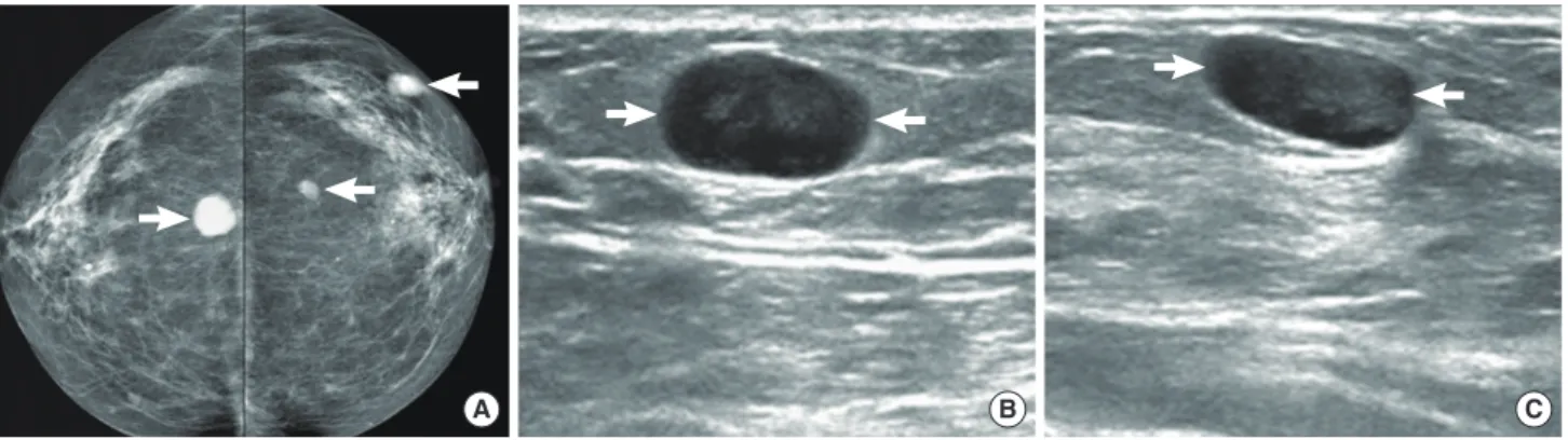
Figure 1. A 61-year-old woman with diffuse large B-cell lymphoma. (A) Both craniocaudal mammograms show multiple circumscribed oval or round masses (arrows) in both breasts. On (B) right and (C) left breast ultrasonography, the masses (arrows) are circumscribed and very low echoic, mimick- ing cysts.
A B C
A Figure 2. A 43-year-old woman with diffuse large B-cell lymphoma. (A) Both craniocaudal and mediolateral oblique mammograms demonstrate bilat- eral indistinct oval or round masses (arrows) and axillary lymph node enlargements (white arrowheads). (Continued to the next page)
Figure 2. (Continued from the previous page) (B) Ultrasonography (US) scan of the right breast shows an indistinct oval hypoechoic mass (ar- rows) with onion peel-like rims (black arrowheads). (C) US scan of the left breast shows an indistinct irregular hypoechoic mass (arrows). (D) US scan of the left axilla demonstrates an enlarged lymph node (white arrowheads). The node has indistinct margins and is very hypoechoic.
(E, F) Enhanced breast computed tomography images show bilateral masses (arrows) with homogeneous enhancement. (G, H) Enhanced T1-weighted transverse magnetic resonance images show bilateral masses (arrows) with peripheral rim enhancement. (I) A time-enhance- ment curve obtained from the mass in left breast reveals rapid initial and washout delayed phase enhancement.
B C D
E F
Immediate 1 2 3 4 5 I
Time (min) 700
600 500 400 300 200 100 0
G H
US is often more informative (Figures 4, 5), as breast lympho- ma usually appears as a hypoechoic solid mass with circum- scribed or indistinct margins (Figures 1-4) [28,29,33-36]. Het- erogeneous echo patterns, hypoechogenicity, and hyperecho- genicity are also seen frequently in breast lymphoma, and were present in 23% of cases in a study by Yang et al. (Figures 6-8) [13]. Posterior acoustic enhancement is another common fea- ture, as is an echogenic rim or onion peel-like rim surround- ing the mass that may represent lymphedema (Figure 2). The value of MRI of breast lymphoma is not firmly established, but inhomogeneous enhancement is seen in most cases on T1- weighted images after the administration of contrast media (Figure 2) [12,37,38]. Kinetic analysis on MRI shows that most breast lymphomas exhibit a rapid initial enhancement and plateau or a washout delayed phase enhancement (Figure 2) [12,13]. On CT, most lymphomas present as a circumscribed round or oval mass with various degrees of enhancement (Fig- ures 2-4) [12].
Subcutaneous or skin change
US, CT, and MRI facilitate the evaluation of diffuse infiltra- tive lesions, tiny nodules, or superficial lesions involving the
skin or subcutis in breast tissue (Figures 3-8). Radiological re- ports of T-cell lymphoma in the breast are extremely rare com- pared to those of B-cell lymphoma, although T-cell lymphoma, especially subcutaneous panniculitis-like T-cell or peripheral T-cell lymphoma, preferentially infiltrate the subcutaneous tis- sues (Figures 6-8) [21,39,40]. US shows indistinct and irregu- lar hyperechoic masses in the subcutis, and the hyperechoic areas may contain internal tubular branching hypoechogenici- ty (Figures 6, 7) [40,41]. Hyperechogenicity in breast lympho- ma probably reflects the cellularity of these tumors [42]. On MRI, irregular masses with a rim or heterogeneous enhance- ment in the subcutis may be present. No characteristic CT findings of breast T-cell lymphoma have been reported to date, although in our study, we observed circumscribed or indistinct isodense masses with skin thickening on dynamic CT scans (Figures 6-8).
Lymphadenopathy
Routine breast imaging does not usually help to distinguish between primary and secondary lymphoma, although bilater- al axillary lymphadenopathy or breast edema could indicate a secondary lymphoma. US, CT, and MRI are all more useful
A B
C D
Figure 3. A 46-year-old woman with diffuse large B-cell lymphoma. (A) Both craniocaudal and mediolateral oblique mammograms are negative. (B) Ultrasonography scans of the right breast show multiple circumscribed oval hypoechoic masses (arrows). (C) Enhanced breast computed tomogra- phy (CT) image shows enlargement of right breast with multiple small enhancing foci (arrowheads). (D) Enhanced breast CT image obtained after che- motherapy shows the disappearance of enhancing foci in the right breast.
techniques than mammography for the evaluation of enlarg- ing lymph nodes (Figures 4-6), and CT may be useful for the evaluation of intrathoracic and extrathoracic lymphadenopa- thy and involvement of the chest wall or lung in lymphoma patients (Figure 4).
DIFFERENTIAL DIAGNOSIS FROM OTHER BREAST MALIGNANCIES AND INFLAMMATORY
BREAST DISEASES
It is difficult to distinguish breast lymphoma from various benign or malignant breast diseases on the basis of clinical and radiological findings. However, it is important to distin- guish between breast lymphoma and other malignant diseases (for example, invasive breast carcinoma), inflammatory breast carcinoma, or metastasis, so that the appropriate treatment can be selected. The diagnostic strategy for lymphoma affect-
ing the breast is determined by the presence or absence of known systemic lymphoma. If a patient is known to have sys- temic lymphoma, changes in the breasts must raise the possi- bility of a lymphomatous involvement [43]. However, in cases where lymphoma is not otherwise suspected, lymphoma of the breast is usually only suggested as one possibility in the differential diagnosis.
The most common symptom of breast malignancy is a painless palpable lump. Local pain, edema, or subcutaneous or skin nodules are frequent in lymphoma, especially the T- cell type, whereas nipple retraction or discharge is extremely rare [12,17,18]. With respect to radiological findings, the characteristic features of more common breast carcinomas, including calcifications, spiculations, or architectural distor- tion, are distinctively absent in lymphoma [17,33,34]. Inflam- matory breast carcinoma manifests as breast edema with a mass on breast imaging that is usually of irregular shape and
A
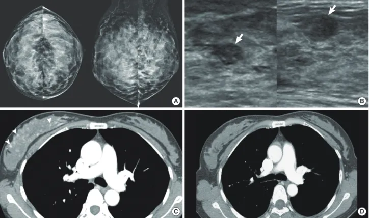
B
C Figure 4. A 52-year-old woman with precursor T-lymphoblastic lymphoma. (A) Right mediolateral oblique mammogram shows a circumscribed and partially obscured marginated round hyperdense mass (arrows). (B) Ultrasonography scan of the right breast demonstrates a circumscribed oval hy- poechoic mass (arrows). (C) Enhanced computed tomography scan demonstrates an oval isodense mass (arrow) in right breast and mediastinal wid- ening (arrowheads) due to lymphadenopathy.
has indistinct margins or is spiculated, characteristics that are somewhat different from those of breast lymphoma. Metasta- sis to the breast manifests as a localized lesion or diffuse infil- tration [43]. The former presents as a circumscribed oval or round mass and the latter appears as breast edema with dilat- ed dermal lymphatics on breast imaging. These are very simi- lar findings to those of lymphoma affecting the breast. There are currently no reliable criteria for distinguishing breast lym- phoma from metastasis, although a history of a known metas- tasis or lymphoma in another organ is important in the diag- nosis of a breast lesion. The findings of our study revealed that because a subcutaneous breast mass or nodule on radiological examination is rare in invasive breast carcinoma, inflammato- ry breast carcinoma, or metastasis, a change in the subcutis might aid in the diagnosis of breast lymphoma. Although there are some indicative clinical and radiological features of breast lymphoma, imaging-guided core needle biopsy or exci- sional biopsy should be performed for confirmative diagnosis.
If a patient has a skin or subcutaneous lesion, a full thickness excisional skin biopsy should be performed in order to differ- entiate breast lymphoma from diffuse infiltrative metastasis and inflammatory carcinoma.
Differentiating breast lymphoma from inflammatory dis- eases is challenging, especially in patients with a painful breast lump, erythema, or skin thickening [44,45]. Inflammatory breast diseases include infectious mastitis, abscess, or idio- pathic granulomatous lobular mastitis. Mammography and US often reveal unilateral breast edema, possibly with accom- panying masses, in both breast lymphoma and inflammatory diseases. However, on breast US, the echo pattern of masses and associated findings differ between lymphoma and inflam- mation. In the latter, the masses are complicated cysts that have movable echoes or sedimentations, complex cysts, which have cystic and solid contents or are hypoechoic [43]. Dilated ducts or fistulous tracts to the skin are also often associated with inflammatory disease [43]. Conversely, a complex cyst is
A B C
D E
Figure 5. A 43-year-old woman with diffuse large B-cell lymphoma. (A) Left mediolateral oblique mammogram shows axillary lymphadenopathy (ar- rows) and trabecular thickening (arrowheads). (B) Ultrasonography (US) scans of the left breast show skin thickening (arrows) and dilated dermal lym- phatics (arrowheads). (C, D) US scans of the left axilla show enlarged lymph nodes (arrows). One node is indistinct and irregular in shape (C) and the other is circumscribed and ovoid (D). The nodes have cortical thickening. (E) Enhanced breast computed tomography image shows multiple-rim en- hancing lymph nodes (arrows) in left axilla.
extremely rare and dilated ducts or fistulas are never associated with breast lymphoma. If a breast lesion is located in the sub- cutis, breast lymphoma needs to be distinguished from pan- niculitis or fat necrosis. Subcutaneous panniculitis, like T-cell lymphoma of the breast, is indistinguishable from inflamma- tory panniculitis and fat necrosis because of their clinical and
pathological similarities [21,40], as both present as solitary or multiple subcutaneous nodules. Fat lobules in the subcutane- ous layer are surrounded by neoplastic T-cells in cases of sub- cutaneous panniculitis like T-cell lymphoma, and inflamma- tory cells and some atypical cells are present in inflammatory panniculitis and fat necrosis on pathological examination, re- Figure 6. A 65-year-old woman with diffuse large B-cell lymphoma. (A) Ultrasonography (US) scan of the right breast shows an indistinct oval hyper- echoic mass (arrows) in the subcutaneous fat layer. (B) US scan of the right axilla shows enlarged lymph nodes with loss of internal fatty hila (arrow- heads). (C) Pre-enhanced and (D) enhanced breast computed tomography (CT) images demonstrate an indistinct oval isodense mass (arrows) in right breast. The mass in right breast reveals 39 Hounsfield unit (HU) on pre-enhanced image and 45 HU on enhanced images. (E) Multiple enlarged axillary lymph nodes (arrowheads) are seen on enhanced CT scan.
A
B E
D C
sulting in radiologically similar features. Clinically, inflamma- tory panniculitis spontaneously regresses without treatment within 1 to 4 weeks and frequently occurs in the lower extrem- ities [46]. For the diagnosis of fat necrosis, it is important know the history of breast surgery, trauma, and biopsy [47].
CONCLUSION
Here we have described the clinical aspects and multimodal imaging findings of lymphoma affecting the breast. Breast lymphoma manifests as a breast mass, changes in the subcuta- neous tissue or the skin, or enlargement of the associated
A
C
B
Figure 7. A 21-year-old woman with NK/T-cell lymphoma. (A) Ultraso- nography (US) scan of the right breast shows an indistinct oval hyper- echoic mass (arrows) with internal tubular hypoechogenicities and thickening of overlying skin. (B) US scan of the left breast shows in- creased echotexture of subcutaneous fat layer (arrows). (C) Enhanced breast computed tomography scan shows multiple indistinct isodense masses (arrows) in both breasts and back, and diffuse skin thickening in both breasts.
A B
Figure 8. A 62-year-old woman with peripheral T-cell lymphoma. (A) Ultrasonography (US) scans of the right breast show multiple oval or round masses (arrows) in skin and subcutaneous fat layers. (B) Enhanced computed tomography scan of right breast shows multiple isodense or hyper- dense masses (arrows) with skin thickening (arrowheads).
lymph node on radiological examination. We also have illus- trated the key clinical and radiological findings that can dis- tinguish breast lymphoma from various breast malignancies and benign diseases. In breast lymphoma, usual malignant ra- diological features such as calcifications, spiculations, or archi- tectural distortions are extremely rare, and skin and subcuta- neous changes frequently accompany T-cell lymphoma. Mul- timodal breast imaging characteristics may aid the diagnosis of breast lymphoma.
CONFLICT OF INTEREST
The authors declare that they have no competing interests.
REFERENCES
1. Swerdlow SH; International Agency for Research on Cancer. WHO Classification of Tumours of Haematopoietic and Lymphoid Tissues.
4th ed. Lyon: IARC Press; 2008.
2. Tavassoli FA, Devilee P; International Agency for Research on Cancer.
Pathology and Genetics of Tumours of the Breast and Female Genital Organs. Lyon: IARC Press; 2003. p.9-112.
3. Giardini R, Piccolo C, Rilke F. Primary non-Hodgkin’s lymphomas of the female breast. Cancer 1992;69:725-35.
4. Dao AH, Adkins RB Jr, Glick AD. Malignant lymphoma of the breast: a review of 13 cases. Am Surg 1992;58:792-6.
5. Brogi E, Harris NL. Lymphomas of the breast: pathology and clinical behavior. Semin Oncol 1999;26:357-64.
6. Mainiero MB, Lourenco A, Mahoney MC, Newell MS, Bailey L, Barke LD, et al. ACR Appropriateness Criteria Breast Cancer Screening. Res- ton: American College of Radiology; 2006. p.217-25.
7. Lee WJ, Seo BK, Cho PK, Yie A, Cho KR, Woo OH, et al. The clinical use of low-dose multidetector row computed tomography for breast cancer patients in the prone position. J Breast Cancer 2010;13:357-65.
8. Yi A, Seo BK, Cho PK, Pisano ED, Lee KY, Je BK, et al. Optimal multi- detector row CT parameters for evaluations of the breast: a phantom and specimen study. Acad Radiol 2010;17:744-51.
9. Seo BK, Pisano ED, Cho KR, Cho PK, Lee JY, Kim SJ. Low-dose multi- detector dynamic CT in the breast: preliminary study. Clin Imaging 2005;29:172-8.
10. American College of Radiology, BI-RADS Committee. ACR BI-RADS Breast Imaging and Reporting Data System: Breast Imaging Atlas. 4th ed. Reston: American College of Radiology; 2003.
11. Wiseman C, Liao KT. Primary lymphoma of the breast. Cancer 1972;
29:1705-12.
12. Surov A, Holzhausen HJ, Wienke A, Schmidt J, Thomssen C, Arnold D, et al. Primary and secondary breast lymphoma: prevalence, clinical signs and radiological features. Br J Radiol 2012;85:e195-205.
13. Yang WT, Lane DL, Le-Petross HT, Abruzzo LV, Macapinlac HA. Breast lymphoma: imaging findings of 32 tumors in 27 patients. Radiology 2007;245:692-702.
14. Arber DA, Simpson JF, Weiss LM, Rappaport H. Non-Hodgkin’s lym- phoma involving the breast. Am J Surg Pathol 1994;18:288-95.
15. Caon J, Wai ES, Hart J, Alexander C, Truong PT, Sehn LH, et al. Treat- ment and outcomes of primary breast lymphoma. Clin Breast Cancer 2012;12:412-9.
16. Avilés A, Delgado S, Nambo MJ, Neri N, Murillo E, Cleto S. Primary breast lymphoma: results of a controlled clinical trial. Oncology 2005;
69:256-60.
17. Domchek SM, Hecht JL, Fleming MD, Pinkus GS, Canellos GP. Lym- phomas of the breast: primary and secondary involvement. Cancer 2002;94:6-13.
18. Gualco G, Chioato L, Harrington WJ Jr, Weiss LM, Bacchi CE. Primary and secondary T-cell lymphomas of the breast: clinico-pathologic fea- tures of 11 cases. Appl Immunohistochem Mol Morphol 2009;17:301-6.
19. Talwalkar SS, Miranda RN, Valbuena JR, Routbort MJ, Martin AW, Medeiros LJ. Lymphomas involving the breast: a study of 106 cases comparing localized and disseminated neoplasms. Am J Surg Pathol 2008;32:1299-309.
20. Portlock C, Vose MJ, Cheson BD. T-cell lymphomas. 2008. Lymphoma Research Foundation. http://www.lymphoma.org/atf/cf/%7B0363 CDD6-51B5-427B-BE48-E6AF871ACEC9%7D/T-CELL%20LYM- PHOMAS.PDF. Accessed July 24th, 2013.
21. Uematsu T, Kasami M. 3T-MRI, elastography, digital mammography, and FDG-PET CT findings of subcutaneous panniculitis-like T-cell lymphoma (SPTCL) of the breast. Jpn J Radiol 2012;30:766-71.
22. Aladily TN, Medeiros LJ, Amin MB, Haideri N, Ye D, Azevedo SJ, et al.
Anaplastic large cell lymphoma associated with breast implants: a re- port of 13 cases. Am J Surg Pathol 2012;36:1000-8.
23. Sahoo S, Rosen PP, Feddersen RM, Viswanatha DS, Clark DA, Chad- burn A. Anaplastic large cell lymphoma arising in a silicone breast im- plant capsule: a case report and review of the literature. Arch Pathol Lab Med 2003;127:e115-8.
24. Aguilera NS, Tavassoli FA, Chu WS, Abbondanzo SL. T-cell lymphoma presenting in the breast: a histologic, immunophenotypic and molecu- lar genetic study of four cases. Mod Pathol 2000;13:599-605.
25. Jewell M, Spear SL, Largent J, Oefelein MG, Adams WP Jr. Anaplastic large T-cell lymphoma and breast implants: a review of the literature.
Plast Reconstr Surg 2011;128:651-61.
26. Smith TJ, Ramsaroop R. Breast implant related anaplastic large cell lym- phoma presenting as late onset peri-implant effusion. Breast 2012;21:
102-4.
27. Thompson PA, Lade S, Webster H, Ryan G, Prince HM. Effusion-asso- ciated anaplastic large cell lymphoma of the breast: time for it to be de- fined as a distinct clinico-pathological entity. Haematologica 2010;95:
1977-9.
28. de Jong D, Vasmel WL, de Boer JP, Verhave G, Barbé E, Casparie MK, et al. Anaplastic large-cell lymphoma in women with breast implants.
JAMA 2008;300:2030-5.
29. Friis S, McLaughlin JK, Mellemkjaer L, Kjøller KH, Blot WJ, Boice JD Jr, et al. Breast implants and cancer risk in Denmark. Int J Cancer 1997;71:
956-8.
30. Jeanneret-Sozzi W, Taghian A, Epelbaum R, Poortmans P, Zwahlen D, Amsler B, et al. Primary breast lymphoma: patient profile, outcome and prognostic factors: a Multicentre Rare Cancer Network study. BMC Cancer 2008;8:86.
31. Yhim HY, Kang HJ, Choi YH, Kim SJ, Kim WS, Chae YS, et al. Clinical outcomes and prognostic factors in patients with breast diffuse large B
cell lymphoma: Consortium for Improving Survival of Lymphoma (CISL) study. BMC Cancer 2010;10:321.
32. Ryan G, Martinelli G, Kuper-Hommel M, Tsang R, Pruneri G, Yuen K, et al. Primary diffuse large B-cell lymphoma of the breast: prognostic factors and outcomes of a study by the International Extranodal Lym- phoma Study Group. Ann Oncol 2008;19:233-41.
33. Lyou CY, Yang SK, Choe DH, Lee BH, Kim KH. Mammographic and sonographic findings of primary breast lymphoma. Clin Imaging 2007;
31:234-8.
34. Irshad A, Ackerman SJ, Pope TL, Moses CK, Rumboldt T, Panzegrau B.
Rare breast lesions: correlation of imaging and histologic features with WHO classification. Radiographics 2008;28:1399-414.
35. Yang WT, Metreweli C. Sonography of nonmammary malignancies of the breast. Am J Roentgenol 1999;172:343-8.
36. Yang WT, Muttarak M, Ho LW. Nonmammary malignancies of the breast: ultrasound, CT, and MRI. Semin Ultrasound CT MR 2000;21:
375-94.
37. Mussurakis S, Carleton PJ, Turnbull LW. MR imaging of primary non- Hodgkin’s breast lymphoma: a case report. Acta Radiol 1997;38:104-7.
38. Naganawa S, Endo T, Aoyama H, Ichihara S. MR imaging of the prima- ry breast lymphoma: a case report. Breast Cancer 1996;3:209-213.
39. Sy AN, Lam TP, Khoo US. Subcutaneous panniculitislike T-cell lym- phoma appearing as a breast mass: a difficult and challenging case ap- pearing at an unusual site. J Ultrasound Med 2005;24:1453-60.
40. Lim HJ, Cho KR, Kim I, Hwang KW, Seo BK, Woo OH, et al. Primary
peripheral T-cell lymphoma of the breast: radiologic and pathologic findings. J Breast Cancer 2010;13:318-22.
41. Ko ES, Seol H, Shin JH, Ko EY. Primary anaplastic lymphoma kinase- negative anaplastic large-cell lymphoma of the breast in a male patient.
Br J Radiol 2012;85:e79-82.
42. Adrada B, Wu Y, Yang W. Hyperechoic lesions of the breast: radiologic- histopathologic correlation. Am J Roentgenol 2013;200:W518-30.
43. Heywang-Köbrunner SH, Dershaw DD, Schreer I. Diagnostic Breast Imaging: Mammography, Sonography, Magnetic Resonance Imaging, and Interventional Procedures. 2nd ed. New York: Thieme; 2001. p.236- 51, 325-38.
44. Sun LM, Huang EY, Meng FY, Chang NJ, Chung LM, Liang JA, et al.
Primary breast lymphoma clinically mimicking acute mastitis: a case report. Tumori 2011;97:233-5.
45. Grubstein A, Givon-Madhala O, Morgenstern S, Cohen M. Extranodal primary B-cell non-Hodgkin lymphoma of the breast mimicking acute mastitis. J Clin Ultrasound 2005;33:140-2.
46. Pinho MC, Souza F, Endo E, Chala LF, Carvalho FM, de Barros N.
Nonnecrotizing systemic granulomatous panniculitis involving the breast: imaging correlation of a breast cancer mimicker. Am J Roent- genol 2007;188:1573-6.
47. Taboada JL, Stephens TW, Krishnamurthy S, Brandt KR, Whitman GJ.
The many faces of fat necrosis in the breast. Am J Roentgenol 2009;
192:815-25.