© 2016 Korean Breast Cancer Society. All rights reserved. http://ejbc.kr | pISSN 1738-6756
INTRODUCTION
Breast cancer is a heterogeneous and complex genetic dis- ease that is characterized by altered expression of several cod- ing and noncoding genes. Despite recent advances in early detection and therapies, breast cancer is still the leading cause of cancer related death among women worldwide. For that reason, unraveling the molecular mechanisms underlying the initiation and progression of breast tumors may lead to prom-
ising translations towards better diagnoses, prognoses, and targeted therapies [1].
The accumulation of a variety of genetic and epigenetic al- terations in somatic cells can drive tumorigenesis in different tissues. Among the genetic changes, gene amplification and deletions are most frequently observed in several kinds of cancers. Specifically, the amplification of oncogenes and dele- tion of tumor suppressor genes have been investigated in ge- nome-wide analyses, which have revealed some of the mech- anisms of carcinogenesis. Similar to other cancers, breast can- cer is initiated by a progressive accumulation of genetic and epigenetic aberrations [2,3].
Loss of heterozygosity (LOH) on the short arm of chromo- some 17 (17p) is a frequent event in breast cancer, where dele- tions in 17p are observed in 49% of breast cancers [2]. Partic-
Aberrant Expression of Breast Development-Related MicroRNAs, miR-22, miR-132, and miR-212, in Breast Tumor Tissues
Zahra Damavandi, Safoora Torkashvand1,2, Mohammad Vasei3, Bahram M. Soltani, Mahmood Tavallaei4, Seyed Javad Mowla
Department of Genetics, Faculty of Biological Sciences, Tarbiat Modares University, Tehran; 1Department of Genetics, Faculty of Biological Sciences, Science and Research Branch, Islamic Azad University, Tehran; 2Razi Drug Research Center, Iran University of Medical Sciences, Tehran; 3Pathology Laboratory, Shariati Hospital, Tehran University of Medical Sciences, Tehran; 4Human Genetic Research Center, Baqiyatallah University of Medical Sciences, Tehran, Iran
ORIGINAL ARTICLE
Purpose: MicroRNAs (miRNAs) are a major class of small en- dogenous RNA molecules that posttranscriptionally regulate the expression of most genes in the human genome. miRNAs are often located in chromosomal fragile sites, which are suscept- ible to amplification or deletion. Chromosomal deletions are fre- quent events in breast cancer cells. Deletion and loss of hetero- zygosity at 17p13.3 have been reported in 49% of breast can- cers. The aim of the current study was to evaluate potential ex- pression alterations of miR-22, miR-132, and miR-212, which are located on the 17p13.3 locus and are required for mammary gland development. Methods: A matched case-control study was conducted, which included 36 pairs of tumor and matched nontumor surgical specimens from patients diagnosed with breast invasive ductal carcinoma. Formalin-fixed paraffin-em- bedded samples from archival collections at the pathology de- partment of Shariati Hospital were prepared for RNA extraction using the xylene-ethanol method before total RNA was isolated with TRIzol Reagent. Specific primers were designed for cDNA
synthesis and miRNA amplification. The expression of miRNAs was then evaluated by real-time polymerase chain reaction (RT- PCR). Results: According to our RT-PCR data, the miR-212/
miR-132 family was downregulated in breast cancer (0.328-fold, p<0.001), and this reduced expression was the most prominent in high-grade tumors. In contrast, miR-22 exhibited a significant upregulation in breast tumor samples (2.183-fold, p= 0.040).
Conclusion: Consistent with the frequent deletion of the 17p13.3 locus in breast tumor cells, our gene expression data demon- strated a significant downregulation of miR-212 and miR-132 in breast cancer tissues. In contrast, we observed a significant upregulation of miR-22 in breast tumor samples. The latter con- flicting result may have been due to the upregulation of miR-22 in stromal/cancer-associated fibroblasts, rather than in the tu- mor cells.
Key Words: Biomarkers, Breast neoplasms, Chromosome deletion, MicroRNAs
Correspondence to: Seyed Javad Mowla
Department of Molecular Genetics, Faculty of Biological Sciences, Tarbiat Modares University, Jalal Al Ahmad Street, Tehran 14115-154, Iran Tel: +98-21-8288-3464, Fax: +98-21-8288-4717
E-mail: [email protected]
Received: October 13, 2015 Accepted: March 18, 2016
Cancer
ularly, in several LOH studies, a high frequency of deletions in two distinct regions of 17p (17p13.1 and 17p13.3) have been reported. The p53 tumor suppressor gene is located on chro- mosome band 17p13.1, and it is thought that the 17p13.3 re- gion encodes other tumor suppressor genes with potential regulatory roles in tumorigenesis [2,4,5].
Three microRNAs (miRNAs), namely miR-22 and the miR- 212/132 family, have been identified in the chromosomal re- gion 17p13.3, and are involved in normal breast tissue devel- opment. These miRNAs have vital roles in normal develop- mental processes such as epithelial-stromal interactions and expansion of mammary progenitor cell populations [6-8].
miRNAs are a new class of endogenous, small (19–25 nucleo- tide length), noncoding RNAs, which posttranscriptionally regulate gene expression. miRNAs are evolutionarily con- served, and are involved in several developmental and cellular processes including the cell cycle, cell proliferation, and tu- morigenesis [9]. The discovery of miRNAs has also led to new strategies for disease diagnosis and therapy [10].
miRNAs exert their functions primarily by targeting the 3´
untranslated region of their target messenger RNAs (mRNAs), by which they induce translational silencing, either by cleav- ing the mRNA or blocking translation. Therefore, miRNAs may act as either tumor suppressor genes or oncogenic miRNAs (oncomiRs) in various types of cancers [11,12]. Several studies have revealed that there is aberrant expression of miRNAs in breast cancer initiation and progression. Moreover, several miRNAs have been considered as potential tumor markers for breast cancer [13,14].
In the present study, we evaluated the expression of miR-22, miR-132, and miR-212 in tumor and nontumor breast tissues.
Moreover, we investigated whether expression alterations in these miRNAs were correlated with the malignant state of the tumors.
METHODS
Human clinical samples
Formalin-fixed paraffin-embedded (FFPE) tissues were obtained from the pathology center of Shariati Hospital in Tehran, Iran. The tissues were collected from female patients with invasive ductal carcinoma as determined through evalu- ation of histopathological parameters according to the World Health Organization (WHO) criteria for histologic grade and the TNM system for stage classification. A total of 36 tumor samples and 36 matched nontumor samples from the same patients were obtained for this study. This study was approved (#91715) by the Ethics Committee of Tarbiat Modares Uni- versity, Tehran, Iran.
RNA extraction, complimentary DNA synthesis, and real-time polymerase chain reaction
RNA extraction from FFPE samples can be troublesome because of different chemical modifications and degradation over time. Specifically, in FFPE samples, the RNA is heavily degraded during the fixation process, fragment lengths vary from single bases to several hundred bases, and thus the yield of RNA extracted from FFPE tissues is much lower than that from fresh tissues. Paraffin from FFPE tissue sections was re- moved by the xylene-ethanol method, and then total RNA was isolated using TRIzol Reagent (Invitrogen, Paisley, UK) according to the manufacturer’s instructions. The RNA yield and A260/280 ratio were determined by a NanoDrop spectro- photometer (Thermo Fisher Scientific, Waltham, USA).
Subsequently, the samples were treated with RNase-free DNase to remove possible traces of genomic DNA. Complimentary DNA (cDNA) synthesis was then performed on 2 µg total RNA using the MiR-Amp Kit (ParsGenome, Tehran, Iran), in which the polyA tailing method with syn primers was used to synthesize specific cDNA from miRNAs. Briefly, the samples were incubated for 10 minutes at 37°C to form the polyA tails.
Next, the samples were incubated for 60 minutes at 43°C.
Lastly, heat-inactivation of reverse transcriptase was carried out by incubation for 1 minute at 85°C.
Real-time polymerase chain reaction (RT-PCR) was per- formed using 1 μL cDNA product, miR-specific primers, and EvaGreen dye (Biotium, Hayward, USA). The 5s rRNA gene was used as an internal control. RT-PCR was carried out in an ABI-7500 real-time quantitative PCR instrument (Applied Biosystems, Foster City, USA) with the following conditions:
95°C for 15 minutes, followed by 40 cycles of 95°C for 15 sec- onds, 63°C for 25 seconds, and 72°C for 35 seconds. All RT- PCR amplifications were performed in duplicate, and a nega- tive control was included for each reaction for quality control.
Product verification
The authenticity of the amplified RT-PCR products was evaluated by the following two methods: agarose gel electro- phoresis and DNA sequencing. Due to the small length of miRNAs, cloning is necessary for accurate sequencing, and thus TA cloning was carried out using the InsTAclone PCR Cloning Kit (Fermentas, Hanover, USA) prior to sequencing.
Statistical analyses
The reaction efficiencies for each primer pair were deter- mined by LinRegPCR software (Academic Medical Centre, Amsterdam, The Netherlands). RT-PCR data analyses were performed with REST 2009 software (Qiagen, Hilden, Germany) and GraphPad Prism 6 software (GraphPad Software Inc., San
Diego, USA). The miRNA expression levels in each sample were normalized to those of 5s rRNA. Then, normalized miRNA expression levels in the tumor samples were normal- ized to those of the matched nontumor samples, by using the 2-ΔΔCt method. The results were analyzed by performing Stu- dent t-tests, with p<0.05 considered statistically significant.
All experiments were carried out at least two times. Samples with a two threshold cycle difference across duplicates were discarded from gene expression analyses.
RESULTS
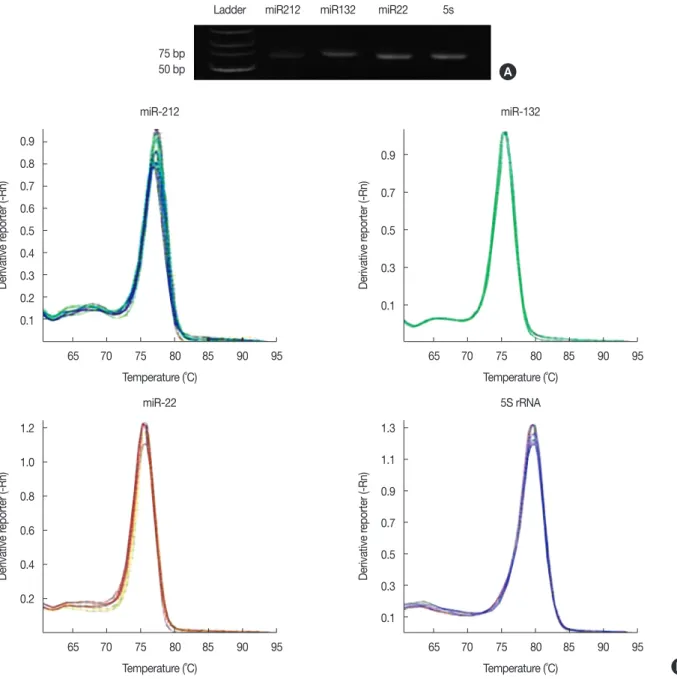
We used specific primers and a quantitative RT-PCR ap- proach to amplify miRNAs located in the 17p13.3 chromo- somal region, which is frequently deleted in breast cancer. Af- ter the addition of polyA tails to the given miRNAs, universal and miR-specific primers amplified ~70 base pair products.
The uniqueness and authenticity of the amplified products were confirmed by agarose gel electrophoresis, the presence of
Figure 1. Uniqueness and authenticity of the real-time polymerase chain reaction (RT-PCR) products. (A) The size and uniqueness of miR-22, miR- 132, miR-212, and 5s rRNA PCR products following agarose gel electrophoresis. (B) The authenticity of the PCR amplified products is shown by sin- gle, sharp melting curves.
0.9 0.8 0.7 0.6 0.5 0.4 0.3 0.2 0.1
1.2 1.0 0.8 0.6 0.4 0.2
0.9 0.7 0.5 0.3 0.1
1.3 1.1 0.9 0.7 0.5 0.3 0.1 65 70 75 80 85 90 95
65 70 75 80 85 90 95
65 70 75 80 85 90 95
65 70 75 80 85 90 95 Temperature (˚C)
Temperature (˚C)
Temperature (˚C) Temperature (˚C) miR-212
miR-22
miR-132
5S rRNA
Derivative reporter (-Rn)Derivative reporter (-Rn) Derivative reporter (-Rn)Derivative reporter (-Rn)
A Ladder miR212 miR132 miR22 5s
75 bp 50 bp
B
single, sharp melting curves, and direct DNA sequencing (Figure 1).
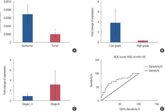
miR-132 is significantly downregulated in breast tumors We determined the relative expression of miR-132 in 36 paired tumor/nontumor FFPE breast tissue samples. The rela- tive expression levels of miR-132, adjusted to those of the 5s rRNA internal control, in tumor samples were normalized to those of the nontumor controls obtained from the same pa- tient. Our data revealed that there was a significant downreg- ulation of miR-132 expression levels in tumor samples (0.328- fold decrease in expression) compared to those of their matched nontumor controls (p <0.001) (Figure 2A). As shown in Figure 2B, a noticeable decrease in miR-132 expres- sion was also observed in high histologic grade tumors com- pared to that of tumor samples with low histologic grade.
However, this observed difference was not statistically signifi-
cant, which may have been due to the small sample size (20 samples of high-grade tumors and 16 samples of low-grade tumors). Similarly, the levels of miR-132 expression in stage III tumors were higher than those in stages Ӏ and II tumors (Figure 2C). Nevertheless, this observed difference was also not statistically significant. We also employed a receiver oper- ating characteristic (ROC) curve analysis to evaluate the sen- sitivity and specificity of miR-132 expression to discriminate between tumor and nontumor breast tissue samples. The total area under the curve (AUC) for miR-132 expression was 73%
(p<0.001), suggesting that it may be suitable as a potential tu- mor marker for breast cancer (Figure 2D).
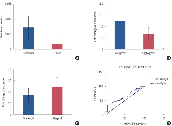
miR-212 is significantly downregulated in breast tumors We determined the relative expression of miR-212 in 31 paired tumor/nontumor FFPE breast tissue samples. The rela- tive expression levels of miR-212, adjusted to those of 5s rRNA,
Figure 2. miR-132 expression in tumor versus nontumor breast tissue samples. The expression levels of miR-132 were normalized to those of the 5s rRNA internal control. (A) Histograms show the mean values of relative miR-132 expression in tumor and nontumor samples, with confidence intervals as the error bars. Note that the expression of miR-132 was significantly lower in tumor tissues than that in the corresponding nontumor tissues ob- tained from the same patients (p=0.001). Comparative expression levels of miR-132 are presented for different grades (B) and stages (C) of breast tumor samples. Note that the observed differences in expression were not statistically significant. (D) Receiver operating characteristic (ROC) curve analysis of the specificity and sensitivity of miR-132 expression in discriminating between tumor and nontumor breast tissue samples. The calculated area under the curve (73%) suggests that miR-132 may be suitable as a tumor marker for breast cancer. *Represents p<0.05.
0.0010 0.0008 0.0006 0.0004 0.0002 0
8 6 4 2 0
8 6 4 2 0
150
100
50
0
Nontumor Tumor
Stage I, II Stage III
Low grade High grade
50 100 150
100%-Sensitivity%
ROC curve: ROC of miR-132
Relative expression Fold change of expression Fold change of expressionSensitivity%
A
C
B
D Sensitivity%
Identity%
*
in tumor samples were normalized to those of nontumor specimens obtained from the same patient. A significant downregulation of miR-212 expression levels was observed in tumor samples (0.281-fold decrease in expression) compared to those of their matched nontumor controls (p=0.043) (Fig- ure 3A). Similar to miR-132, the apparent expression altera- tions of miR-212 were not statistically associated with tumor grades (Figure 3B) or stages (Figure 3C). Compared to miR- 132, the ROC curve analysis revealed a lower sensitivity and specificity for miR-212 expression (AUC=63%, p=0.061) to discriminate between tumor and nontumor breast tissue sam- ples (Figure 3D).
miR-22 is significantly upregulated in breast tumors
Our data revealed that there was a significant upregulation of miR-22 expression levels in tumor samples (2.183-fold in-
crease in expression) compared to those of their matched nontumor controls (p=0.040) (Figure 4A). As shown in Fig- ure 4B, a noticeable decrease in the expression of miR-22 was observed in high-grade tumors compared to that in low-grade tumors. However, this observed difference was not statistically significant, which may have been due to the small sample size (20 samples of high-grade tumors and 16 samples of low- grade tumors). In contrast, miR-22 expression levels in stage III tumors were higher than those in stages I and II tumors (Figure 4C). Nevertheless, this observed difference was also not statistically significant. ROC curve analysis revealed an AUC of 64% (p=0.047) for miR-22 expression in discrimi- nating between tumor and nontumor breast tissue samples, suggesting that it may be moderately suitable as a potential marker for breast cancer (Figure 4D).
Figure 3. miR-212 expression in tumor versus nontumor breast tissue samples. The expression levels of miR-212 were normalized to those of the 5s rRNA internal control. (A) Histograms show the mean values of relative miR-212 expression in tumor and nontumor samples, with confidence intervals as the error bars. As shown, the expression of miR-212 is significantly downregulated in tumor samples (p=0.043) compared to that in nontumor controls. Comparative expression levels of miR-212 are presented for different grades (B) and stages (C) of breast tumor samples. Note that the ob- served differences in expression were not statistically significant. (D) Receiver operating characteristic (ROC) curve analysis revealed an area under the curve of 63% for miR-212, suggesting it has moderate sensitivity and specificity to discriminate between tumor and nontumor breast tissue samples.
*Represents p<0.05.
0.015
0.010
0.005
0
2.0 1.5 1.0 1.5 0
2.0 1.5 1.0 1.5 0
150
100
50
0
Nontumor Tumor
Stage I, II Stage III
Low grade High grade
50 100 150
100%-Sensitivity%
ROC curve: ROC of miR-212
Relative expression Fold change of expression Fold change of expressionSensitivity%
A
C
B
D Sensitivity%
Identity%
*
Figure 4. miR-22 expression in tumor versus nontumor breast tissue samples. The expression levels of miR-22 were normalized to those of the 5s rRNA internal control. (A) Histograms show the mean values of relative miR-22 expression in tumor and nontumor samples, with confidence intervals as the error bars. Note that the expression of miR-22 was significantly higher than that in the corresponding nontumor tissues from the same patients (p=0.04). Comparative expression levels of miR-22 are presented for different grades (B) and stages (C) of breast tumor samples. The observed dif- ferences in expression were not statistically significant between groups. (D) Receiver operating characteristic (ROC) curve analysis revealed an area under the curve of 64% for miR-22, indicating that miR-22 is a potential tumor marker with moderate specificity and sensitivity to discriminate be- tween tumor and nontumor breast tissue samples. *Represents p<0.05.
0.004 0.003 0.002 0.001 0
60
40
20
0
60
40
20
0
150
100
50
0
Nontumor Tumor
Stage I, II Stage III
Low grade High grade
50 100 150
100%-Sensitivity%
ROC curve: ROC of miR-22
Relative expression Fold change of expression Fold change of expressionSensitivity%
A
C
B
D Sensitivity%
Identity%
DISCUSSION
Deletions or LOH are frequent events in cancers. For in- stance, frequent deletions in 17p13.3 have been reported for breast cancer [2,4,5]. It is postulated that at least one or two tumor suppressor genes reside within this region [4]. As three breast development-related miRNAs, namely miR-132, miR- 212, and miR-22, are located within this region, we hypothe- sized that theses miRNAs may have tumor suppressor roles in breast tumorigenesis. Thus, we expected to observe downreg- ulation of these miRNAs in breast tumor tissues.
Recently, both downregulated and upregulated expression of miR-132, miR-212, and miR-22 have been reported for dif- ferent malignancies. These prior investigations have suggested that the aforementioned miRNAs can act as either oncogenes or tumor suppressor genes by targeting different mRNAs in
multiple cellular pathways [15-21]. The versatile expression patterns of these miRNAs in various cancers point to their di- verse cellular functions and the difficulty in determining their regulation and potential targets.
In the current study, we found that the miRNAs of interest were expressed in all tumor and nontumor samples. As ex- pected, a significant downregulation of miR-132 and miR-212 was observed in breast tumor samples, suggesting that they have potential tumor-suppressor roles in breast tumorigenesis.
These results are in contrast with those in a report by Park et al. [19], in which increased expression of both these miRNAs was observed in pancreatic cancer. However, our data are con- sistent with those of Zhang et al. [22], whose findings demon- strated the downregulation of miR-132 in pancreatic cancer.
One logical explanation for the conflicting reports on miRNA expression in similar cancer types may be that they were the
*
result of using samples with different stages or grades of ma- lignancy. As we have shown here, the expression levels of these miRNAs varied greatly in different grade and stage sub- groups. Interestingly, the pattern of expression in the different grade and stage subgroups was very similar for miR-132 and miR-212, which both differed from that of miR-22. This ob- servation seems to be associated with the chromosomal loca- tions of these miRNAs. Specifically, while miR-132 (17:
1953202–1953302) and miR-212 (17:1953565–1953674) are closely packed together, miR-22 is located ~336 kb away from them (17:1617197–1617281). Therefore, the distance between them could lead to their involvement in separate genetic and epigenetic events during the initiation and progression of breast tumorigenesis.
As mentioned above, we found a different pattern of expres- sion for miR-22 in breast tumors. In contrast to what we hy- pothesized and to what we observed for miR-132 and miR- 212, we found a significant upregulation of miR-22 expression in breast tumors compared to that in their marginal nontu- mor counterparts. Nevertheless, miR-22 expression levels in different grades and stages of malignancy were similar to those of miR-132 and miR-212. Interestingly, a conflicting role for miR-22 as an oncomiR [23] and as a tumor suppres- sor [24-26] has been reported. In addition to the possible ef- fects of sample grade and stage on the outcome of results, an- other possibility should be taken into account that may ex- plain these conflicting reports. As we have already reported for miR-21, the stromal content of samples can affect the out- come of expression results [27]. This may be explained as miRNA expression is not confined to tumor cells, and thus some altered expression may be attributed to stromal cells within the tumor microenvironment, including cancer-asso- ciated fibroblasts [28]. Therefore, the amount of stromal con- tent in tumor samples may affect the outcome of results [28].
Our data revealed a consistent and noticeable downregul- ation of miR-132, miR-212, and miR-22 in high-grade samples, as well as a noticeable upregulation in stage III breast tumors.
The latter finding is in agreement with a recent report by Hanieh [29], in which antimetastatic properties of the miR- 212/132 cluster through SOX4 suppression in human tumor cells were demonstrated. In the case of miR-132, our data are consistent with those in a report by Zhang et al. [30], which indicated a critical role for miR-132 in breast cancer by sup- pressing cell proliferation, invasion, migration, and metastasis via negative regulation of HN1.
Taken together, our data revealed significant expression al- terations of miR-132, miR-212, and miR-22 in breast tumors, and that miR-132 may be utilized to differentiate between tu- mor and nontumor states of breast tissue samples.
CONFLICT OF INTEREST
The authors declare that they have no competing interests.
ACKNOWLEDGMENTS
We are grateful to the personnel in the pathology depart- ment of Shariati Hospital for their valuable help in collecting clinical samples.
REFERENCES
1. Iorio MV, Casalini P, Piovan C, Braccioli L, Tagliabue E. Breast cancer and microRNAs: therapeutic impact. Breast 2011;20 Suppl 3:S63-70.
2. Lerebours F, Lidereau R. Molecular alterations in sporadic breast can- cer. Crit Rev Oncol Hematol 2002;44:121-41.
3. Kashiwagi H, Uchida K. Genome-wide profiling of gene amplification and deletion in cancer. Hum Cell 2000;13:135-41.
4. Cornelis RS, van Vliet M, Vos CB, Cleton-Jansen AM, van de Vijver MJ, Peterse JL, et al. Evidence for a gene on 17p13.3, distal to TP53, as a tar- get for allele loss in breast tumors without p53 mutations. Cancer Res 1994;54:4200-6.
5. Isomura M, Tanigami A, Saito H, Harada Y, Katagiri T, Inazawa J, et al.
Detailed analysis of loss of heterozygosity on chromosome band 17p13 in breast carcinoma on the basis of a high-resolution physical map with 29 markers. Genes Chromosomes Cancer 1994;9:173-9.
6. Piao HL, Ma L. Non-coding RNAs as regulators of mammary develop- ment and breast cancer. J Mammary Gland Biol Neoplasia 2012;17:33- 42.
7. Ucar A, Vafaizadeh V, Jarry H, Fiedler J, Klemmt PA, Thum T, et al.
miR-212 and miR-132 are required for epithelial stromal interactions necessary for mouse mammary gland development. Nat Genet 2010;42:1101-8.
8. Ibarra I, Erlich Y, Muthuswamy SK, Sachidanandam R, Hannon GJ. A role for microRNAs in maintenance of mouse mammary epithelial progenitor cells. Genes Dev 2007;21:3238-43.
9. Yu Z, Baserga R, Chen L, Wang C, Lisanti MP, Pestell RG. MicroRNA, cell cycle, and human breast cancer. Am J Pathol 2010;176:1058-64.
10. Lee RC, Feinbaum RL, Ambros V. The C. elegans heterochronic gene lin-4 encodes small RNAs with antisense complementarity to lin-14.
Cell 1993;75:843-54.
11. Lee YS, Dutta A. MicroRNAs: small but potent oncogenes or tumor suppressors. Curr Opin Investig Drugs 2006;7:560-4.
12. Gregory RI, Shiekhattar R. MicroRNA biogenesis and cancer. Cancer Res 2005;65:3509-12.
13. Wu Q, Li H, Lu J, Ge Q, Lu Z. Aberrant microRNA expression in the development of breast carcinoma. Chin Sci Bull 2010;55:3517-26.
14. Iorio MV, Ferracin M, Liu CG, Veronese A, Spizzo R, Sabbioni S, et al.
MicroRNA gene expression deregulation in human breast cancer. Can- cer Res 2005;65:7065-70.
15. Zhang J, Yang Y, Yang T, Liu Y, Li A, Fu S, et al. MicroRNA-22, downreg- ulated in hepatocellular carcinoma and correlated with prognosis, sup- presses cell proliferation and tumourigenicity. Br J Cancer 2010;103:
1215-20.
16. Anand S, Majeti BK, Acevedo LM, Murphy EA, Mukthavaram R, Scheppke L, et al. MicroRNA-132-mediated loss of p120RasGAP acti- vates the endothelium to facilitate pathological angiogenesis. Nat Med 2010;16:909-14.
17. Bar N, Dikstein R. miR-22 forms a regulatory loop in PTEN/AKT pathway and modulates signaling kinetics. PLoS One 2010;5:e10859.
18. Pandey DP, Picard D. miR-22 inhibits estrogen signaling by directly tar- geting the estrogen receptor alpha mRNA. Mol Cell Biol 2009;29:3783- 90.
19. Park JK, Henry JC, Jiang J, Esau C, Gusev Y, Lerner MR, et al. miR-132 and miR-212 are increased in pancreatic cancer and target the retino- blastoma tumor suppressor. Biochem Biophys Res Commun 2011;406:
518-23.
20. Chang TC, Yu D, Lee YS, Wentzel EA, Arking DE, West KM, et al.
Widespread microRNA repression by Myc contributes to tumorigene- sis. Nat Genet 2008;40:43-50.
21. He L, Hannon GJ. MicroRNAs: small RNAs with a big role in gene reg- ulation. Nat Rev Genet 2004;5:522-31.
22. Zhang S, Hao J, Xie F, Hu X, Liu C, Tong J, et al. Downregulation of miR-132 by promoter methylation contributes to pancreatic cancer de- velopment. Carcinogenesis 2011;32:1183-9.
23. Song SJ, Ito K, Ala U, Kats L, Webster K, Sun SM, et al. The oncogenic microRNA miR-22 targets the TET2 tumor suppressor to promote he-
matopoietic stem cell self-renewal and transformation. Cell Stem Cell 2013;13:87-101.
24. Xu D, Takeshita F, Hino Y, Fukunaga S, Kudo Y, Tamaki A, et al. miR-22 represses cancer progression by inducing cellular senescence. J Cell Biol 2011;193:409-24.
25. Tsuchiya N, Izumiya M, Ogata-Kawata H, Okamoto K, Fujiwara Y, Nakai M, et al. Tumor suppressor miR-22 determines p53-dependent cellular fate through post-transcriptional regulation of p21. Cancer Res 2011;71:4628-39.
26. Jazbutyte V, Fiedler J, Kneitz S, Galuppo P, Just A, Holzmann A, et al.
MicroRNA-22 increases senescence and activates cardiac fibroblasts in the aging heart. Age (Dordr) 2013;35:747-62.
27. Nouraee N, Van Roosbroeck K, Vasei M, Semnani S, Samaei NM, Naghshvar F, et al. Expression, tissue distribution and function of miR- 21 in esophageal squamous cell carcinoma. PLoS One 2013;8:e73009.
28. Nouraee N, Mowla SJ, Calin GA. Tracking miRNAs’ footprints in tu- mor–microenvironment interactions: insights and implications for tar- geted cancer therapy. Genes Chromosomes Cancer 2015;54:335-52.
29. Hanieh H. Aryl hydrocarbon receptor-microRNA-212/132 axis in hu- man breast cancer suppresses metastasis by targeting SOX4. Mol Can- cer 2015;14:172.
30. Zhang ZG, Chen WX, Wu YH, Liang HF, Zhang BX. MiR-132 prohib- its proliferation, invasion, migration, and metastasis in breast cancer by targeting HN1. Biochem Biophys Res Commun 2014;454:109-14.