© 2012 Korean Breast Cancer Society. All rights reserved. http://ejbc.kr | pISSN 1738-6756
INTRODUCTION
Breast carcinoma is one of the most common neoplasms in women and is a leading cause of cancer-related deaths world- wide [1]. In Egypt breast cancer is the most common cancer among women, representing 18.9% of total cancer cases (35.1%
in women and 2.2% in men) among the Egypt National Cancer Institute (NCI) series of 10,556 patients for 2001 (World Health Organization Regional Office for the Eastern Mediterranean, Cairo, Egypt), with an age-adjusted rate of 49.6 per 100,000 in the population. However, this represents the hospital-based data of referral tertiary centers and does not represent all breast cancer cases in Egypt.
Significant breast cancer risk factors include: age, menarche at an early age, menopause at late age, late age for the first pregnancy, obesity, dense breast tissue, oral contraception use, hormone replacement therapy, alcohol, tobacco smoke, diet, family history, lactation, and prior history of benign breast disease [2]. A number of genes, including BRCA1 and BRCA2, HER2/neu, and p53, have been linked to breast cancer suscep- tibility and development [3].
Oxidative stress is due to a disturbance in the balance between the production of a reactive oxygen species (ROS) and the efficiency of the antioxidant defense. In other words, oxidative stress results in an excessive production of ROS which over- whelms the antioxidant defense system or when there is a significant decrease or lack of antioxidant defense [4].
Experimental evidence revels that ROS are involved in the initiation and promotion of carcinogenesis, where inactivation or loss of certain tumor suppressor genes have occurred [5].
The levels of free radical molecules are controlled by various cellular defense mechanisms, consisting of enzymatic compo-
Impact of Breast Cancer and Combination Chemotherapy on Oxidative Stress, Hepatic and Cardiac Markers
Kamal Adel Amin, Basant Mahmoud Mohamed1, Mohamed Aly M. El-wakil2, Sanaa Omar Ibrahem1
Biochemistry Department, Faculty of Veterinary Medicine, 1Chemistry Department, Faculty of Science, and 2Clinical Oncology Department, Faculty of Medicine, Beni-Suef University, Beni-Suef, Egypt
ORIGINAL ARTICLE
Purpose: Carcinoma of the breast is the most prevalent cancer among Egyptian women and constitutes 29% of National Cancer Institute cases. The aim of this study was to determine the effect of breast cancer on oxidative stress, cardiac markers and liver function tests, moreover the role of 5-fluorouracil, doxorubicin, and cyclophosphamide (FAC) in the treatment of breast cancer and its mechanism through changing the measured markers.
Methods: Forty female breast cancer patients who were admitted to the Department of Oncology of the Beni-Suef University Hospital were enrolled in the study. This study included three arms: a control group of healthy age-matched females (n=20), breast cancer patients who weren’t receiving treatment (n=20), and patients undergoing treatment with anticancer combination drugs FAC (n=20). Blood samples collected from the control subjects and patients were analysed to determine levels of cata- lase, reduced glutathione (GSH), uric acid, nitric oxide (NO),
malondialdehyde, creatine kinase (CK), lactate dehydrogenase (LDH), liver enzymes (alanine aminotransferase and aspartate aminotransferase), and creatinine. Results: The levels of catalase and GSH were significantly reduced (p<0.05) in breast carcino- ma and FAC treated breast cancer patients. The lipid peroxida- tion and NO levels were significantly enhanced in both untreated and FAC treated breast cancer patients. The CK and LDH were significantly enhanced (p<0.05) in the FAC group. Conclusion:
The results from the present study show that oxidative stress is implicated in breast carcinoma and chemotherapy aggravates this oxidative stress which causes damage to many cellular targets and has the main side effect of cardiotoxicity.
Key Words: Breast neoplasms, Cardiac function, Chemotherapy, Liver function, Oxidative stress
Correspondence: Kamal Adel Amin
Biochemistry Department, Faculty of Veterinary Medicine, Beni-Suef University, Beni-Suef 62511, Egypt
Tel: +20-822327982, Fax: +20-822327982 E-mail: [email protected]
Received: May 18, 2012 Accepted: August 13, 2012
Cancer
nents (catalase, glutathione peroxidase, superoxide dismutase) and non-enzymatic components (vitamin E, vitamin C, and glutathione components) [6].
Anthracyclines rank among the most effective anticancer drugs ever developed [7]. Most patients with breast cancer are treated with a combination of the anticancer chemotherapy drugs of 5-fluorouracil, doxorubicin, and cyclophosphamide (FAC). These antineoplastic agents cause a reduction in anti- oxidant levels because their toxicity increases the peroxidation of the unsaturated fatty acids of membrane phospholipids [8].
Their main adverse effects may be heart damage (cardiotoxicity) and vomiting which considerably limits their usefulness. The molecular pathogenesis of anthracycline cardiotoxicity remains highly controversial, although the oxidative stress-based hypothesis involving the intramyocardial production of ROS has gained the widest acceptance [9]. Therefore the current study aimed to investigate the effect and association of breast cancer on oxidative stress, as well as hepatic and cardiac bio- markers in patients receiving combination chemotherapy treatment.
METHODS
Drugs and instruments
Doxorubicin for intravenous administration was obtained from Ebwe Pharma Company (Cairo, Egypt) and cyclophos- phamide and 5-flurouracil from EIMC Pharmaceuticals Company (Cairo, Egypt). Chemicals for sensitive biochemical assays were obtained from Randox Laboratories Ltd., Crumlin Company, Antrim United Kingdom, Bio-diagnostic Company (Giza, Egypt) and Spectrum Company for Biotechnology (Cairo, Egypt). Distilled water was used for the biochemical assays. Spectrophotometric measurements were done with T80 UV/Vis spectrophotometer (PG Instruments Ltd., Wibtoft, UK).
Patients and experimental protocol
This study included 40 breast cancer patients from the Department of Oncology of Beni-Suef University Hospital.
The inclusion criteria for this study were: the presentation of a palpable mass in the breast that was observed with mam- mography that was later histologicaly diagnosed. Most of the patients were clinically categorized as stage II and some were in stage I, stage III according to the classification of the Union for International Cancer Control (UICC 1997). The cases were classified according to TNM classification and tumors were histologicaly diagnosed in most cases as invasive ductal and invasive lobular and a few other types are presented in a Table 1 according to Rajneesh et al. [10].
The patients were classified into two groups. The first were recent breast cancer patients that had clinical and histopato- logical evidence of breast cancer and were receiving neither chemotherapy nor hormonal treatments. The second group was FAC treated breast cancer patients who mostly received 2 FAC cycles (5-flurouracil 500 mg/m2, doxorubicin 50 mg/m2, and cyclophosphamide 500 mg/m2).
Twenty healthy women served as control normal group for the breast cancer group. Cases with illness that are associated with altering free radical levels in healthy and cancer patients (such as diabetes, hypertension, myocardial ischemia, myocar- dial infarction, renal disorders, pancreatic disorders, pulmo- nary disease, and pregnancy) and patients with fibroadenoma or with any previous treatment were excluded from this study.
Both the patients and control normal groups had a similar socioeconomic status and dietary pattern. A medical history, physical examination and laboratory tests were obtained from all subjects. None of the subjects included in the study had a history of substance abuse or dependence, serious medical conditions, severe head injury or seizure disorders.
Ethics approval
The materials in our study are human blood sample from normal, breast cancer and treated with FAC, which are giving during breast cancer treatment.
Moreover, our study contains no private information on patients. So, our work has no problems in causing any ethical issue or violation of human rights. Informed consent was obtained from all participants and the protocol used in our study was approved by the Committee of the Beni-Suef Uni- versity (Beni-Univ- MSc 171-2010).
Serum collection and preparation
Venous blood samples were collected by arm puncture in patients, controls and the FAC treated group 24 hours after the administration of the last dose. Blood (3 mL) was collected, allowed to clot, and centrifuged at 3,000×g for 15 minutes.
The separated serum was then stored at -4°C and the clarified serum preparations were used for biochemical analysis.
Biochemical analysis
Catalase was assayed by the Cohen et al. [11] method based on the reaction of catalase with a known quantity of H2O2. The remaining H2O2 reacts with a dye to form a chromophore with an intensity of color inversely proportional to the amount of catalase in the original sample. The glutathione reduced (GSH) estimated by Beutler et al. [12], this estimation based on the reduction of 5, 5´ dithiobis (2-nitrobenzoic acid) (DTNB) with GSH. The reduced chromogen is directly pro-
portional to the GSH concentration and its absorbance can be measured at 405 nm. Nitric oxide (NO) was measured accord- ing to Miranda et al. [13], and lipid peroxide (malondialde- hyde) was measured according to Satoh [14], by an estimation of thiobarbituric acid reactive product which measured color- imetrically at 534 nm. Analysis of liver enzymes alanine ami- notransferase (ALT) and aspartate aminotransferase (AST) were based on the measurement of pyruvate hydrazone and oxaloacetate hydrazone colorimetrically at 546 nm. Serum creatine kinase (CK) was determined using the Burtis and Ashwood [15] method. The catalytic activity was determined from the rate of NADPH formation, measured at 340 nm by means of the hexokinase (HK) and glucose-6-phosphate de- hydrogenase (G6PDH) coupled Reactions. Determination of serum lactate dehydrogenase (LDH) via catalyzes the reaction between pyruvate and NADH to produce NAD and L-lactate.
It is determined by measuring a decrease in the absorbance of 340 nm. Serum creatinine was assessed by a reaction with pic-
ric acid in alkaline solution to form a colored complex. Uric acid was measured according to Fossati et al. [16].
Statistical analysis was carried out using Graph Pad Instat software version 5 (ISS-Rome, Rome, Italy). One-way analysis of variance followed by Tukey-Kramer multiple comparisons post-test were used in an analysis of the data. Values of p<0.05 were regarded as significant. Data were expressed in tables and figures as mean±SEM. The number of patients for each group was 12. Means that shared the same letter were not significantly different. Means that have different letters were significantly different (p<0.05).
RESULTS
Results are summarized in tables and figures as follow: Table 1 shows the classification of breast cancer patients who partic- ipated in our study. Data shows that MDA level increased significantly in breast cancer patients compared to normal control. FAC chemotherapy triggered an enhancement in the Table 1. General characteristics of breast cancer patients
Characteristic No. of patients
Total No. of subjects 40
Menopausal status
Premenopausal 20
Postmenopausal 20
Cancer site
Left breast 23
Right breast 17
Clinical status
Invasive ductal carcinoma 34
Invasive lobular carcinoma 4
Mucinous adenocarcinoma 1
Medullary carcinoma 1
Clinical stage
Grade 1 2
T1N0M0
Grade 2 36
T1N1M0 15
T2N0M0 9
T2N1M0 10
T3N0M0 2
Grade 3
T3N1M0 2
ER, PR status
Negative 18
Positive 22
HER2 status
Negative 25
Positive 15
T=tumor size, T1: <2 cm, T2: 2-4 cm, T3: >4 cm; N=nodal metastasis, N0:
no regional lymph node metastasis, N1: metastasis in a single ipsilateral node of <3 cm diameter; M =distant metastasis, M0: no distant metastasis;
ER=estrogen receptors; PR=progesterone receptors; HER2=human epider- mal growth factor receptor-2.
Table 2. The levels of catalase, GSH, serum NO and lipid peroxidation in breast cancer patients before and after chemotherapy treatment
Group Catalase GSH NO MDA
Control 739±27.5 34.1±2.2 0.35±0.03 7.0±0.31
Breast cancer
patients 455±36.9* 24.7±1.4* 3.6±0.4* 9.5 ±0.2*
Breast cancer patients after chemotherapy
270±18.23*,† 15.4±0.55*,† 6.5±0.6*,†11.3±0.72*,†
Values are presented as mean±SE. Number of patients is 20 for each group.
Values (among the three groups) followed by different letters are significantly different (p<0.05).
GSH=reduced glutathione; NO=nitric oxide; MDA=malondialdehyde.
*Significantly different from the control group; †Significantly different from the breast cancer patients before chemotherapy group.
Figure 1. The malondialdehyde (MDA) levels in the control group, breast cancer patients before treatment and chemotherapy treated groups.
FAC=5-fluorouracil, doxorubicin, and cyclophosphamide.
*Significantly different from the control group; †Significantly different from the breast cancer patients before chemotherapy group.
Concentration of MDA (nmol/mL)
Control Patients before FAC Patients after FAC Different experimental groups
20
15
10
5
0
*
*,†
lipid peroxidation compared to healthy controls (Table 2, Figure 1).
The serum NO increased in breast cancer patients before treatment as compared to normal control. FAC chemotherapy caused an increase in NO compared to patients before treat-
Table 3. The activity of cardiac enzymes in breast cancer patients be- fore and after chemotherapy treatment
Group LDH CK
Control 259.7±18.9 13.6±3.2
Breast cancer patients 303.2±7.9 17.4±1.8
Breast cancer patients after chemotherapy 368.9±19.6*,† 32.1 ±2.6*,†
Values are presented as mean±SE. Number of patients is 20 for each group.
Values (among the three groups) followed by different letters are significantly different (p≤0.05).
LDH=lactate dehydrogenase; CK=creatine kinase.
*Significantly different from the control group; †Significantly different from the breast cancer patients before chemotherapy group.
Figure 2. The nitric oxide concentration in the control group, breast cancer patients before treatment and chemotherapy treated groups.
FAC=5-fluorouracil, doxorubicin, and cyclophosphamide.
*Significantly different from the control group; †Significantly different from the breast cancer patients before chemotherapy group.
Concentration of nitric oxide (μmol/mL)
Control Patients before FAC Patients after FAC Different experimental groups
10
8
6
4
2
0
*
*,†
Figure 4. The catalase activity in the control group, breast cancer pa- tients before treatment and chemotherapy treated groups.
FAC=5-fluorouracil, doxorubicin, and cyclophosphamide.
*Significantly different from the control group; †Significantly different from the breast cancer patients before chemotherapy group.
Catalase activity (U/L)
Control Patients before FAC Patients after FAC Different experimental groups
1,000
800
600
400
200
0
*
*,†
Figure 3. The reduced glutathione (GSH) concentration in the control group, breast cancer patients before treatment and chemotherapy treated groups.
FAC=5-fluorouracil, doxorubicin, and cyclophosphamide.
*Significantly different from the control group; †Significantly different from the breast cancer patients before chemotherapy group.
Concentration of GSH (mg/dL)
Control Patients before FAC Patients after FAC Different experimental groups
50
40
30
20
10
0
*
*,†
Figure 5. The creatine kinase (CK) activity in the control group, breast cancer patients before treatment and chemotherapy treated groups.
FAC=5-fluorouracil, doxorubicin, and cyclophosphamide.
*Significantly different from the control group; †Significantly different from the breast cancer patients before chemotherapy group.
Activity of CK (U/L)
Control Patients before FAC Patients after FAC Different experimental groups
50
40
30
20
10
0
*,†
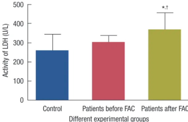
Figure 6. The lactate dehydrogenase (LDH) activity in the control group, breast cancer patients before treatment and chemotherapy treated groups.
FAC=5-fluorouracil, doxorubicin, and cyclophosphamide.
*Significantly different from the control group; †Significantly different from the breast cancer patients before chemotherapy group.
Activity of LDH (U/L)
Control Patients before FAC Patients after FAC Different experimental groups
500
400
300
200
100
0
*,†
ment and to healthy controls (Table 2, Figure 2).
Serum antioxidants catalase activity and the level of GSH were significantly diminished in breast carcinoma patients be- fore treatment as compared to normal control. These changes more declined with FAC chemotherapy as compared to breast cancer patients and healthy controls (Table 2, Figures 3 and 4).
FAC chemotherapy induced a significant increase in the activities of CK compared to patients before treatment and healthy controls. Moreover the activity of LDH was elevated in FAC treated patients as compared to healthy controls (Table 3, Figures 5, and 6).
The liver enzymes ALT and AST activities and renal creati- nine and uric acid levels show no change in patients before treatment or after receiving FAC in comparison to healthy control (Table 4).
DISCUSSION
In our study we investigated the effect of breast cancer on the levels of various enzymes with antioxidant activities and oxidative stress markers NO and lipid peroxidation and the effect of FAC treatment on these parameters.
Several reports have presented evidence that ROS are involved in the etiology and progression of breast cancer [17]. However, most studies are based on breast cancer culture cells or experi- mental data. Moreover, few studies have investigated the change of oxidative parameters in patients with breast cancer after chemotherapy and the results are also controversial. Therefore, our study provides the most significant data.
ROS plays an important role in tumor initiation, and in a healthy organism ROS levels are controlled by endogenous mechanisms including glutathione and enzymes like catalase.
Elevated ROS levels can initiate DNA damage, and might ulti- mately lead to carcinogenesis [18].
The present results showed an increased NO and lipid per- oxidation in breast cancer patients with a decreased antioxidant defense of catalase and GSH. This may be due to increased Table 4. The levels of renal function and liver enzymes in breast cancer patients before and after chemotherapy treatment
Group Creatinine Uric acid ALT AST
Control 0.65±0.04 5.23±0.26 7.7±0.44 8.3±0.44
Breast cancer patients 0.73±0.032 5.7±0.24 7.62±0.8 8.7±0.47 Breast cancer patients 0.74±0.045 5.6±0.32 7.65±0.47 7.9±0.6 after chemotherapy
Values are presented as mean±SE. Number of patients is 20 for each group.
ALT=alanine aminotransferase; AST=aspartate aminotransferase.
Values (among the three groups) followed by different letters are significantly different (p≤0.05).
*Significantly different from the control group; †Significantly different from the breast cancer patients before chemotherapy group.
free radical generation that damage several cellular molecules.
These results are in accordance with Prabasheela et al. [19], and Kasapović et al. [20] who found that catalase and superoxide dismutase activity were found to decrease indicating enhanced free radical activity in breast cancer patients while the antioxi- dant defense mechanism is weakened. Cancer cells generate a ROS, and biochemically have low levels of antioxidant enzyme in most animal and human cancers.
The high levels of NO production in breast cancer and che- motherapy may result in high cytotoxic activity. Consequently the mechanism of inhibiting the production of NO is impor- tant for the improvement of breast cancer and chemotherapy.
NO promotes cancer progression by activating several onco- genic signaling pathways such as extracellular signal-regulated kinases and phosphoinositide 3-kinases [21].
NOS2 upregulation and elevated NO production affect the redox state of cells and can induce protein, lipid, and DNA modifications. A release of variable amounts of NO into the tumor microenvironment can activate oncogenic pathways, including Akt, epidermal growth factor receptor, and c-Myc signaling pathways, and stimulate tumor microvasculariza- tion. More recent findings suggest that NO induces stem cell- like tumor characteristics in breast cancer [22]. The significantly elevated NO in our data was associated with breast cancer as compared with control and that increment was aggravated with chemotherapy.
This work provides novel evidence that NO modulates the progression of breast cancer in association with oxidative stress markers in patients with breast cancer.
Our data elucidates that FAC chemotherapy increases the already existing oxidative stress in breast cancer patients.
Alshabanah et al. [23] reported a decrease in the gene expres- sion levels of glutathione peroxidase (GSHPx), catalase, gluta- thione reductase (GR), and glutathione transferase (GST) in liver tissue with the cumulative dose of doxorubicin with a decrease in their activity in the serum. The data showed that doxorubicin not only increased free radical formation but also decreased its ability to detoxify a ROS. The formation of super- oxide radicals together with NO might form peroxynitrite induced by doxorubicin which causes tissue damage leading to an increase in the levels of thiobarbituric acid substance.
These chemotherapeutic drugs are hydrophilic and cannot penetrate the inner membrane of cells where they would be reduced by NADH located on the inner membrane surface [24,25]. Chemotherapeutic drugs, particularly doxorubicin used in FAC treatment are able to enter the outer mitochondrial membrane and enter the cytosol. Intramolecular rearrange- ments result in the formation of a lipophilic deoxyaglycone that can penetrate the inner membrane of the mitochondria.
There doxorubicin competes with coenzyme Q10 as an electron acceptor and diverts electrons to molecular oxygen resulting in the formation of super oxide radicals [25]. Doxorubicin intercalates DNA coils and interferes with normal cellular metabolism through a diverse set of biochemical mechanisms that may explain its toxicity. It causes an increase in the perox- idation of unsaturated fatty acids of membrane phospholipids which lead to a decrease in the level of antioxidants and gen- erate a high level of oxidative stress. In addition, doxorubicin is able to divert electrons from the mitochondrial electron transport system in addition to generating ROS at cellular sites.
Our results indicated that enhanced cardiac enzymes CK and LDH after FAC treatment which evidenced that cardiotoxicity is the main side effect of chemotherapy particularly doxorubicin used in FAC treatment. These results are in agreement with the results by Kaithwas et al. [26]. The increment in serum LDH level suggests an increased leakage of this enzyme from mito- chondria as a result of toxicity induced by treatment with doxorubicin [27]. It is recently reported that the doxorubicin- induced free radical generation triggers membrane peroxida- tion and the disruption of cardiac myocytes, which can led to an increased release of CK in the serum [27].
The heart is more susceptible than other tissues to the oxi- dative stress produced by ANTs. Several responses have been proposed; ANTs have been shown to be retained within car- diomyocytes more than other cell types. ANTs are thought to enter the mitochondria and to inhibit the respiratory chain by binding to cardiolipin, which is a relatively cardiospecific phospholipid that is rich in polyunsaturated fatty acids and that is found in the inner mitochondrial membrane. Cardio- lipin has a high affinity for ANTs [28].
Cardiac tissue has weak antioxidant activity, since it lacks catalase, and some researcher have shown that doxorubicin selectively down-regulates glutathione peroxidase suggesting that cardiomyocytes are exposed to high levels of hydrogen peroxide. In addition, cardiomyocytes are rich in mitochondria, which represent up to 50% of cardiomyocyte mass which serve as both source and target of ROS [29,30]. Moreover, the important role has been attributed to exogenous NADH dehydrogenase.
Unlike cardiac mitochondria, liver mitochondria lack the NADH-related pathway of reducing equivalents from cytosol to the respiratory chain. As a result liver mitochondria do not generate significant amounts of ANT semiquinones [30].
The current study provide novel data about the relation of breast cancer, oxidative stress, cardiac and hepatic function biomarkers with and without the combined formula used in chemotherapy for an Egyptian breast cancer patient.
It could be concluded that breast carcinoma leads to oxida-
tive stress evidenced by an increased NO level and decreased catalase and glutathione which enhance lipid peroxidation level.
The treatment by FAC chemotherapy exaggerates oxidative stress leading to cardiotoxicity so monitoring serum oxidative markers and cardiac enzymes is highly recommended.
The data revealed indicated that oxidative stress and cardio- toxicity development may ensure breast cancer progression, possibly mediated through catalase, GSH, MDA, NO, LDH, and CK activity.
Future studies needs to investigate the uses of potent anti- oxidant compounds to compete the oxidative stress of breast cancer and chemotherapy.
ACKNOWLEDGEMENTS
Authors’ contributions: S.I., K.A., B.M., and M.E. carried out experimental work; biochemical and statistical analysis, inter- pretation and discussion of the results wrote the paper, related to their part of the work. All authors perform drafting, read and approved the final manuscript.
CONFLICT OF INTEREST
The authors declare that they have no competing interests.
REFERENCES
1. Polyak K. On the birth of breast cancer. Biochim Biophys Acta 2001;
1552:1-13.
2. Gönenç A, Erten D, Aslan S, Akinci M, Simşek B, Torun M. Lipid per- oxidation and antioxidant status in blood and tissue of malignant breast tumor and benign breast disease. Cell Biol Int 2006;30:376-80.
3. Yeh CC, Hou MF, Tsai SM, Lin SK, Hsiao JK, Huang JC, et al. Superox- ide anion radical, lipid peroxides and antioxidant status in the blood of patients with breast cancer. Clin Chim Acta 2005;361:104-11.
4. Kang DH. Oxidative stress, DNA damage, and breast cancer. AACN Clin Issues 2002;13:540-9.
5. Birnboim HC. DNA strand breakage in human leukocytes exposed to a tumor promoter, phorbol myristate acetate. Science 1982;215:1247-9.
6. Matés JM, Sánchez-Jiménez FM. Role of reactive oxygen species in apoptosis: implications for cancer therapy. Int J Biochem Cell Biol 2000;
32:157-70.
7. Weiss RB. The anthracyclines: will we ever find a better doxorubicin?
Semin Oncol 1992;19:670-86.
8. Conklin KA. Chemotherapy-associated oxidative stress: impact on chemotherapeutic effectiveness. Integr Cancer Ther 2004;3:294-300.
9. Simůnek T, Stérba M, Popelová O, Adamcová M, Hrdina R, Gersl V.
Anthracycline-induced cardiotoxicity: overview of studies examining the roles of oxidative stress and free cellular iron. Pharmacol Rep 2009;
61:154-71.
10. Rajneesh CP, Manimaran A, Sasikala KR, Adaikappan P. Lipid peroxi- dation and antioxidant status in patients with breast cancer. Singapore
Med J 2008;49:640-3.
11. Cohen G, Dembiec D, Marcus J. Measurement of catalase activity in tis- sue extracts. Anal Biochem 1970;34:30-8.
12. Beutler E, Duron O, Kelly BM. Improved method for the determination of blood glutathione. J Lab Clin Med 1963;61:882-8.
13. Miranda KM, Espey MG, Wink DA. A rapid, simple spectrophotomet- ric method for simultaneous detection of nitrate and nitrite. Nitric Oxide 2001;5:62-71.
14. Satoh K. Serum lipid peroxide in cerebrovascular disorders determined by a new colorimetric method. Clin Chim Acta 1978;90:37-43.
15. Burtis CA, Ashwood ER. Tietz Textbook of Clinical Chemistry. 3rd ed.
Philadelphia: W.B. Saunders Company; 1998. p.1034.
16. Fossati P, Prencipe L, Berti G. Use of 3,5-dichloro-2-hydroxybenzene- sulfonic acid/4-aminophenazone chromogenic system in direct enzy- mic assay of uric acid in serum and urine. Clin Chem 1980;26:227-31.
17. Acharya A, Das I, Chandhok D, Saha T. Redox regulation in cancer: a double-edged sword with therapeutic potential. Oxid Med Cell Longev 2010;3:23-34.
18. Gerhäuser C, Klimo K, Heiss E, Neumann I, Gamal-Eldeen A, Knauft J, et al. Mechanism-based in vitro screening of potential cancer chemo- preventive agents. Mutat Res 2003;523-524:163-72.
19. Prabasheela B, Singh AK, Fathima A, Pragulbh K, Deka NJ, Kumar R.
Association between antioxidant enzymes and breast cancer. Recent Res Sci Technol 2011;3:93-5.
20. Kasapović J, Pejić S, Stojiljković V, Todorović A, Radošević-Jelić L, Saičić ZS, et al. Antioxidant status and lipid peroxidation in the blood of breast cancer patients of different ages after chemotherapy with 5-fluorouracil, doxorubicin and cyclophosphamide. Clin Biochem 2010;43:1287-93.
21. Switzer CH, Glynn SA, Ridnour LA, Cheng RY, Vitek MP, Ambs S, et
al. Nitric oxide and protein phosphatase 2A provide novel therapeutic opportunities in ER-negative breast cancer. Trends Pharmacol Sci 2011;
32:644-51.
22. Ambs S, Glynn SA. Candidate pathways linking inducible nitric oxide synthase to a basal-like transcription pattern and tumor progression in human breast cancer. Cell Cycle 2011;10:619-24.
23. Alshabanah OA, Hafez MM, Al-Harbi MM, Hassan ZK, Al Rejaie SS, Asiri YA, et al. Doxorubicin toxicity can be ameliorated during antioxi- dant L-carnitine supplementation. Oxid Med Cell Longev 2010;3:428- 33.
24. Nohl H. Demonstration of the existence of an organo-specific NADH dehydrogenase in heart mitochondria. Eur J Biochem 1987;169:585-91.
25. Kono Y, Fridovich I. Superoxide radical inhibits catalase. J Biol Chem 1982;257:5751-4.
26. Kaithwas G, Dubey K, Pillai KK. Effect of aloe vera (Aloe barbadensis Miller) gel on doxorubicin-induced myocardial oxidative stress and calcium overload in albino rats. Indian J Exp Biol 2011;49:260-8.
27. Koti BC, Vishwanathswamy AH, Wagawade J, Thippeswamy AH. Car- dioprotective effect of lipistat against doxorubicin induced myocardial toxicity in albino rats. Indian J Exp Biol 2009;47:41-6.
28. Goormaghtigh E, Huart P, Praet M, Brasseur R, Ruysschaert JM. Struc- ture of the adriamycin-cardiolipin complex. Role in mitochondrial tox- icity. Biophys Chem 1990;35:247-57.
29. Deng S, Kulle B, Hosseini M, Schlüter G, Hasenfuss G, Wojnowski L, et al. Dystrophin-deficiency increases the susceptibility to doxorubicin- induced cardiotoxicity. Eur J Heart Fail 2007;9:986-94.
30. Nohl H, Gille L, Staniek K. The exogenous NADH dehydrogenase of heart mitochondria is the key enzyme responsible for selective cardio- toxicity of anthracyclines. Z Naturforsch C 1998;53:279-85.