INTRODUCTION
Over the last decade, the classic concept of three- step bonding to dental tissues has developed rapidly to more user-friendly, simplified adhesive systems.
These comprise the two-step etch-and-rinse and the self-etching system. The use of self-etching system eliminates the conditioning, rinsing and drying steps
that have been shown to be both critical and difficult to standardize in operative conditions, because of the instability of the demineralized dentin matrix.1Two- step self-etching primers combine the acid and the primer in one solution to form an acidic monomer, followed by the application of a resin monomer.2One- step self-etching adhesives were developed in order to shorten the bonding procedure and reduce the
Influence of application methods of one-step self-etching adhesives on microtensile bond strength
Chul-Kyu Choi1, Sung-Ae Son1, Jin-Hee Ha1, Bock Hur1, Hyeon-Cheol Kim1, Yong-Hun Kwon2, Jeong-Kil Park1*
1Department of Conservative Dentistry, 2Department of Dental Materials, Pusan National University School of Dentistry, Yangsan, Korea
Objectives: The purpose of this study was to evaluate the effect of various application methods of one-step self-etch adhesives to microtensile resin-dentin bond strength.
Materials and Methods: Thirty-six extracted human molars were used. The teeth were assigned randomly to twelve groups (n = 15), according to the three different adhesive systems (Clearfil Tri-S Bond, Adper Prompt L-Pop, G-Bond) and application methods. The adhesive systems were applied on the dentin as fol- lows: 1) The single coating, 2) The double coating, 3) Manual agitation, 4) Ultrasonic agitation. Following the adhesive application, light-cure composite resin was constructed. The restored teeth were stored in dis- tilled water at room temperature for 24 hours, and prepared 15 specimens per groups. Then microtensile bond strength was measured and the failure mode was examined.
Results: Manual agitation and ultrasonic agitation of adhesive significantly increased the microtensile bond strength than single coating and double coating did. Double coating of adhesive significantly increased the microtensile bond strength than single coating did and there was no significant difference between the manual agitation and ultrasonic agitation group. There was significant difference in microtensile bonding strength among all adhesives and Clearfil Tri-S Bond showed the highest bond strength.
Conclusions: In one-step self-etching adhesives, there was significant difference according to application methods and type of adhesives. No matter of the material, the manual or ultrasonic agitation of the adhe- sive showed significantly higher microtensile bond strength. [J Kor Acad Cons Dent 2011;36(3):203-210.]
Key words:Adhesive application methods; Microtensile bond strength; One-step self-etching adhesives -Received 22 March 2011; revised 2 May 2011; accepted 2 May 2011- ABSTRACT
1Choi CK, DDS, Graduate student; Son SA, DDS, MSD, Graduate student; Ha JH, DDS, MSD, Graduate student; Hur B, DDS, PhD, Professor;
Kim HC, DDS, PhD, Associate Professor; Park JK, DDS, PhD, Associate Professor, Department of Conservative Dentistry, Pusan National University School of Dentistry
2Kwon YH, PhD, Associate Professor, Department of Dental Materials, Pusan National University School of Dentistry, Yangsan, Korea
*Correspondence to Jeong-Kil Park, DDS, PhD.
Associate Professor, Department of Conservative Dentistry, Pusan National University School of Dentistry, Beom-a-li, Mul-gem-up, Yangsan, Korea 626-770 TEL, +82-55-360-5221; FAX, +82-55-360-5214; E-mail, [email protected]
* This work was supported by a 2-Year Research Grant of Pusan National University.
sensitivity of the technique,3 since all of the compo- nents are blended in one solution. And these advan- tages have been responsible for the increased popu- larity of this system in daily practice.4
However, a poor bonding performance has been reported for some of these simplified adhesives,5,6the main reasons being: 1) the presence of highly hydrophilic monomers that are sensitive to water sorption from the underlying dentin,7 increasing hybrid layer permeability and nanoleakage3,8,9; 2) a differential infiltration gradient through the dentin related to differences in the molecular weight of the adhesive system compounds10; 3) a high concentra- tion of protic solvents and dissolved molecular oxygen within the adhesive layer as a result of poor evapora- tion11,12; 4) the limited thickness of the adhesive layer12, which may also magnify the oxygen inhibition effect on adhesive polymerization13 and 5) their low degree of cure.11
To overcome these limitations of one-step self-etch adhesive system, altered adhesive application meth- ods that increase resin-dentin bond quality were sug- gested. The alternative bonding strategies include the multiple application of adhesive and the applica- tion with manual agitation or ultrasonic agitation of adhesive.
The double coating, one of the alternative adhesive application methods, has been hypothesized that more uniform adhesive infiltration and greater sol- vent/water evaporation would be achieved13,14 and improve the bonding performance.15 Nara et al.
reported that the double coating of a single-step self- etch adhesive increases the tensile bond strength to sound dentin16, and Hashimoto et al. stated that the multiple consecutive coating of another single-step self-etch adhesive can reduce nanoleakage.17Ito et al.
also, reported that improvements in bond strength and reduction in nanoleakage and water tree forma- tion may be achieved when multiple coats of one-step self-etch adhesives were used on sound dentin, instead of the standard number of coats recommend- ed by manufacturers.18
Another alternative approach to improve the bond- ing strength of one-step self-etch system is the active agitation application. Miyazaki et al. reported that the active primer application may be helpful in
removing the smear layer, thus improving the micro- mechanical interlocking and chemical interaction with the underlying dentin, regardless of adhesive acidity.19 Chan et al. observed complete dissolution and dispersion of the smear layer after agitation in mild self-etch adhesives.20Similarly, Velasquez et al.
reported significant improvement in dentin shear bond strength after agitation in all three self-etch adhesives.21
Ultrasonic agitation has also been suggested as an application method for increasing bonding strength, as reported in the literature, although the number of studies is still limited. Lee et al. showed that the shear bond strengths of the ultrasonic vibration groups were significantly higher than those of the non-vibration groups.22 Kim et al. reported that ultrasonic vibration resulted in longer and thicker resin tag with more lateral branched than the non- agitated control group.23 In contrast, Finger et al.
and Bora et al. reported that ultrasonic agitation had no effect on bonding performance of self-etching adhesives.24,25
Although these approaches were shown to improve the performance of one-step self-etch systems, there is limited information regarding the effect on the bond strength of adhesives according to the different solvent (ethanol, water and acetone). Therefore, the current study evaluated the effect of double coating, active agitation and ultrasonic agitation of ethanol-, water-, and acetone- based one-step self-etch adhe- sives to microtensile resin-dentin bond strength.
MATERIALS AND METHODS Tooth preparation
Thirty-six extracted, caries-free human molars were used in the current study and the teeth were stored in distilled water. A plastic mold was filled with an autopolymerizing resin (Tokuso Curefast, Tokuyama Dental, Tokyo, Japan) and the root was embedded in the acrylic resin, leaving the clinical crown exposed. After removal from the plastic mold, the teeth were horizontally-sectioned at mid-coronal level using a diamond-saw sectioning (Accutom-50, Struers, R∅dovre, Denmark) under continuous water
cooling. A flat dentin surface was exposed using a
#600 grit silicone carbide paper to provide uniform dentin surfaces. The teeth were then randomly assigned to 12 groups, according to the adhesive sys- tem and the application methods.
Dentin bonding and resin composite buildups
Three different solvent-based, one-step self-etch adhesive systems were tested: Clearfil Tri-S Bond (Kuraray Medical, Tokyo, Japan), an ethanol/water- based system, Adper Prompt L-Pop (3M ESPE, St.
Paul, MN, USA), a water-based system and G-Bond (GC, Tokyo, Japan), an acetone-based system. Their compositions and manufactures are given in Table 1.
A single operator applied all the adhesive systems on the dentin as follows:
1) Single coating: The adhesive was applied once and wait for approximately 10 seconds for G- Bond, 15 seconds for Adper Prompt L-Pop and 20 seconds for Clearfil Tri-S Bond according to the manufacturers’instruction. An air stream was applied at a distance of 20 cm for 5 seconds.
2) Double coating: The adhesive was applied twice;
for each coating, the application time and air drying were followed according to the manufac- turers’instructions. An air stream was applied at a distance of 20 cm for 5 seconds at each coating. The second adhesive layer was applied
without photo curing the first layer.
3) Manual agitation: The adhesive was vigorously agitated on the entire dentin surface for same time with single coating groups. The microbrush was scrubbed on the dentin surface under man- ual pressure (equivalent to approximately 34.5
± 6.9 g).26,27Then, an air stream was applied at a distance of 20 cm for 5 seconds.
4) Ultrasonic agitation: The adhesive was first transferred to tooth surfaces with a microbrush to just cover them, then application proceeded with a therapeutic ultrasonic device (FS-262 Instrument P, EMS, Nyon, Switzerland) on low- est power for 15 seconds, after which all the manufacturers’instructions were followed.
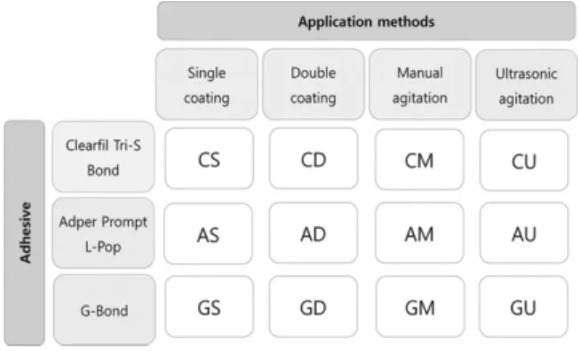
Then, an air stream was applied at a distance of 20 cm for 5 seconds. The groups used in this study according to the type of adhesive and application methods are presented in Figure 1.
Light emitting diode (LED) visible light-polymeriz- ing unit (Bluephase, Ivoclar vivadent, Schann, Liechtenstein) was used throughout the restorative procedure. Following the adhesive application, light- cure composite resin (Premise, Kerr, Orange, CA, USA) were constructed in 1.0 mm increments and light-cured for 20 seconds each. The height of total resin build up was approximately 5 mm. The restored teeth were then stored in distilled water at room temperature for 24 hours.
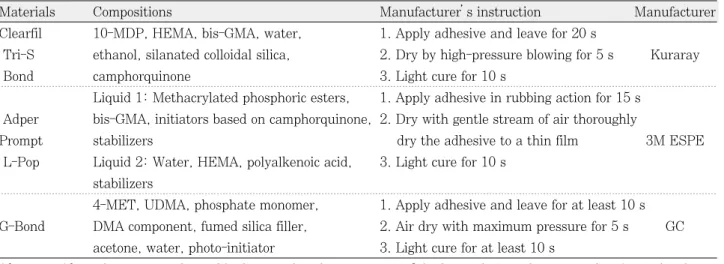
Table 1.The compositions of materials used in this study
Materials Compositions Manufacturer’s instruction Manufacturer
Clearfil 10-MDP, HEMA, bis-GMA, water, 1. Apply adhesive and leave for 20 s
Tri-S ethanol, silanated colloidal silica, 2. Dry by high-pressure blowing for 5 s Kuraray
Bond camphorquinone 3. Light cure for 10 s
Liquid 1: Methacrylated phosphoric esters, 1. Apply adhesive in rubbing action for 15 s Adper bis-GMA, initiators based on camphorquinone, 2. Dry with gentle stream of air thoroughly
Prompt stabilizers dry the adhesive to a thin film 3M ESPE
L-Pop Liquid 2: Water, HEMA, polyalkenoic acid, 3. Light cure for 10 s stabilizers
4-MET, UDMA, phosphate monomer, 1. Apply adhesive and leave for at least 10 s G-Bond DMA component, fumed silica filler, 2. Air dry with maximum pressure for 5 s GC
acetone, water, photo-initiator 3. Light cure for at least 10 s
10-MDP, 10-methacryloyloxydecyl dihydrogen phosphate; HEMA, 2-hydroxyethyl methacrylate; bis-GMA, bisphenol A-glycidyl methacrylate; 4-MET, 4-methacryloxyethyl trimellitic acid; UDMA, urethane dimethacrylate; DMA, dimethacrylate.
Microtensile bond strength testing
The restored teeth were longitudinally sectioned in both the “X”and “Y”directions across the bonded interface with a diamond saw (Accutom-50) under water cooling to obtain bonded sticks with a cross- sectional area of approximately 1.0 mm2. Three teeth were used per each group and we obtained 5 speci- mens per each tooth (n = 15). Every specimen was glued to the jig of custom-made microtensile testing machine manufactured from BISCO Inc. (BISCO, Schaumburg, IL, USA). The force was applied at a 1.0 mm/min cross head speed until bonding failure was occurred and the maximum load at failure was recorded.
Failure mode investigation
Fractured test specimens were examined using operating microscope (OPMI pico, Carl zeiss,
Obercohen, Germany) under 25 times magnification and the failure mode was classified as follows: adhe- sive, if the composite resin cone had fractured at the adhesive-tooth interface; cohesive, if the composite resin cone had fractured inside the composite resin or dentin; or mixed, a combination of adhesive and cohesive.
Statistical analysis
Statistical analysis of the microtensile bond strength between application methods were per- formed with two-way ANOVA and Tukey’s test was used for post-hoc multiple comparison. The level of significance was set at p < 0.05.
RESULTS Microtensile Bond Strength
The means and standard deviations of microtensile bond strength for all subgroups are shown in Table 2.
The result of two-way ANOVA indicates that man- ual agitation and ultrasonic agitation of adhesive sig- nificantly increased the microtensile bond strength than single coating and double coating did (p < 0.05).
Double coating of adhesive significantly increased the microtensile bond strength than single coating did (p
< 0.05). There was no significant difference between the manual agitation and ultrasonic agitation (p >
0.05). Among all adhesives, there was significant dif- ference in microtensile bonding strength and Clearfil Tri-S Bond showed the highest bond strength (p <
0.05).
Figure 1.Groups used in this study are noted according to the type of adhesive and application methods.
Table 2.Mean microtensile bond strength (MPa) and standard deviation (n = 15) Application methods
Single coatinga� Double coatingb Manual agitationc Ultrasonic agitationc p-values� Clearfil Tri-S Bond1� 17.97 ± 3.29 26.45 ± 3.03 26.31 ± 4.01 24.15 ± 2.64 α< 0.001 β< 0.001 Adper Prompt L-Pop2 10.33 ± 3.09 13.19 ± 2.75 23.56 ± 2.74 22.02 ± 2.58 α*β< 0.001
G-Bond3 17.99 ± 2.83 18.85 ± 2.14 23.22 ± 3.88 23.79 ± 3.86
�Statistically significant difference on application methods is shown by superscript lettersa,b,c, on adhesives by super- script number1,2,3. Same letters or numbers are not significantly different (p > 0.05). �On p-values, the letters α, β, and α*βdenote application methods, adhesives, and interaction of two factors, respectively.
Failure mode
Failure mode was presented in Table 3. Adhesive failure was predominantly observed in all groups.
DISCUSSION
The clinical success of dentin bonding may be affected by the structure and composition of the den- tal hard tissue and the contents of the adhesive material. The system of the adhesives (etch-and- rinse, self-etching/one bottle or two bottles) is one of the most important factors and the clinical procedure can also affect the bonding performance of adhesives.
The differences in clinical procedure are related to several factors, such as preparation of the dentinal surface, acid etching, drying of dentin, application methods (number of application, inactive/active application), removal of excess adhesive (compressed air or clean brush), and the use of different light sources. In the current study, we evaluated some of these factors, the effect of application methods of one-step self-etching adhesives on resin-dentin microtensile bond strength.
The first factor investigated in this study was the effect of double coating of adhesives. In the current
study, the double coating of adhesives significantly increased the bond strength than single coating did.
This result is in agreement with previous stud- ies.13,18,28 It is likely that the increased bond strength when multiple coatings are applied is due to several mechanisms operating simultaneously. As the first layers of the adhesive begins to etch the dentin sub- strate, it might become rapidly buffered by the hydroxiapatite,29 so that the additional layers of unpolymerized acidic monomers may improve the etching ability of these adhesives by increasing the concentration of acidic reagents. Simultaneously to this process, more impregnation of resin might occur by the additional supply of adhesive resin, as hypothesized by Ito et al..18 The solvent also evapo- rates between coats, thus the concentration of co- monomers that exist after each coat can be increased. Therefore, doubling the layer would facili- tate co-monomer infiltration with a further increase in the hybrid layer thickness.
In this study, we also used the manual pressure and ultrasonic vibration method for agitation applica- tion of adhesives. Previously, this application was suggested to improve the bonding strength on self- etching adhesives by some authors.19-21 According to previous research, the significantly higher bond strength in the agitation group could be related to better solvent evaporation and dispersion of water.30 They suggested that the effect of evaporation of the remaining solvent would have been apparent in the agitation group. Solvents in adhesives, such as ace- tone and ethanol, are necessary to dissolve both hydrophilic and hydrophobic monomers into one phase within the one-step self-etching solution,31but they may also help water to evaporate upon comple- tion of the self-etching process. Because remaining solvents may adversely affect adhesive performance that is decreasing polymerization efficacy and alter- ing the mechanical properties,32 it is important to evaporate solvent in one-step self-etching adhesives, and there are two ways to increase solvent evapora- tion from the primer mixture after its placement on the dentin surface: either by use of compressed air or by agitation application.
The use of compressed air can be highly variable, depending on how distant it is from the tooth sub- Table 3. The number of the specimen according to
the failure mode
Groups Adhesive Cohesive Mixed
failure failure failure
CS 12 2 1
CD 11 3 1
CM 14 1 0
CU 13 0 2
AS 14 1 0
AD 13 1 1
AM 14 0 1
AU 13 0 2
GS 12 2 1
GD 14 1 0
GM 15 0 0
GU 11 2 2
strate.30 This is why the distance from the dentin surface (20 cm) and the air-syringe was standard- ized in the current study. Although the sufficient evaporation of solvent is important, when the strong air-drying is used, water and solvents are evaporated quickly, resulting in a viscous resinous material with entrapped air bubbles remaining on the dentin sur- face.33 Also, this procedure reduces the thickness of the adhesive layer, making it more susceptible to polymerization inhibition by oxygen.34
On the other hands, the agitation application may speed up solvent evaporation, while at the same time causing a higher rate of monomer incorporation inside the smear layer. This method is likely to carry fresh acidic resin monomers to the basal part of the etched dentin, producing a more aggressive deminer- alization, facilitating diffusion of the monomers and promoting a better interaction with the smear layer and underlying dentin.19 And Reis et al. explained that the agitation method can improve the kinetics and allow for better monomer diffusion inward, while solvents are diffusing outward.27
From the smear layer point of view, with continu- ous agitation, smear layers were completely dis- persed or dissolved, and thicker hybrid layers with upstanding collagen fibrils were observed.20 Chan et al. found bond strength to dentin with a thick smear layer increased significantly with agitation and, on SEM evaluation, they noted passive application resulted in a hybridized smear layer, while agitation resulted in the smear layer being completely dis- solved or dispersed into the adhesive.20 The result of current study indicated that application with manual agitation of Clearfil Tri-S Bond, Adper Prompt L-Pop and G-Bond significantly increased the bonding strength than single and double coating and this is in agreement with previous research that demonstrated the effect of active agitation application on self-etch- ing adhesives.19-21
In some studies about the effect of the agitation methods, the ultrasonic agitation application was also used to improve the bonding strength of self- etching adhesives, but the ones that have evaluated the effect of ultrasonic agitation have reached contro- versial results.24,25,35This study demonstrated that the bonding strength of Clearfil Tri-S Bond, Adper
Prompt L-Pop and G-Bond to dentin increased sig- nificantly after ultrasonic agitation than single coat- ing and double coating.
Ultrasonic effect may facilitate the evaporation of water by increasing temperature which improves infiltration and possibly decomposes water to radicals35 and help the adhesive to flow into the free dentinal spaces more easily and facilitate tag forma- tion.25Additionally, the chemical change of the bond- ing agent after ultrasonic energy application, such as pH and related aggressiveness of the bonding agent, and possible temperature increase at the time of ultrasonic agitation with the ultrasonically assisted microstream effect of the bonding agent over a pro- longed time period also offer other possible reasons for the higher bond strength.35
But there was no significant difference between manual agitation and ultrasonic agitation on all adhesive systems. This result indicated the similarity of the ultrasonic effect to that of manual agitation methods of one-step self-etching adhesive. Therefore, manual or ultrasonic agitation is the recommended application method regardless of the adhesives.
Considering the findings of the current study, the manual agitation and ultrasonic agitation application were significantly effective than double coating in all adhesives. No matter of the material, application of manual or ultrasonic agitation may be recommended step for one-step self-etching adhesives. However, further studies are still required to extend the use of these methods to other one-step self-etching adhe- sives available on the market.
REFERENCES
1. Tay FR, Gwinnett AJ, Pang KM, Wei SH. Resin per- meation into acid-conditioned, moist, and dry dentin: a paradigm using water-free adhesive primers. J Dent Res 1996;75:1034-1044.
2. Watanabe I, Nakabayashi N, Pashley DH. Bonding to ground dentin by a phenyl-P self-etching primer. J Dent Res 1994;73:1212-1220.
3. Tay FR, Pashley DH, Yoshiyama M. Two modes of nanoleakage expression in single-step adhesives. J Dent Res 2002;81:472-476.
4. Tay FR, Pashley DH. Have dentin adhesives become too hydrophilic? J Can Dent Assoc 2003;69:726-731.
5. Osorio R, Toledano M, de Leonardi G, Tay F.
Microleakage and interfacial morphology of self-etching adhesives in class V resin composite restorations. J
Biomed Mater Res B Appl Biomater 2003;66:399-409.
6. Toledano M, Osorio R, de Leonardi G, Rosales-Leal JI, Ceballos L, Cabrerizo-Vilchez MA. Influence of self- etching primer on the resin adhesion to enamel and dentin. Am J Dent 2001;14:205-210.
7. Wang Y, Spencer P. Continuing etching of an all-in- one adhesive in wet dentin tubules. J Dent Res 2005;
84:350-354.
8. Tay FR, Pashley DH. Water treeing-a potential mecha- nism for degradation of dentin adhesives. Am J Dent 2003;16:6-12.
9. Carvalho RM, Chersoni S, Frankenberger R, Pashley DH, Prati C, Tay FR. A challenge to the conventional wisdom that simultaneous etching and resin infiltra- tion always occurs in self-etch adhesives. Biomaterials 2005;26:1035-1042.
10. Eliades G, Vougiouklakis G, Palaghias G. Heterogeneous distribution of single-bottle adhesive monomers in the resin-dentin interdiffusion zone. Dent Mater 2001;17:
277-283.
11. Nunes TG, Garcia FC, Osorio R, Carvalho R, Toledano M. Polymerization efficacy of simplified adhesive sys- tems studied by NMR and MRI techniques. Dent Mater 2006;22:963-972.
12. Tay FR, Pashley DH, Garcl′a-Godoy F, Yiu CK. Single- step, self-etch adhesives behave as permeable mem- branes after polymerization. Part II. Silver tracer pen- etration evidence. Am J Dent 2004;17:315-322.
13. Son SA, Hur B. The effect of multiple application on microtensile bond strength of all-in-one dentin adhe- sive systems. J Kor Acad Cons Dent 2004;29:423-429.
14. King NM, Tay FR, Pashley DH, Hashimoto M, Ito S, Brackett WW, Garcl′a-Godoy F, Sunico M. Conversion of one-step to two-step self-etch adhesives for improved efficacy and extended application. Am J Dent 2005;18:126-134.
15. Park JG, Cho KH, Cho YG. Effect of application meth- ods of a self-etching primer adhesive system on enamel bond strength. J Kor Acad Cons Dent 2008;33:90-97.
16. Nara Y, Nagakura Y, Ito T, Suzuki T, Kimishima T, Maseki T, Dogon IL. Bond strength of self-etching adhesives to cervical lesion dentin. J Dent Res 2004;83(Special Issue B):Abstract #0445.
17. Hashimoto M, Sano H, Yoshida E, Hori M, Kaga M, Oguchi H, Pashley DH. Effects of multiple adhesive coatings on dentin bonding. Oper Dent 2004;29:416- 423.
18. Ito S, Tay FR, Hashimoto M, Yoshiyama M, Saito T, Brackett WW, Waller JL, Pashley DH. Effects of mul- tiple coatings of two all-in-one adhesives on dentin bonding. J Adhes Dent 2005;7:133-141.
19. Miyazaki M, Hinoura K, Kanamaru T, Koga K, Iwauchi H, Onose H. Study on light cured composite resin-influence of pretreated procedures for dentin sur- faces on shear bond strength to dentin. Japan J Cons Dent 1991;34:734-741.
20. Chan KM, Tay FR, King NM, Imazato S, Pashley DH.
Bonding of mild self-etching primers/adhesives to dentin with thick smear layers. Am J Dent 2003;16:
340-346.
21. Velasquez LM, Sergent RS, Burgess JO, Mercante DE.
Effect of placement agitation and placement time on the shear bond strength of 3 self-etching adhesives.
Oper Dent 2006;31:426-430.
22. Lee J, Jang KT, Kim JW, Lee SH, Hahn SH, Kim CC.
Effect of ultrasonic vibration on dentin bond strength and resin infiltration. Am J Dent 2003;16:404-408.
23. Kim JW, Jang KT, Lee SH, Kim CC, Hahn SH. Effect of ultrasonicvibration on resin-dentin bonding: SEM study. J Dent Res 2002;81:Abstract #1905, pA-248.
24. Finger WJ, Tani C. Effect of application mode on bond- ing performance of self-etching adhesives. Am J Dent 2005;18:41-44.
25. Bagis B, Turkarslan S, Tezvergil-Mutluay A, Uctasli S, Vallittu PK, Lassila LV. Effect of ultrasonic agitation on bond strength of self-etching adhesives to dentin.J Adhes Dent 2008;10:441-445.
26. Dal-Bianco K, Pellizzaro A, Patzlaft R, de Oliveira Bauer JR, Loguercio AD, Reis A. Effects of moisture degree and rubbing action on the immediate resin- dentin bond strength. Dent Mater 2006;22:1150-1156.
27. Reis A, Pellizzaro A, Dal-Bianco K, Gones OM, Patzlaff R, Loguercio AD. Impact of adhesive applica- tion to wet and dry dentin on long-term resin-dentin bond strengths. Oper Dent 2007;32:380-387.
28. Toledano M, Proença JP, Erhardt MC, Osorio E, Aguilera FS, Osorio R, Tay FR. Increases in dentin- bond strength if doubling application time of an ace- tone-containing one-step adhesive. Oper Dent 2007;
32:133-137.
29. Camps J, Pashley DH. Buffering action of human dentin in vitro. J Adhes Dent 2000;2:39-50.
30. Tewari S, Goel A. Effect of placement agitation and drying time on dentin shear bond strength: anin vivo study. Oper Dent 2009;34:524-530.
31. Furukawa M, Shigetani Y, Finger WJ, Hoffmann M, Kanehira M, Endo T, Komatsu M. All-in-one self-etch model adhesives: HEMA-free and without phase sepa- ration. J Dent 2008;36:402-408.
32. Park SI, Kim YM, Roh BD. The effect of dentin bond- ing agent thickness on stress distribution of composite- tooth interface : Finite element method. J Kor Acad Cons Dent 2009;34:442-449.
33. Shinkai K, Suzuki S, Katoh Y. Effect of air-blowing variables on bond strength of all-in-one adhesives to bovine dentin. Dent Mater J 2006;25:664-668.
34. Rueggeberg FA, Margeson DH. The effect of oxygen inhibition on an unfilled/filled composite system. J Dent Res 1990;69:1652-1658.
35. Bagis B, Turkaslan S, Vallittu PK, Lassila LV. Effect of high frequency ultrasonic agitation on the bond strength of self-etching adhesives. J Adhes Dent 2009;
11:369-374.
국문초록
한 단계 자가 산부식 접착제의 적용 방식이 미세인장 결합강도에 미치는 효과
최철규1∙손성애1∙하진희1∙허 복1∙김현철1∙권용훈2∙박정길1*
부산대학교 치의학전문대학원1치과보존학교실, 2치과재료학교실
연구목적: 이 연구의 목적은 다양한 방식의 한 단계 자가 부식 접착제의 적용이 직접 복합 레진 수복의 미세 인장 강도에 미 치는 영향을 평가하는 것이었다.
연구 재료 및 방법: 36개의 발거된 대구치를 사용하여, 3종류의 한 단계 자가 부식 접착제(Clearfil Tri-S Bond, Adper Prompt L-Pop, G-Bond)와 적용 방식에 따라 12개의 군으로 나누었다. 접착제를 다음의 방식으로 상아질에 적용하였다.
1) single coating, 2) double coating, 3) manual agitation, 4) ultrasonic agitation. 접착제 적용 후, 광중합 복합 레진 으로 수복하였다. 24시간 동안 실온의 증류수에 보관한 후, 각 군당 15개의 시편을 준비하여 미세인장 결합 강도를 측정하였다.
결과: Manual agitation, ultrasonic agitation군은 single coating, double coating군에 비해 유의하게 높은 미세인장 결 합 강도를 보였다. Double coating군은 single coating군에 비해 유의하게 높은 미세인장 결합 강도를 보였으며, manual agitation군과 ultrasonic agitation군 간에는 미세인장 결합 강도의 유의한 차이가 없었다. 접착제 종류에 따라 미세인장 결 합 강도의 유의한 차이가 있었고, 그 중 Clearfil Tri-S Bond가 가장 높았다.
결론: 한 단계 자가 산부식 접착제의 적용 방법과 재료에 따라 미세인장 결합 강도에 유의한 차이가 있었다. 접착제 종류에 관계없이 manual agitation과 ultrasonic agitation은 유의하게 높은 미세인장 결합강도를 보였다.
주요단어: 미세인장 결합강도; 접착제 적용 방식; 한 단계 자가 산부식 접착제