ORIGINAL ARTICLE J Cardiovasc Ultrasound 2017;25(2):47-56
• Received: January 5, 2017 • Revised: May 9, 2017 • Accepted: May 23, 2017
• Address for Correspondence: Sung-Ji Park, Division of Cardiology, Department of Internal Medicine, Cardiovascular Imaging Center, Heart Vascular Stroke Institute, Samsung Medical Center, Sungkyunkwan University School of Medicine, 81 Irwon-ro, Gangnam-gu, Seoul 06351, Korea Tel: +82-2-3410-0887, Fax: +82-2-3410-3849, E-mail: tyche.park@gmail.com
• This is an Open Access article distributed under the terms of the Creative Commons Attribution Non-Commercial License (http://creativecommons.org/licenses/by-nc/4.0) which permits unrestricted non-commercial use, distribution, and reproduction in any medium, provided the original work is properly cited.
Effects of Decreased Annular Height and Annular Saddle-Shaped Non-Planarity in Degenerative Severe Mitral
Regurgitation with Normal Left
Ventricular Ejection Fraction: Real-Time 3D Transesophageal Echocardiography
Eun Jeong Cho, MD, PhD1,2, Sung-Ji Park, MD, PhD1, Ga Yeon Lee, MD, PhD1, Eun Kyoung Kim, MD, PhD1, Sung-A Chang, MD, PhD1, Jin-Oh Choi, MD, PhD1,
Sang-Chol Lee, MD, PhD1, Seung Woo Park, MD, PhD1, and Pyo Won Park, MD, PhD3
1Division of Cardiology, Department of Internal Medicine, Cardiovascular Imaging Center,
Heart Vascular Stroke Institute, Samsung Medical Center, Sungkyunkwan University School of Medicine, Seoul, Korea
2Division of Cardiology, Department of Internal Medicine, National Cancer Center, Goyang, Korea
3Department of Thoracic Surgery, Heart Vascular Stroke Institute, Samsung Medical Center, Sungkyunkwan University School of Medicine, Seoul, Korea
Background: The extent of mitral annular (MA) remodeling and dysfunction is correlated with the severity of mitral regurgi- tation (MR) as well as left atrial (LA) and left ventricular (LV) dilation. MA dysfunction may be a useful prognostic factor for opera- tive timing and MR recurrence after successful mitral valve (MV) repair. The aim of this study was to evaluate additive prognostic factors of MA non-planarity using real-time 3D transesophageal echocardiography (RT3D-TEE) analysis in patients with chronic severe MR and preserved LV systolic function.
Methods: Forty-seven patients with chronic severe MR and preserved LV systolic function scheduled for MV repair were prospec- tively enrolled. Echocardiographic studies were performed before surgery and postoperatively within 2 weeks and at least 6 months after surgery. RT3D-TEE was performed before the operation and immediately post-operative.
Results: Mean age was 55.4 ± 15.1 years and 24 were male. Annulus height/body surface area (BSA) obtained via RT3D-TEE was correlated with the degree of postoperative LA remodeling. Patients were divided into two groups by average baseline annu- lus height/BSA. Patients with normal annular height had a smaller postoperative LV end-diastolic dimension, LV end-systolic di- mension and LA volume index than patients with decreased annular height. Preoperative annulus height/BSA values strongly pre- dicted postoperative LA remodeling.
Conclusion: MA height may be a useful prognostic factor for determining the timing of surgery in patients with chronic pri- mary MR. Annulus height/BSA assessed via RT3D-TEE may provide additional information predictive of postoperative LA re- modeling after successful MV repair.
KEY WORDS: Mitral regurgitation · Mitral annular · Annulus height · Real-time 3D transesophageal echocardiography.
Introduction
Degenerative mitral regurgitation (MR) is the most common form of organic MR disease in developed countries.1) Surgical repair of the mitral valve (MV) remains the best treatment op- tion for patients with severe degenerative MR,2) which is asso- ciated with good long-term follow-up.3) However, degenerative MR disease still carries a sizable risk of recurrent MR after sur- gery. This seems to be related to the progression of left ven- tricular (LV) and mitral annular (MA) disease.4) Both left atrial (LA) enlargement and remodeling are compensatory mecha- nisms in patients with severe MR. In patients with severe MR, LA size is an important predictor of outcome in both conserva- tive treatment and after MV surgery.5)6) Moreover, one of the predictors of successful MV repair is the extent of MA disease.7) A previous study using a Laplace’s law model indicated that curvature of the leaflets is a beneficial feature. Thus, saddle shape preservation decreases leaflet and annular strain and also in- creases leaflet coaptation.8)9) In another study, a late systolic de- crease in MA non-planarity can additionally increase leaflet stress in patients with MR, resulting in elongation and secondary chordae rupture.9) Consequently, the extent of MA remodeling and dysfunction is thought to be correlated with MR severity.
MA remodeling and contractile dysfunction are also associated with LA dilation and LV dilation. Generally, a decrease in LA size, or reverse remodeling, has been observed after MV surgery.
LA reverse remodeling occur after surgery of MR with preserved LV ejection fraction (LVEF).10) One previous study concluded that MA dysfunction might be a prognostic factor for increased repair durability and MR recurrence after successful MV repair.11) Therefore, thorough assessment of MA geometry and function as well as of the determinants of MA remodeling are pivotal to understanding the pathophysiology and severity of MR, and to planning effective reparative surgery.
Two-dimensional echocardiography (2DE) guides the timing of surgical intervention by providing information on MR sever- ity, monitoring LV and LA remodeling, and estimating pulmo- nary artery systolic pressure.12) Currently, the only recommen- dation for the use of echocardiography in MA assessment before surgery is for the measurement of its anteroposterior diameter on LV 2DE long axis view.13) In addition, MA quantitative as- sessment that completely changed our way of assessing MA shape and function by the advent of 3-dimensional echocardiog- raphy has been used for a better understanding of MR patho- physiology,14) and on following up patients.15) However, the dy- namics of the MA in MR remains controversial.16) The relationship between the function of the MA and MR severity has not been reported, and the connection between MA remodeling and left heart chamber size and function remains to be defined. The aim of this study was to identify additive prognostic factors that could be used for timing surgical assessment of MA non-planar- ity using real-time 3D transesophageal echocardiography (RT3D- TEE) in patients with severe chronic primary MR and preserved LV systolic function.
Methods
Patient selection
Forty-seven patients with severe chronic primary MR and preserved LVEF (> 60%) scheduled for MV repair were en- rolled between March 2010 and March 2012. Serial 2D TTEs were performed before surgery, 2 weeks after surgery and at least 6 months postoperatively. RT3D-TEE was also performed just before operation and in the operating room immediately post-operative. The severity of MR was determined via inte- grated echocardiography evaluation using the following mea- surements: LV dimension, effective regurgitant orifice (ERO) and regurgitant volume. Severe MR was defined as an ERO > 40 mm or a regurgitant volume > 60 mL.17)18) Exclusion criteria included secondary MR due to either a distortion of the sub- valvular apparatus or LV enlargement and remodeling (idio- pathic cardiomyopathy or ischemic heart disease); other con- comitant valvular disease of moderate or severe severity; coro- nary artery disease (defined as > 50% narrowing in at least one coronary artery in a previous angiogram); history of myocardial infarction; coronary artery bypass graft; acute coronary syndrome;
atrial fibrillation; chronic renal failure or suboptimal imaging.
The regional ethics committee approved the study. The pa- tients provided informed consent prior to enrollment.
Two-dimensional transthoracic echocardiography
2DE was performed using commercially available equipment (Vivid 9; GE Medical Systems, Milwaukee, WI, USA). End di- astole was defined as the frame with the largest cavity area im- mediately before the onset of QRS, while end systole was the frame with the smallest cavity area. The LV end-diastolic di- mension (LVEDD), LV end-systolic dimension (LVESD), dia- stolic interventricular septum thickness and diastolic LV pos- terior wall thickness were all obtained from the parasternal views, according to standard guidelines.19) LV mass was calcu- lated from the linear dimensions using the American Society of Echocardiography recommended formula. LA volume was measured at end-systole from the frame just preceding MV opening using the biplane area length method in the apical 4- and 2-chamber views, and was indexed to body surface area (BSA).19) LV diastolic function was assessed by the early (E wave) and late (A wave) transmitral velocities, the correspond- ing E/A ratio, and the E wave deceleration time using pulsed- wave Doppler. Tissue Doppler imaging was used to measure peak early diastolic MA velocity (e’) at the septal mitral annulus in the apical 4-chamber view. An E/e’ ratio was calculated to noninvasively estimate LV filling pressure.20) We used the aver- age of three consecutive Doppler signals to take these measure- ments.
Quantitative and qualitative measures of MR severity were taken according to the American Society of Echocardiography guidelines.18) MR regurgitant volume was calculated using the
proximal isovelocity surface area (PISA) method. The ERO area was determined by dividing the regurgitant flow rate (calcu- lated as 2 πr2 × aliasing velocity, where r is the PISA radius) by the peak MR velocity.12)
Real-time three-dimensional transesophageal echocardiography
The RT3D-TEE images of the MV were obtained in full-vol- ume mode, in which electrocardiographically triggered wedge- shaped subvolumes were obtained over seven consecutive car- diac cycles using an iE33 system equipped with a matrix probe (X7-2t; Philips Medical Systems, Andover, MA, USA). And, the RT3D-TEE images of patients with atrial fibrillation were ob- tained during a single cardiac cycle.
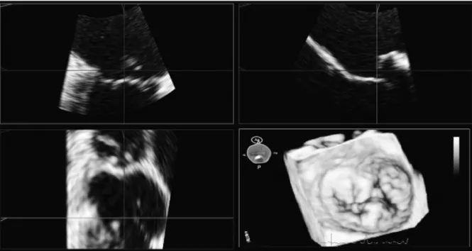
Full-volume 3D data sets were digitally stored and transferred to a workstation with Q-Laboratory Mitral-Valve-Quantifica- tion Software (Philips Medical Systems, Bothell, WA, USA) for offline analysis. Three orthogonal mitral annulus images were displayed and subsequently modified to optimize visualization of the entire annulus (Fig. 1). MA measurements were performed six times during the cardiac cycle in early, middle, and late dias- tole and early, middle, and late systole. Measurements of annu- lar diameter, annular perimeter, annular area and annular height were performed during late systole. Early diastole was identified with MV opening, late diastole before mitral closure, and middle diastole as midway between these frames. Early systo- le was identified just before mitral closure, late systole on the frame preceding aortic closure, and middle systole midway be- tween these frames.
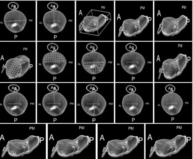
The resulting 3D representation was adjusted to visually match the anatomy viewed in 3D and 2D cut-plane views. From this model, several parameters were calculated (Fig. 2): 1) annular geometry: annular area, as the area of the minimal surface span-
ning the annulus and the anteroposterior, anterolateral and posteromedial diameters; annular height, as the distance along the atrial-ventricular direction between the lowest and highest point of the annulus; and the planarity index, defined as the ra- tio of the height to anterolateral-posteromedial diameter, equal to zero for a flat MV and increasing when the MV’s saddle shape was more pronounced; 2) leaflet size: exposed 3D area of the anterior or posterior leaflet, as well as the 3D total exposed leaf- let area, as the sum of the two previous measurements; 3) coap- tation geometry: the length of the coaptation line projected to approximate the leaflet surface, the area of the region where the anterior and posterior leaflets overlap and the mean height of the same region; and 4) the aortic-to-mitral plane angle.
Measurement variability was determined by repeating the measurements on stored 3D data sets at least 1 week after the initial measurements by the same observer (intraobserver) and a different observer (interobserver).
The average value of baseline annulus height/BSA was 4.
And, we defined definitions of decreased annular height/BSA to be less than 4.
Follow-up
Data were obtained until August 2012 (mean follow-up du- ration: 5.43 months) within 2 weeks after regular outpatient visits and at least 6 months postoperatively. Particular care was taken to obtain information regarding the development of symp- toms, eventual MV repair or replacement, and deterioration of LV function.
Statistical analyses
Continuous variables are listed as mean values. Categorical variables are presented as frequencies and group percentages.
Continuous variables were compared using the Student’s t-test.
Fig. 1. Screenshot of the Mitral-Valve-Quantification software showing the volume-rendered 3D data set (bottom right) as well as the three cut planes used to improve the visualization of the mitral valve. Ao: aorta, A: anterior, P: posterior, AL: anterolateral, PM: posteromedial.
The chi-square or Fisher’s exact test was used for comparison of categorical variables. A 2-tailed p value < 0.05 was consid- ered statistically significant. Pearson’s correlation coefficient (r) and intraclass correlation coefficient (ICC) were calculated to express agreement between LA volume index (LAVI) and the annular height/BSA obtained using RT3D-TEE. Interobserver agreement was demonstrated by calculating the coefficient of variation of repeated measurements and the ICC. All p values
< 0.05 were considered significant. Data analysis was performed utilizing SPSS version 18.0 (SPSS Inc., Chicago, IL, USA).
Results
Pre-LAVI, delta LAVI (pre-post) and annular height/BSA on RT3D-TEE correlation
Mean patient age was 55.4 ± 15.1 years and 24 (51.1%) were male. The annular height/BSA obtained from RT3D-TEE was correlated with delta LAVI (pre-post) [r = 0.519, p < 0.001, 95% confidence interval (CI) 0.273–0.702] (Fig. 3).
Baseline characteristics and echocardiography in severe MR with decreased or normal annular height/BSA
The study subjects were then divided into 2 groups based on average annular height/BSA: normal annular height/BSA and decreased annular height/BSA. Baseline characteristics and echocardiographic parameters are shown in Table 1 and 2. None of the baseline characteristics except for sex and BSA were sig- nificantly different when comparing the normal and decreased annular height/BSA groups. The decreased annular height/
BSA group had a higher number of men than normal annular height/BSA group (p = 0.017). Additionally, the decreased an- nular height/BSA group had a higher BSA than the normal annular height/BSA group (p = 0.003). Echocardiography pa- rameters of the normal and decreased annular height/BSA groups were also compared and were not significantly differ- ent between the two groups.
Analysis of postoperative echocardiography After at least 6 months of postoperative follow-up, patients
Fig. 2. Three-dimensional reconstruction of the mitral valve, from which several parameters were automatically calculated. From top to bottom, left to right: anterolateral to posteromedial diameter of annulus; anterior to posterior diameter of annulus; mitral annular height, defined as the height of the bounding box of the mitral valve in the atrial-ventricular direction; maximal prolapse height; maximal tenting height; area of annulus in projection plane; exposed area of anterior leaflet; exposed area of posterior leaflet; perimeter of annulus; aortic orifice to mitral plane angle; length of coaptation in projection plane; exposed length of A2; exposed length of P2; volume of leaflet prolapse; volume of the leaflets tent; angle of anterior leaflet; non- planar angle of leaflets; angle of posterior leaflet; annular height to commissural width ratio. Ao: aorta, A: anterior, P: posterior, AL: anterolateral, PM:
posteromedial.
with decreased annular height/BSA had larger LVEDDs and LVESDs than patients with normal annular height/BSA (p = 0.002 and 0.038). Patients in the decreased annular height/
BSA group also had higher LAVIs than patients in the normal annular height/BSA group (p = 0.047).
In addition, patients with decreased annular height/BSA had larger pre-post2 LVEDDs and pre-post2 LAVIs than pa- tients with normal annular height/BSA (p = 0.045 and 0.040) (Table 3).
Real-time 3D transesophageal echocardiography analysis
None of the preoperative baseline characteristics except for annular height to commissural width ratio (AHCWR) were significantly different when the normal and decreased annular
height/BSA groups were compared. The decreased annular height/BSA group had a smaller AHCWR than the normal an- nular height/BSA group (p < 0.001) (Table 4).
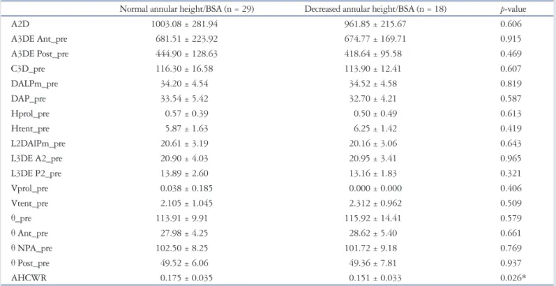
Evaluation of the postoperative RT3D-TEE results revealed that none of the parameters aside from the AHCWR were sig- nificantly different in the normal and decreased annular height/
BSA groups. The decreased annular height/BSA group had a smaller AHCWR than the normal annular height/BSA group (p = 0.026) (Table 5).
Independent predictors of postoperative LA remodeling in patients with severe MR
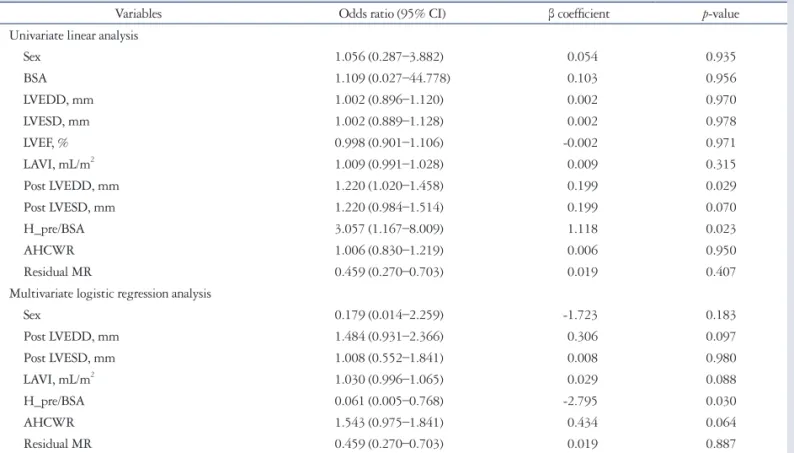
Univariate linear analysis revealed that the preoperative an- nular height/BSA (95% CI, 1.167–8.009; p = 0.023) indepen- dently predicted postoperative LA remodeling (postoperative LAVI reduction of less than 34 mL/m2). Furthermore, post- operative LVEDD (95% CI, 1.020–1.458; p = 0.029) was also independently predictive of postoperative LA remodeling (postoperative LAVI reduction of less than 34 mL/m2).
In multivariable analysis, preoperative annular height/BSA (95% CI, 0.005–0.768; p = 0.030) independently predicted postoperative LA remodeling (postoperative LAVI reduction of less than 34 mL/m2) (Table 6). To summarize, we found that LA reverse remodeling occurs frequently after surgery for severe MR with preserved LVEF, and that preoperative annulus height is the only variable significantly associated with a postoperative LAVI reduction of less than 34 mL/m2.
Observer agreement
ICC was used to determine intra- and inter-rater reliability with 95% CIs. The ICC showed excellent intra- and inter-rater correlations for RT3D-TEE measurements (intra-rater, ICC 0.942, 95% CI, 0.879–0.973; inter-rater, ICC 0.927, 95% CI, 0.846–0.965).
Fig. 3. Scatter diagram with Pearson’s correlation of the annular height/BSA by RT3D-TEE and the degree of LAVI decrease between echocardiography obtained at baseline and at least 6 months postoperatively. LAVI: left atrial volume index, BSA: body surface area, RT3D-TEE: real-time 3D transesophageal echocardiography.
250
200
150
100
50
0
-50
2 4 6 8 10
The annular height/BSA
r = 0.519 p < 0.001
Degree of decrease of LAVI
Table 1. Baseline characteristics of patients with severe mitral regurgitation and decreased or normal annular height/BSA
Normal annular height/BSA (n = 29) Decreased annular height/BSA (n = 18) p-value
Age, years 58.40 ± 13.98 51.56 ± 15.91 0.126
Sex, M/F 11/18 13/5 0.017*
BSA, m2 1.60 ± 0.15 1.76 ± 0.19 0.003*
Systolic BP, mm Hg 118.43 ± 16.35 117.67 ± 17.04 0.878
Diastolic BP, mm Hg 72.90 ± 9.61 69.22 ± 9.60 0.206
HR 77.03 ± 13.15 75.72 ± 15.65 0.757
HTN, % 14 (46.7) 10 (55.6) 0.551
DM, % 5 (16.7) 3 (16.7) 1.000
Dyslipidemia, % 7 (23.3) 5 (27.8) 0.733
Atrial fibrillation, % 6 (20.7) 8 (44.4) 0.071
Annuloplasty ring size, mm 28.90 ± 1.37 29.73 ± 1.83 0.095
Data are listed as mean value (percentage). The p value denotes statistical significance when comparing the non-remodeling and remodeling groups. *p < 0.05 on Student’s t-test (continuous variables) or chi-square test (categorical variables). BSA: body surface area, BP: blood pressure, HR: heart rate, HTN: hyperten- sion, DM: diabetes mellitus
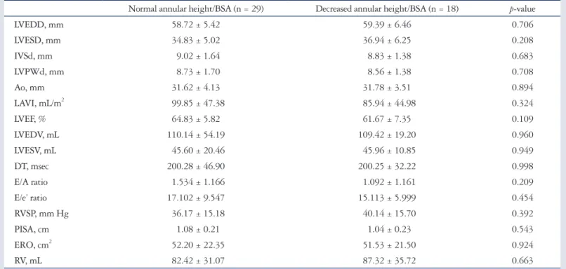
Table 2. Preoperative echocardiographic parameters in severe mitral regurgitation with decreased or normal annular height/BSA Normal annular height/BSA (n = 29) Decreased annular height/BSA (n = 18) p-value
LVEDD, mm 58.72 ± 5.42 59.39 ± 6.46 0.706
LVESD, mm 34.83 ± 5.02 36.94 ± 6.25 0.208
IVSd, mm 9.02 ± 1.64 8.83 ± 1.38 0.683
LVPWd, mm 8.73 ± 1.70 8.56 ± 1.38 0.708
Ao, mm 31.62 ± 4.13 31.78 ± 3.51 0.894
LAVI, mL/m2 99.85 ± 47.38 85.94 ± 44.98 0.324
LVEF, % 64.83 ± 5.82 61.67 ± 7.35 0.109
LVEDV, mL 110.14 ± 54.19 109.42 ± 19.20 0.960
LVESV, mL 45.60 ± 20.46 45.96 ± 10.85 0.949
DT, msec 200.28 ± 46.90 200.25 ± 32.22 0.998
E/A ratio 1.534 ± 1.166 1.092 ± 1.161 0.209
E/e’ ratio 17.102 ± 9.547 15.113 ± 5.999 0.454
RVSP, mm Hg 36.17 ± 15.18 40.14 ± 15.70 0.392
PISA, cm 1.08 ± 0.21 1.04 ± 0.23 0.543
ERO, cm2 52.20 ± 22.35 51.53 ± 21.50 0.924
RV, mL 82.42 ± 31.07 87.32 ± 35.72 0.663
Data are listed as mean value (percentage). The p value denotes statistical significance when comparing the non-remodeling and remodeling groups. p < 0.05 on Student’s t-test (continuous variables) or chi-square test (categorical variables). BSA: body surface area, LVEDD: left ventricular end-diastolic dimension, LVESD: left ventricular end-systolic dimension, IVSd: diastolic interventricular septum thickness, LVPWd: diastolic left ventricular posterior wall thickness, Ao: aorta, LAVI: left atrial volume index, LVEF: left ventricular ejection fraction, LVEDV: left ventricular end-diastolic volume, LVESV: left ventricular end- systolic volume, DT: deceleration time, RVSP: right ventricular systolic pressure, PISA: proximal isovelocity surface area, ERO: effective regurgitant orifice, RV: regurgitant volume
Table 3. Postoperative echocardiographic parameters in severe MR with decreased or normal annular height/BSA
Normal annular height/BSA (n = 29) Decreased annular height/BSA (n = 18) p-value
LVEDD, mm 47.67 ± 3.89 51.78 ± 4.89 0.002*
LVESD, mm 29.67 ± 3.36 32.78 ± 6.75 0.038*
IVSd, mm 8.93 ± 1.44 9.28 ± 1.67 0.454
LVPWd, mm 8.53 ± 1.20 8.89 ± 1.49 0.368
Ao, mm 31.70 ± 3.99 32.44 ± 4.12 0.540
LAVI, mL/m2 42.56 ± 12.30 52.49 ± 21.47 0.047*
LVEF, % 61.70 ± 6.10 60.72 ± 9.15 0.659
DT, msec 315.86 ± 84.56 314.82 ± 70.82 0.966
E/A ratio 1.095 ± 0.663 1.486 ± 0.923 0.145
E/e’ ratio 19.871 ± 5.331 22.444 ± 8.216 0.205
RVSP, mm Hg 25.23 ± 6.20 24.08 ± 5.98 0.537
Residual MR 0.173
Minimal, % 21 (72.4) 16 (88.9)
Mild, % 8 (27.6) 2 (11.1)
Pre-post
LVIDd (pre-post) 10.97 ± 5.91 7.61 ± 4.51 0.045*
LVIDs (pre-post) 5.14 ± 5.05 4.17 ± 4.20 0.499
LVEF (pre-post) 3.00 ± 6.97 0.94 ± 8.56 0.373
LAVI (pre-post) 57.05 ± 47.46 33.45 ± 28.99 0.040*
Data are listed as mean value (percentage). The p value denotes statistical significance when comparing non-remodeling and remodeling groups. *p < 0.05 on Student’s t-test (continuous variables) or chi-square test (categorical variables). BSA: body surface area, LVEDD : left ventricular end-diastolic dimension, LVESD: left ventricular end-systolic dimension, IVSd: diastolic interventricular septum thickness, LVPWd: diastolic left ventricular posterior wall thickness, Ao: aorta, LAVI: left atrial volume index, LVEF: left ventricular ejection fraction, DT: deceleration time, RVSP: right ventricular systolic pressure, MR: mitral regurgitation, pre-post: difference between echocardiography parameters at baseline and at least 6 months post-operation
Discussion
The main findings of our study of patients with severe degen- erative MR and preserved LVEF were: 1) after at least 6 months of postoperative follow-up, patients with the decreased annular height/BSA had larger LVEDDs and LVESDs than patients with normal annular height/BSA. Patients with decreased annular height/BSA had larger LAVIs, pre-post LVEDDs, and pre-post LAVIs than those with normal annular height/BSA. 2) The de- creased annular height/BSA group had a lower AHCWR than the normal annular height/BSA group. 3) LA reverse remodel- ing occurs after surgery for severe degenerative MR with pre- served LVEF, and preoperative annulus height/BSA was the only variable significantly associated with postoperative LAVI reduction less than 34 mL/m2.
LAVI changes and postoperative LA remodeling or dysfunction in patients with severe
degenerative MR and preserved LVEF
Both LA enlargement and remodeling are compensatory mechanisms in patients with severe MR. In patients with se- vere MR, LA size seems to be an important predictor of out- comes after conservative treatment or MV surgery.5)6)21) In a pre- vious study, Grewal et al.22) showed that the patients with MR
present a loss of early MA contraction, despite the same mag- nitude of LV contraction, and suggested ventriculo-annular de- coupling. Another study suggested that MA size and reduced fractional area shortening were more related to LA rather than to LV size and dysfunction, at least in cases of degenerative se- vere MR with preserved LVEF.22) A decrease in LA size, or re- verse remodeling, has been observed after MV surgery.10) In the present study, the LAVI obtained using echocardiography at least 6 months postoperatively was smaller in the normal an- nular height/BSA group than in the decreased annular height/
BSA group. Moreover, the degree of LAVI decrease between baseline and at least 6 months postoperatively was greater in the normal annular height/BSA group. Thus, annular height/BSA might be an additive predictive factor for postoperative LA dysfunction or remodeling. In this way, assessment of MA ge- ometry and function and the determinants of MA remodeling in MR have become pivotal to understanding the pathophysi- ology of severe MR and to planning effective reparative surgery.
Early detection of postoperative LA remodeling or dysfunction in patients with severe
degenerative MR and preserved LVEF
Surgical intervention for severe MR is usually triggered by the
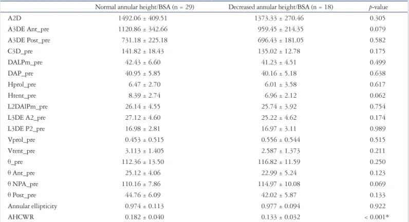
Table 4. Preoperative RT3D-TEE parameters in severe mitral regurgitation with decreased or normal annular height/BSA at baseline on 3D echo- cardiography
Normal annular height/BSA (n = 29) Decreased annular height/BSA (n = 18) p-value
A2D 1492.06 ± 409.51 1373.33 ± 270.46 0.305
A3DE Ant_pre 1120.86 ± 342.66 959.45 ± 214.35 0.079
A3DE Post_pre 731.18 ± 225.18 696.43 ± 181.05 0.582
C3D_pre 141.82 ± 18.43 135.02 ± 12.78 0.175
DALPm_pre 42.43 ± 6.60 41.23 ± 4.51 0.499
DAP_pre 40.95 ± 5.85 40.16 ± 5.18 0.638
Hprol_pre 6.47 ± 2.70 6.01 ± 3.58 0.617
Htent_pre 8.39 ± 2.74 6.96 ± 2.12 0.062
L2DAlPm_pre 26.14 ± 4.55 25.74 ± 3.92 0.754
L3DE A2_pre 27.12 ± 4.60 25.22 ± 4.62 0.174
L3DE P2_pre 16.98 ± 2.81 16.97 ± 3.11 0.989
Vprol_pre 0.453 ± 0.515 0.556 ± 0.544 0.515
Vtent_pre 3.113 ± 1.405 2.587 ± 1.373 0.211
θ_pre 112.36 ± 13.50 116.82 ± 11.59 0.250
θ Ant_pre 25.12 ± 4.06 22.99 ± 5.24 0.123
θ NPA_pre 110.16 ± 7.86 114.97 ± 10.08 0.069
θ Post_pre 44.76 ± 6.09 42.02 ± 5.87 0.133
Annular ellipticity 0.974 ± 0.113 0.977 ± 0.094 0.922
AHCWR 0.182 ± 0.040 0.133 ± 0.032 < 0.001*
Data are listed as mean value (percentage). The p value denotes statistical significance when comparing non-remodeling and remodeling groups. *p < 0.05 on Student’s t-test (continuous variables), chi-square test (categorical variables). RT3D-TEE: real-time 3D transesophageal echocardiography, BSA: body surface area, A2D: area of annulus in projection plane, A3DE Ant_pre: exposed area of anterior leaflet, A3DE Post_pre: exposed area of the posterior leaflet, C3D_pre:
perimeter of the annulus, DALPm_pre: anterolateral to posteromedial diameter of the annulus = intercommissural diameter, DAP_pre: anterior to posterior diameter of the annulus, Hprol_pre: maximal prolapse height, Htent_pre: maximal tenting height, L2DAlPm_pre: length of coaptation in the projection plane, L3DE A2_pre: exposed length of A2, L3DE P2_pre: exposed length of P2, Vprol_pre: volume of leaflet prolapse, Vtent_pre: volume of the leaflet tent, θ_pre: aortic orifice to mitral plane angle, θ Ant_pre: angle of the anterior leaflet, θ NPA_pre: non-planar angle of leaflets, θ Post_pre: angle of the posterior leaflet, AHCWR: annular height to commissural width ratio
occurrence of symptoms, declining LV function, significant LV enlargement, or by the development of atrial fibrillation or severe pulmonary hypertension.23) The extent of MA remodel- ing and dysfunction has recently been correlated with the se- verity of MR. A previous study found lower MA displacement in patients with MR than in normal patients.16) Unique annu- lar shape is considered to be important in reducing leaflet stress and enhancing valve competence during systole.24)
A previous study using Laplace’s law model indicated that curvature of the leaflets is a beneficial feature. Saddle shape preservation decreases leaflet and annular strain while increasing leaflet coaptation.8)9) In another study, MA non-planarity was re- ported to have a role in reducing MV leaflet stress.24) Late systol- ic decrease in MA non-planarity can additionally increase leaflet stress in patients with MR, causing elongation and secondary chordae rupture.9) As expected, MV tenting height and volume decreased from mid- to end-systole in patients with MR due to progressive mitral leaflet prolapse. This also supports the hypoth- esis that MA non-planarity further reduces peak leaflet stress.25) To summarize, annular flattening and enlargement has been shown to decrease leaflet curvature, resulting in increased leaflet stress and strain, which over time may cause MV degeneration, chordal rupture, and MR.11)
The mitral annulus of patients with severe degenerative MR
is not only greater in area, but also flatter, with a decreased AHC- WR. In a previous study, this ratio was shown to be strongly as- sociated with chordal rupture, the prevalence of which progres- sively increased from 7% in those with the most saddle-shaped annuli to 42% in those with the most planar. Annular flattening is also associated with increased leaflet billowing volume. Tak- en together, these variables determine MR severity.25)
In previous studies, when used to quantify the changes in MA non-planarity in chronic MR, the AHCWR and the non- planarity angle have shown a similar inverse relation. And, the AHCWR showed a favorable correlation with the non-planar- ity angle. Contrariwise, an increase in the saddle configuration of the mitral annulus is demonstrated as an increase in the AHCWR. The geometric description of the non-planarity an- gle made it intuitive that changes in the AHCWR will be as- sociated with an inverse change of the non-planarity angle. And an increase in the annular height was associated with a decrease of the non-planarity angle.26)
MA non-planarity has been already reported to have a role in reducing MV leaflet stress. So, the previous study also revealed that annulus flattening and contractile dysfunction progressed in parallel with severe enlargement of the LA and MA.27)
Previous studies showed that RT3D-TEE was both more accurate and more reproducible than 2D imaging for the assess-
Table 5. Immediate postoperative RT3D-TEE parameters in severe mitral regurgitation with decreased or normal annular height/BSA Normal annular height/BSA (n = 29) Decreased annular height/BSA (n = 18) p-value
A2D 1003.08 ± 281.94 961.85 ± 215.67 0.606
A3DE Ant_pre 681.51 ± 223.92 674.77 ± 169.71 0.915
A3DE Post_pre 444.90 ± 128.63 418.64 ± 95.58 0.469
C3D_pre 116.30 ± 16.58 113.90 ± 12.41 0.607
DALPm_pre 34.20 ± 4.54 34.52 ± 4.58 0.819
DAP_pre 33.54 ± 5.42 32.70 ± 4.21 0.587
Hprol_pre 0.57 ± 0.39 0.50 ± 0.49 0.613
Htent_pre 5.87 ± 1.63 6.25 ± 1.42 0.419
L2DAlPm_pre 20.61 ± 3.19 20.16 ± 3.06 0.643
L3DE A2_pre 20.90 ± 4.03 20.95 ± 3.41 0.965
L3DE P2_pre 13.89 ± 2.60 13.16 ± 1.83 0.321
Vprol_pre 0.038 ± 0.185 0.000 ± 0.000 0.406
Vtent_pre 2.105 ± 1.045 2.312 ± 0.962 0.509
θ_pre 113.91 ± 9.91 115.92 ± 14.41 0.579
θ Ant_pre 27.98 ± 4.25 28.62 ± 5.40 0.661
θ NPA_pre 102.50 ± 8.25 101.72 ± 9.18 0.769
θ Post_pre 49.52 ± 6.06 49.36 ± 7.81 0.937
AHCWR 0.175 ± 0.035 0.151 ± 0.033 0.026*
Data are listed as mean value (percentage). The p value denotes statistical significance when comparing non-remodeling and remodeling groups. *p < 0.05 on Student’s t-test (continuous variables), chi-square test (categorical variables). RT3D-TEE: real-time 3D transesophageal echocardiography, BSA: body surface area, A2D: area of the annulus in the projection plane, A3DE Ant_pre: exposed area of the anterior leaflet, A3DE Post_pre: exposed area of the posterior leaflet, C3D_pre: perimeter of the annulus, DALPm_pre: anterolateral to posteromedial diameter of the annulus = intercommissural diameter, DAP_pre: anterior to posterior diameter of the annulus, Hprol_pre: maximal prolapse height, Htent_pre: maximal tenting height, L2DAlPm_pre: length of coaptation in the pro- jection plane, L3DE A2_pre: exposed length of A2, L3DE P2_pre: exposed length of P2, Vprol_pre: volume of leaflet prolapse, Vtent_pre: volume of the leaf- let tent, θ_pre: Aortic orifice to mitral plane angle, θ Ant_pre: angle of the anterior leaflet, θ NPA_pre: non-planar angle of leaflets, θ Post_pre: angle of the pos- terior leaflet, AHCWR: annular height to commissural width ratio
Table 6. Univariate linear analysis and multivariate logistic regression analysis of determinants of postoperative LAVI remodeling
Variables Odds ratio (95% CI) β coefficient p-value
Univariate linear analysis
Sex 1.056 (0.287–3.882) 0.054 0.935
BSA 1.109 (0.027–44.778) 0.103 0.956
LVEDD, mm 1.002 (0.896–1.120) 0.002 0.970
LVESD, mm 1.002 (0.889–1.128) 0.002 0.978
LVEF, % 0.998 (0.901–1.106) -0.002 0.971
LAVI, mL/m2 1.009 (0.991–1.028) 0.009 0.315
Post LVEDD, mm 1.220 (1.020–1.458) 0.199 0.029
Post LVESD, mm 1.220 (0.984–1.514) 0.199 0.070
H_pre/BSA 3.057 (1.167–8.009) 1.118 0.023
AHCWR 1.006 (0.830–1.219) 0.006 0.950
Residual MR 0.459 (0.270–0.703) 0.019 0.407
Multivariate logistic regression analysis
Sex 0.179 (0.014–2.259) -1.723 0.183
Post LVEDD, mm 1.484 (0.931–2.366) 0.306 0.097
Post LVESD, mm 1.008 (0.552–1.841) 0.008 0.980
LAVI, mL/m2 1.030 (0.996–1.065) 0.029 0.088
H_pre/BSA 0.061 (0.005–0.768) -2.795 0.030
AHCWR 1.543 (0.975–1.841) 0.434 0.064
Residual MR 0.459 (0.270–0.703) 0.019 0.887
LAVI: left atrial volume index, BSA: body surface area, LVEDD: left ventricular end-diastolic dimension, LVESD: left ventricular end-systolic dimension, LVEF: left ventricular ejection fraction, H_pre: annulus height, AHCWR: annular height to commissural width ratio, MR: mitral regurgitation, CI: confidence interval
ment of MV lesions. In our study, the decreased annular height/
BSA group had a smaller annular height and AHCWR than the normal annular height/BSA group in baseline and 6-month post- operative RT3D-TEE images.
Additionally, a prior study showed that RT3D-TEE provid- ed incremental diagnostic value, particularly in the assessment of complex multisegmental MV disease involving one or both leaflets compared to simple monoleaflet lesions.28) Therefore, RT3D-TEE is a comprehensively balanced technique for de- scribing functional MV anatomy and may be provided addi- tional information predictive of postoperative LA remodeling and postoperative prognosis.
Limitations
Several potential limitations of our study must be noted. First, this was a single-center study that included a relatively selective population of patients with chronic primary severe MR with- out other concomitant valvular diseases. Consequently, the sample size was relatively small. Second, patients with severe MR and decreased annular height had larger BSA compared with those with normal annular height. However, BSA was not significantly associated with annular height variance. Third, only patients with chronic primary severe MR and preserved LV systolic function were enrolled. Therefore, our results cannot be extended to patients with chronic severe MR and LV dysfunc- tion. In the future, large-scale prospective studies are needed to
assess MA height in chronic primary severe MR with LV dys- function. Finally, we did not investigate long-term follow-up echocardiographic data in the current study; only data collected at least 6 months postoperatively were analyzed. Ongoing study of this topic at our institution will include long-term follow-up data, with the intention of elucidating the prognostic role of MA height in the future.
Conclusions
MA height may be a useful prognostic factor for determining the timing of surgery in patients with chronic primary MR. An- nulus height/BSA assessed via RT3D-TEE may provide addi- tional information predictive of postoperative LA remodeling after successful MV repair.
• Acknowledgements
This study was funded by a research grant from the Korean Society of Echo- cardiography in Seoul, Korea.
References
1. Freed LA, Levy D, Levine RA, Larson MG, Evans JC, Fuller DL, Lehman B, Benjamin EJ. Prevalence and clinical outcome of mitral-valve prolapse. N Engl J Med 1999;341:1-7.
2. Enriquez-Sarano M, Sundt TM 3rd. Early surgery is recommended for mitral regurgitation. Circulation 2010;121:804-11; discussion 812.
3. Flameng W, Herijgers P, Bogaerts K. Recurrence of mitral valve regur- gitation after mitral valve repair in degenerative valve disease. Circulation
2003;107:1609-13.
4. Flameng W, Meuris B, Herijgers P, Herregods MC. Durability of mi- tral valve repair in Barlow disease versus fibroelastic deficiency. J Thorac Car- diovasc Surg 2008;135:274-82.
5. Le Tourneau T, Messika-Zeitoun D, Russo A, Detaint D, Topilsky Y, Mahoney DW, Suri R, Enriquez-Sarano M. Impact of left atrial volume on clinical outcome in organic mitral regurgitation. J Am Coll Cardiol 2010;
56:570-8.
6. Rusinaru D, Tribouilloy C, Grigioni F, Avierinos JF, Suri RM, Barb- ieri A, Szymanski C, Ferlito M, Michelena H, Tafanelli L, Bursi F, Mezghani S, Branzi A, Habib G, Modena MG, Enriquez-Sarano M;
Mitral Regurgitation International DAtabase (MIDA) Investigators.
Left atrial size is a potent predictor of mortality in mitral regurgitation due to flail leaflets: results from a large international multicenter study. Circ Car- diovasc Imaging 2011;4:473-81.
7. Adams DH, Anyanwu AC, Rahmanian PB, Abascal V, Salzberg SP, Filsoufi F. Large annuloplasty rings facilitate mitral valve repair in Bar- low’s disease. Ann Thorac Surg 2006;82:2096-100; discussion 2101.
8. Jensen MØ, Jensen H, Nielsen SL, Smerup M, Johansen P, Yogana- than AP, Nygaard H, Hasenkam JM. What forces act on a flat rigid mi- tral annuloplasty ring? J Heart Valve Dis 2008;17:267-75; discussion 275.
9. Jensen MO, Jensen H, Levine RA, Yoganathan AP, Andersen NT, Nygaard H, Hasenkam JM, Nielsen SL. Saddle-shaped mitral valve annuloplasty rings improve leaflet coaptation geometry. J Thorac Cardiovasc Surg 2011;142:697-703.
10. Machado LR, Meneghelo ZM, Le Bihan DC, Barretto RB, Carvalho AC, Moises VA. Preoperative left ventricular ejection fraction and left atri- um reverse remodeling after mitral regurgitation surgery. Cardiovasc Ultra- sound 2014;12:45.
11. Jassar AS, Vergnat M, Jackson BM, McGarvey JR, Cheung AT, Fer- rari G, Woo YJ, Acker MA, Gorman RC, Gorman JH 3rd. Regional annular geometry in patients with mitral regurgitation: implications for an- nuloplasty ring selection. Ann Thorac Surg 2014;97:64-70.
12. Zoghbi WA, Enriquez-Sarano M, Foster E, Grayburn PA, Kraft CD, Levine RA, Nihoyannopoulos P, Otto CM, Quinones MA, Rakowski H, Stewart WJ, Waggoner A, Weissman NJ; American Society of Echocardiography. Recommendations for evaluation of the severity of na- tive valvular regurgitation with two-dimensional and Doppler echocardiog- raphy. J Am Soc Echocardiogr 2003;16:777-802.
13. Lancellotti P, Moura L, Pierard LA, Agricola E, Popescu BA, Tribouil- loy C, Hagendorff A, Monin JL, Badano L, Zamorano JL; European Association of Echocardiography. European Association of Echocardiog- raphy recommendations for the assessment of valvular regurgitation. Part 2:
mitral and tricuspid regurgitation (native valve disease). Eur J Echocardiogr 2010;11:307-32.
14. Lee AP, Hsiung MC, Salgo IS, Fang F, Xie JM, Zhang YC, Lin QS, Looi JL, Wan S, Wong RH, Underwood MJ, Sun JP, Yin WH, Wei J, Tsai SK, Yu CM. Quantitative analysis of mitral valve morphology in mitral valve prolapse with real-time 3-dimensional echocardiography: impor- tance of annular saddle shape in the pathogenesis of mitral regurgitation. Cir- culation 2013;127:832-41.
15. Mahmood F, Subramaniam B, Gorman JH 3rd, Levine RM, Gorman RC, Maslow A, Panzica PJ, Hagberg RM, Karthik S, Khabbaz KR.
Three-dimensional echocardiographic assessment of changes in mitral valve geometry after valve repair. Ann Thorac Surg 2009;88:1838-44.
16. Little SH, Ben Zekry S, Lawrie GM, Zoghbi WA. Dynamic annular geometry and function in patients with mitral regurgitation: insight from three- dimensional annular tracking. J Am Soc Echocardiogr 2010;23:872-9.
17. Joint Task Force on the Management of Valvular Heart Disease of the
European Society of Cardiology (ESC); European Association for Car- dio-Thoracic Surgery (EACTS), Vahanian A, Alfieri O, Andreotti F, Antunes MJ, Barón-Esquivias G, Baumgartner H, Borger MA, Car- rel TP, De Bonis M, Evangelista A, Falk V, Iung B, Lancellotti P, Pie- rard L, Price S, Schäfers HJ, Schuler G, Stepinska J, Swedberg K, Tak- kenberg J, Von Oppell UO, Windecker S, Zamorano JL, Zembala M. Guidelines on the management of valvular heart disease (version 2012).
Eur Heart J 2012;33:2451-96.
18. Nishimura RA, Otto CM, Bonow RO, Carabello BA, Erwin JP 3rd, Guyton RA, O’Gara PT, Ruiz CE, Skubas NJ, Sorajja P, Sundt TM 3rd, Thomas JD; American College of Cardiology/American Heart Association Task Force on Practice Guidelines. 2014 AHA/ACC guide- line for the management of patients with valvular heart disease: executive sum- mary: a report of the American College of Cardiology/American Heart Asso- ciation Task Force on Practice Guidelines. J Am Coll Cardiol 2014;63:2438- 88.
19. Lang RM, Badano LP, Mor-Avi V, Afilalo J, Armstrong A, Ernande L, Flachskampf FA, Foster E, Goldstein SA, Kuznetsova T, Lancel- lotti P, Muraru D, Picard MH, Rietzschel ER, Rudski L, Spencer KT, Tsang W, Voigt JU. Recommendations for cardiac chamber quantifi- cation by echocardiography in adults: an update from the American Society of Echocardiography and the European Association of Cardiovascular Imaging.
J Am Soc Echocardiogr 2015;28:1-39.e14.
20. Sohn DW, Chai IH, Lee DJ, Kim HC, Kim HS, Oh BH, Lee MM, Park YB, Choi YS, Seo JD, Lee YW. Assessment of mitral annulus veloc- ity by Doppler tissue imaging in the evaluation of left ventricular diastolic function. J Am Coll Cardiol 1997;30:474-80.
21. Messika-Zeitoun D, Bellamy M, Avierinos JF, Breen J, Eusemann C, Rossi A, Behrenbeck T, Scott C, Tajik JA, Enriquez-Sarano M. Left atrial remodelling in mitral regurgitation--methodologic approach, physio- logical determinants, and outcome implications: a prospective quantitative Doppler-echocardiographic and electron beam-computed tomographic study.
Eur Heart J 2007;28:1773-81.
22. Grewal J, Suri R, Mankad S, Tanaka A, Mahoney DW, Schaff HV, Miller FA, Enriquez-Sarano M. Mitral annular dynamics in myxomatous valve disease: new insights with real-time 3-dimensional echocardiography.
Circulation 2010;121:1423-31.
23. Adams DH, Rosenhek R, Falk V. Degenerative mitral valve regurgita- tion: best practice revolution. Eur Heart J 2010;31:1958-66.
24. Salgo IS, Gorman JH 3rd, Gorman RC, Jackson BM, Bowen FW, Plappert T, St John Sutton MG, Edmunds LH Jr. Effect of annular shape on leaflet curvature in reducing mitral leaflet stress. Circulation 2002;
106:711-7.
25. Jensen MO, Hagège AA, Otsuji Y, Levine RA; Leducq Transatlantic MITRAL Network. The unsaddled annulus: biomechanical culprit in mi- tral valve prolapse? Circulation 2013;127:766-8.
26. Warraich HJ, Chaudary B, Maslow A, Panzica PJ, Pugsley J, Mah- mood F. Mitral annular nonplanarity: correlation between annular height/
commissural width ratio and the nonplanarity angle. J Cardiothorac Vasc Anesth 2012;26:186-90.
27. Machino-Ohtsuka T, Seo Y, Ishizu T, Sato K, Sugano A, Yamamoto M, Hamada-Harimura Y, Aonuma K. Novel Mechanistic Insights Into Atrial Functional Mitral Regurgitation-3-Dimensional Echocardiographic Study. Circ J 2016;80:2240-8.
28. La Canna G, Arendar I, Maisano F, Monaco F, Collu E, Benussi S, De Bonis M, Castiglioni A, Alfieri O. Real-time three-dimensional trans- esophageal echocardiography for assessment of mitral valve functional anato- my in patients with prolapse-related regurgitation. Am J Cardiol 2011;107:
1365-74.