403 https://icjournal.org
ABSTRACT
Tsutsugamushi disease is caused by the bacterium, Orientia tsutsugamushi and transmitted by chigger mites. In addition to the typical dark eschar, various forms of the eschar, including papules and vesicles, develop at chigger bite sites. Macular lesions were reported only in a human inoculation study; the inoculation lesions relapsed as erythematous macules or erythema-based papules concomitant with fever relapse. Herein, we report an erythematous patch as an inoculation lesion in two patients with tsutsugamushi disease, which,
additionally, displayed a central small circle of 1 mm in diameter, possibly a chigger bite site, and desquamation around the circle during doxycycline therapy.
Keywords: Erythema; Eschar; Orientia tsutsugamushi; Scrub typhus; Tsutsugamushi disease
Tsutsugamushi disease, also widely known as scrub typhus, is a disease caused by Orientia tsutsugamushi, which is mostly transmitted by chigger mites. Theoretically, eschar should be formed at the mite bite site, and it is always or usually observed in patients living in temperate countries (i.e., tsutsugamushi disease), whereas it is uncommon in those living in the tropical areas (i.e., scrub typhus). The reason why eschar is uncommon in scrub typhus has not been resolved for nearly one hundred years since Fletcher proposed “scrub typhus”
for a typhus-like illness exhibiting no eschar in 1926 [1], and Lewthwaite and Savoor proved in 1936 that tsutsugamushi disease and scrub typhus are caused by the same or closely related organism [2].
Previous Japanese studies have described various forms of the eschar. Keisuke Tanaka, a physician who practiced during 1888 – 1940 at Yuzawa-shi, Akita Prefecture (an endemic area for tsutsugamushi disease in Japan), described that the initial papule at chigger bite sites usually progresses to form a vesicle, pustule, ulcer, or necrosis [3]. Factors affecting the morphology of eschars have not been determined precisely, but the virulence of the O.
tsutsugamushi strain, chigger species, and immunity have been suggested to be involved.
In an experimental human study, small erythematous macules developed initially at the inoculation sites, and then 4 – 5 days after the inoculation, the macules enlarged and papules formed at the centers of the macules. On days 8 – 10, fever developed and was easily treated with chloramphenicol for 1 – 3 days. Fever relapsed 3 – 13 days after the beginning of antibiotic therapy; at the time of clinical relapse, the primary lesions also relapsed in Infect Chemother. 2020 Sep;52(3):403-406
https://doi.org/10.3947/ic.2020.52.3.403 pISSN 2093-2340·eISSN 2092-6448
Brief Communication
Received: Jan 6, 2020 Accepted: May 11, 2020 Corresponding Author:
Moon-Hyun Chung, MD, PhD
Department of Internal Medicine, Seogwipo Medical Center, Jangsu-ro 47, Seogwipo 63585, Jeju Special Self-Governing Province, Korea.
Tel: +82-64-730-3196 Fax: +82-64-730-4320 E-mail: mhchungid@daum.net Copyright © 2020 by The Korean Society of Infectious Diseases, Korean Society for Antimicrobial Therapy, and The Korean Society for AIDS
This is an Open Access article distributed under the terms of the Creative Commons Attribution Non-Commercial License (https://
creativecommons.org/licenses/by-nc/4.0/) which permits unrestricted non-commercial use, distribution, and reproduction in any medium, provided the original work is properly cited.
ORCID iDs Moon-Hyun Chung
https://orcid.org/0000-0003-2905-3459 Jae-Seung Kang
https://orcid.org/0000-0003-2730-3709 Conflict of Interest
No conflicts of interest.
Author Contributions
Conceptualization: MHC. Methodology: JSK.
Supervision: JSK. Visualization: MHC. Writing – original draft: MHC, JSK.
Moon-Hyun Chung 1 and Jae-Seung Kang 2
1Department of Internal Medicine, Seogwipo Medical Center, Seogwipo, Korea
2Department of Microbiology, Inha University, Incheon, Korea
Erythematous Patch in Tsutsugamushi
Disease – An Atypical Form of Eschar
4 of 7 patients, which were erythematous areas of about 10 mm in diameter, and papules also reappeared in 2 of the 4 patients [4]. Therefore, because the relapsed primary lesions (macules and papules) developed in patients with clinical relapse, acquired immunity against O. tsutsugamushi partly determines the morphology of eschar. Additional evidence supporting the above finding is that many Korean patients with tsutsugamushi disease are serologically re-infected with O. tsutsugamushi [5, 6]. For example, a study conducted in Jinju City, Korea, reported in 1987 that 59% of patients with tsutsugamushi disease had higher IgG titers than IgM titers [5]. Similarly, 64% of Jeju Island patients with this illness had higher IgG titers than IgM titers [6]. Therefore, it can be said that many Korean patients contracted tsutsugamushi disease despite partial immunity against O. tsutsugamushi.
We observed erythematous patches in Jeju Island patients with tsutsugamushi disease and explained it with respect to the rarity of the eschar in scrub typhus. Consent for using photograms was obtained from the respective patients.
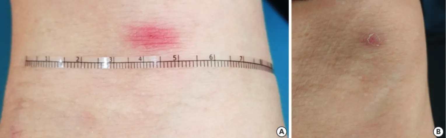
Case 1: A 70-year-old man presented with a 5-day history of fever in November. He had resided in Seogwipo-si, cultivated mushroom for a living, and had no history of tsutsugamushi disease. The patient had an erythematous patch, 8 × 16 mm in diameter, on the right antecubital fossa; this patch had been observed from the day of fever onset and was not accompanied by itching or pain. At the center of the patch, a small 1-mm diameter circle was noticed (Fig. 1A). Tender lymphadenopathy on the right axilla was palpated with additional non-tender lymphadenopathy on both sides of the neck. Sparse erythematous macular rashes developed on the upper chest on the third day from fever onset. Blood test results were as follows: white blood cell count, 4,110/mm3; platelet count, 149,000/
mm3; and aspartate and alanine aminotransferase levels, 68/59 U/L, respectively. Tests for immunofluorescent anti-Orientia antibodies were negative, and nested polymerase chain reaction (PCR) to detect the 56-kDa type-specific antigen gene, which was performed as previously described [7], was positive. On the next day after doxycycline medication, his fever disappeared. On the second visit to our hospital, 5 days after doxycycline treatment, desquamation around the central circle was observed (Fig. 1B).
Case 2: A 42-year-old man presented with a 5-day history of myalgia in November. The patient played golf 9 days ago and had severe myalgia, particularly in the upper extremities,
404 https://icjournal.org https://doi.org/10.3947/ic.2020.52.3.403
Erythematous patch in tsutsugamushi disease
A B
Figure 1. Photographs of Case 1. An erythematous patch measuring 8 × 16 mm in diameter on the right antecubital fossa is noticed, accompanied with a central circle of 1 mm in diameter (A). Five days later, the erythema disappears and desquamation around the site of the central circle develops (B).
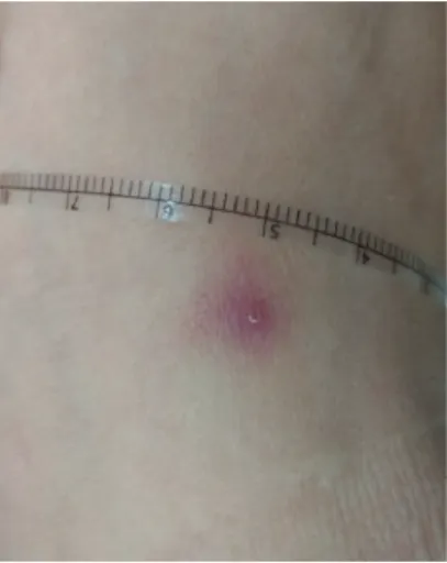
5 days later, and fever 8 days later. Myalgia may indicate the presence of fever; however, the body temperature was not checked. On physical examination, there were sparse rashes on the upper trunk, one palpable cervical lymph node, but no inguinal lymphadenopathy. Eschar was not observed on the first hospital visit, but on the second visit, 3 days after doxycycline treatment, an erythematous macule measuring 9 × 10 mm in diameter with central desquamation of 1 mm in diameter was found on the left ankle (Fig. 2). It is not certain whether the erythematous lesion was a macule or papule initially, but a 3-days treatment with doxycycline would not change a papule to a macule, and therefore the initial lesion was more likely a patch or macule. A day after doxycycline treatment, his fever abated. On the second hospital visit, regional lymphadenopathy was reassessed but not palpated. PCR of his blood specimen tested positive.
We believe that the erythematous patch is first reported in naturally affected patients with tsutsugamushi disease and that this type is described only in experimental human infections.
The erythematous patch might be the initial cutaneous lesion following an infected mite bite, which is static or progress to a papule, vesicle, ulcer, or eschar. It is erythematous and somewhat large (>10 mm in diameter; thus, a patch rather than a macule) and has indistinct margin; therefore, it is similar to the rash of tsutsugamushi disease, but larger and more intensely erythematous than the rash. It is noticed earlier before the appearance of the rash and is usually accompanied with regional lymphadenopathy, as described in Case 1. It is not accompanied with itching or pain, and therefore, if patients have dark skin, this type of inoculation lesion may not be noticed. A small (1 mm in diameter) round circle was observed at the center of the patch, which might indicate a chigger bite site. Nucleic acid amplification of scraped or biopsied materials from erythematous patches will aid in confirming that the patch is an inoculation lesion of tsutsugamushi disease. Regional lymphadenopathy draining from eschar has been underlined as an important clinical finding of tsutsugamushi disease, but we could not sometimes palpate regional lymphadenopathy. Early effective antibiotic therapy may delay the development of the antibodies and result in negative antibody responses in the patients.
It has been explained that the rarity of eschar in scrub typhus is attributed to dark-colored skin because erythematous rashes are difficult to identify in patients with dark skin.
However, cutaneous lesions with altered skin surface, including vesicle, ulcer, or necrosis,
405 https://icjournal.org https://doi.org/10.3947/ic.2020.52.3.403
Erythematous patch in tsutsugamushi disease
Figure 2. A photograph of Case 2. Three days after doxycycline therapy, an erythematous patch of 9 × 10 mm in diameter is noticed on the left ankle with desquamation at the center of the erythema.
can be noticed without difficulty irrespective of the skin color. For example, in a report on imported tsutsugamushi disease in Scandinavia, an eschar was easily identified in a dark- skinned Sri Lankan patient with tsutsugamushi disease [8]. Many reports have described typical eschars in Indian patients, who have also dark skin [9]. However, erythema surrounding the eschars was hardly identified in these cases. Therefore, dark skin might not be a sole cause of failure to identify eschar. We propose that the manifestation of an inoculation lesion as an erythematous patch might be the main reason for the rarity of eschar in scrub typhus in dark-skinned patients owing to difficulty in identifying these lesions.
Furthermore, these lesions might have been dismissed in temperate areas because of their unfamiliarity to most physicians.
REFERENCES
1. Fletcher W, Field JW. The tsutsugamushi disease in the Federated Malay States. Bull Inst Med Res FMS 1927;1:1-26.
2. Lewthwaite R, Savoor SR. The typhus group of diseases in Malaya. Part VIII: the relation of the tsutsugamushi disease (including rural typhus) to urban typhus. Part IX: the relation of the
tsutsugamushi disease (including rural typhus) and urban typhus to Rocky Mountain spotted fever. (with special reference to cross-immunity tests.). Br J Exp Pathol 1936;17:461-72.
3. Tanaka K. Important clinical findings of kedani (tsutsugamushi) disease. Kansenshogaku Zasshi 1929;4:168-70.
4. Smadel JE, Ley HL Jr, Diercks FH, Traub R. Immunity in scrub typhus: resistance to induced reinfection.
AMA Arch Pathol 1950;50:847-61.
PUBMED
5. Lee KS, Chong Y, Chun CH, Suto T. Importance of finding eschar in the early diagnosis of tsutsugamushi disease. J Korean Med Assoc 1987;30:1009-16.
6. Kim YH, Hyun W, Kim DP, Chung MH, Im JH, Baek JH, Lee JS, Kang JS. The eschar size and early inoculation lesion of tsutsugamushi disease on Jeju Island, Korea. Infect Chemother 2019;51:345-54.
PUBMED | CROSSREF
7. Chung MH, Lee JS, Baek JH, Kim M, Kang JS. Persistence of Orientia tsutsugamushi in humans. J Korean Med Sci 2012;27:231-5.
PUBMED | CROSSREF
8. Jensenius M, Montelius R, Berild D, Vene S. Scrub typhus imported to Scandinavia. Scand J Infect Dis 2006;38:200-2.
PUBMED | CROSSREF
9. Sarma N, Chakraborty S. Scrub typhus in Southern districts of West Bengal. Indian J Dermatol 2017;62:512-4.
PUBMED
406 https://icjournal.org https://doi.org/10.3947/ic.2020.52.3.403
Erythematous patch in tsutsugamushi disease