Korean J Hepatobiliary Pancreat Surg 2015;19:75-77
http://dx.doi.org/10.14701/kjhbps.2015.19.2.75
Case Report
Bilateral ovarian metastasis from distal common bile duct carcinoma developing after choledochal cyst excision
Seung Eun Lee1, Yoo Shin Choi1, Mi Kyung Kim2, Hyoung-Chul Oh3, and Jae Hyuk Do3
1Department of Surgery, 2Department of Pathology and 3Devision of Gastroenterology, Chung-Ang University College of Medicine, Seoul, Korea
Ovarian metastases represent about 3-5% of all ovarian malignancies. Most of these tumors originate in the digestive tract and cholangiocarcinoma rarely involves the ovary. A 60-year-old woman was admitted for the investigation of abdominal distension that had lasted 1 week. One and a half years prior, the patient had undergone choledochal cyst excision, Roux-en Y hepaticojejunostomy and cholecystectomy. Computed tomography scans of the abdomen revealed a papillary mass in the remnant distal common bile duct and enlargement of both ovaries with a huge amount of ascites. An explorative laparotomy disclosed no peritoneal seeding with resectable cholangiocarcinoma and bilateral ovarian mass. Pylorus-preserving pancreatoduodenectomy and bilateral salphingo-oophorectomy with hysterectomy were performed. Histologically, it was a well-differentiated adenocarcinoma and all surgical margins were free of tumor.
Both ovarian masses were consistent with metastatic adenocarcinoma from the common bile duct. The patient received six cycles of postoperative adjuvant systemic chemotherapy, dying after 10 months due to pulmonary embolism.
(Korean J Hepatobiliary Pancreat Surg 2015;19:75-77)
Key Words: Common bile duct; Choledochal cyst; Metastasis; Ovarian neoplasm
Received: May 12, 2015; Revised: May 15, 2015; Accepted: May 26, 2015 Corresponding author: Seung Eun Lee
Department of Surgery, Chung-Ang University College of Medicine, 224-1 Heukseok-dong, Dongjak-gu, Seoul 156-755, Korea Tel: +82-2-6299-3121, Fax: +82-2-824-7869, E-mail: [email protected]
Copyright Ⓒ 2015 by The Korean Association of Hepato-Biliary-Pancreatic Surgery
This is an Open Access article distributed under the terms of the Creative Commons Attribution Non-Commercial License (http://creativecommons.org/
licenses/by-nc/4.0) which permits unrestricted non-commercial use, distribution, and reproduction in any medium, provided the original work is properly cited.
Korean Journal of Hepato-Biliary-Pancreatic Surgery ∙ pISSN: 1738-6349ㆍeISSN: 2288-9213
INTRODUCTION
Ovarian metastases represent about 3-5% of all ovarian malignancies.1 Because they occasionally mimic the clin- ical and morphological appearance of primary tumors, di- agnosis is difficult. The majority of these neoplasias origi- nate from the digestive tract, particularly from stomach and biliary tract, cholangiocarcinomas are very rare tu- mors that rarely involve the ovary. We present a very un- usual case of metastatic carcinoma of the common bile duct originating from a choledochal cyst presenting as an ovarian metastasis in a 60-year-old woman.
CASE
A 60-year-old woman was admitted for the investigation of abdominal distension that had lasted for 1 week. One and a half years prior, the patient had been diagnosed with a choledochal cyst and had undergone choledochal cyst
excision, Roux-en Y hepaticojejunostomy and chol- ecystectomy at another hospital. Histopathologic findings from that time showed no evidence of malignancy of chol- edochal cyst. On physical examination, her abdomen was soft and distended severely without a palpable mass.
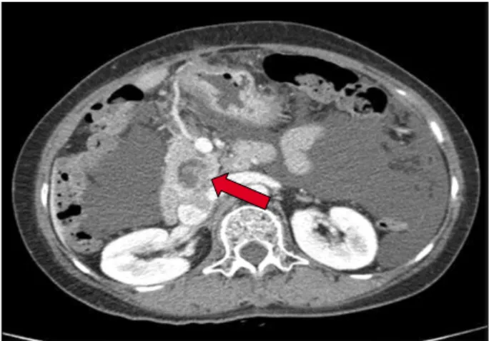
Laboratory evaluation revealed elevation levels of alkaline phosphatase (415 U/L, normal range 50-200 U/L) and CA 19-9 (2,409 U/L, normal range <37 U/L). Computed to- mography scan and magnetic resonance chol- angiopancreatography revealed a papillary mass in the rem- nant distal common bile duct (CBD) (Fig. 1), bilateral en- largement of ovaries with huge amount of ascites (Fig. 2) and no other metastatic lesion. An explorative laparotomy disclosed no peritoneal seeding with resectable chol- angiocarcinoma, bilateral ovarian masses measuring about 10 cm with irregular surface, multilobular cut surface and solid texture and ascites totaling about 1,000 ml. She under- went a pylorus preserving pancreaticoduodenectomy and bilateral salphingo-oophorectomy with hysterectomy. Cross
76 Korean J Hepatobiliary Pancreat Surg Vol. 19, No. 2, May 2015
Fig. 1. Computed tomography scan showing papillary mass in the remnant distal common bile duct (arrow).
Fig. 2. Computed tomography scan showing bilateral enlarge- ment of ovaries (arrows) with huge amount of ascites.
sections of the excised specimen showed CBD dilated to 5 cm in the pancreatic head and papillary mass was seen inside of it. Histologically, it was a well-differentiated ad- enocarcinoma, which extended to the pancreas with peri- neural invasion and regional lymph node metastasis. All surgical margins were free of tumor. Both ovarian masses were consistent with metastatic adenocarcinoma from CBD. The post-operative course was uneventful. The pa- tient received six cycles of postoperative palliative adjuvant systemic chemotherapy with 5-fluorouracil and cisplatin.
After finishing 6 cycles of 5-fluorouracil-based chemo- therapy, severe ascites redeveloped without definite meta- static lesion and with negative cytology. The patient ex- pired at approximately 10 months postoperatively due to pulmonary embolism.
DISCUSSION
The present case featured two unusual findings: cancer deveolpment from remnant distal CBD after choeldochal cyst excision and bilateral ovarian metastases from cholangiocacinoma.
Theoretically, biliary malignancy should not develop af- ter excision of the choledochal cyst because the presumed cause was abrogated by separation-operation. However, al- though the incidence of biliary malignancy after excision of a choledochal cyst was 0.6% in Korean multicenter study2 and 0.7% in a Japanese nationwide study,3 far lower than that in association with unresected choledochal cysts, it is still approximately 200 times higher than the in-
cidence of biliary cancer in the general population in Japan. According to the author’s literature review,4 58 cas- es were identified between 1970 and 2011. Among them, only 9 cases had malignancy at the intrapancreatic remnant bile duct. The mean interval between cyst excision and cancer detection was 9 years with a range of 4 to 17 years.
Because the presenting case developed malignancy only one and half years after cyst excision, it could be a missed lesion but de novo malignancy. However, preoperative en- doscopic retrograde cholangiopancreaticography and aspi- ration biopsy showed no abnormalities. Despite the poor prognosis of the presenting case, since the prognosis of the malignancy associated choledochal cyst was generally comparable with that of cholangiocarcinoma,2 regular fol- low-up should be done through tumor marker such as se- rum level of CA19-9 and imaging modalities such as com- puted tomography or ultrasonography for early detection of subsequent biliary malignancy after cyst excision.
Metastatic cancers in the ovary are relatively uncommon, accounitng for only about 3-5% of ovarian tumors.1,5 Although ovarian metastatic tumors ar typically bilateral, small (<10 cm), nodular, and solid,2,5 it is not easy to differ- entiate primary tumors from metastatic tumors. The present case showed bilateral, small and solid ovaries in preoperative imaging studies suggesting the presence of metastatic ovar- ian tumors. Since there was no guideline for the treatment of metastatic ovarian tumors from cholangiocarcinoma, py- lorus preserving pancreaticoduodenectomy and bilateral sal- phingo-oophorectomy with hysterectomy were performed.
The management of primary ovarian cancer includes max-
Seung Eun Lee, et al. Ovarian metastasis from bile duct carcinoma 77
imum tumor volume reduction, which is advantageous for survival.6 However, very few cases documenting ovarian metastases from cholangiocarcinoma have been reported in the literature,7,8 so the benefit of ovarian metastatectomy remains to be elucidated. Bilateral oophorectomy for ovar- ian metastasis from colorectal cancer has a positive impact on disease-free and overall survival in isolated ovarian metastases patients in an Italian study.9 Also, for patients with gastric cancer, a Korean study suggested that debulk- ing or gastrectomy plus metastasectomy may confer surviv- al benefits on patients with distant metastases who are re- ceiving systemic chemotherapy.10 However, since the prog- nosis of ovarian metastasis is dismal, and the benefit of ovarian metastatectomy is limited, further investigation is needed.
The present case is the first reported case of ovarian metastasis from cholangiocarcinoma associated with a choledochal cyst. Since biliary malignancy could develop within a relatively short period after choledochal cyst ex- cision, regular follow-up should be done for early de- tection of subsequent biliary malignancy after cyst excision. Although most reported cases with ovarian metastasis from cholangiocarcinoma were treated with bi- lateral salphigo-oophorectomy, the benefit of ovarian met- astatectomy is uncertain and should be investigated.
REFERENCES
1. Holtz F, Hart WR. Krukenberg tumors of the ovary: a clin- icopathologic analysis of 27 cases. Cancer 1982;50:2438-2447.
2. Lee SE, Jang JY, Lee YJ, Choi DW, Lee WJ, Cho BH, et al;
Korean Pancreas Surgery Club. Choledochal cyst and associated malignant tumors in adults: a multicenter survey in South Korea.
Arch Surg 2011;146:1178-1184.
3. Watanabe Y, Toki A, Todani T. Bile duct cancer developed after cyst excision for choledochal cyst. J Hepatobiliary Pancreat Surg 1999;6:207-212.
4. Lee SE, Jang JY. Development of biliary malignancy after cyst excision for congenital choledochal cysts: what should we do?
J Gastroenterol Hepatol 2013;28:210-212.
5. Lee KR, Young RH. The distinction between primary and meta- static mucinous carcinomas of the ovary: gross and histologic findings in 50 cases. Am J Surg Pathol 2003;27:281-292.
6. Fader AN, Rose PG. Role of surgery in ovarian carcinoma. J Clin Oncol 2007;25:2873-2883.
7. Young RH, Scully RE. Ovarian metastases from carcinoma of the gallbladder and extrahepatic bile ducts simulating primary tu- mors of the ovary. A report of six cases. Int J Gynecol Pathol 1990;9:60-72.
8. Khunamornpong S, Lerwill MF, Siriaunkgul S, Suprasert P, Pojchamarnwiputh S, Chiangmai WN, et al. Carcinoma of extra- hepatic bile ducts and gallbladder metastatic to the ovary: a re- port of 16 cases. Int J Gynecol Pathol 2008;27:366-379.
9. Erroi F, Scarpa M, Angriman I, Cecchetto A, Pasetto L, Mollica E, et al. Ovarian metastasis from colorectal cancer: prognostic value of radical oophorectomy. J Surg Oncol 2007;96:113-117.
10. Kim KH, Lee KW, Baek SK, Chang HJ, Kim YJ, Park do J, et al. Survival benefit of gastrectomy ± metastasectomy in pa- tients with metastatic gastric cancer receiving chemotherapy.
Gastric Cancer 2011;14:130-138.