S116 Ann Dermatol
Received February 4, 2010, Revised August 25, 2010, Accepted for publication October 26, 2010
Corresponding author: Chun Wook Park, M.D., Department of Dermatology, College of Medicine, Hallym University, 948-1 Daelim 1-dong, Youngdeungpo-gu, Seoul 150-950, Korea. Tel: 82-2-829-5223, Fax: 82-2-832-3237, E-mail: [email protected]
This is an Open Access article distributed under the terms of the Creative Commons Attribution Non-Commercial License (http://
creativecommons.org/licenses/by-nc/3.0) which permits unrestricted non-commercial use, distribution, and reproduction in any medium,
provided the original work is properly cited. Fig. 1. An oval-shaped, whitish papule measuring 0.5×0.5 cm on an erythematous base of the left sole.
Ann Dermatol Vol. 23, Suppl. 1, 2011 http://dx.doi.org/10.5021/ad.2011.23.S1.S116
CASE REPORT
A Case of a Subepidermal Calcified Nodule on the Sole without Trauma
In Su Ahn, M.D., Bo Young Chung, M.D., Hee Bong Lee, M.D., Hye One Kim, M.D., Hye Kyoung Ahn, M.D.
1, Chun Wook Park, M.D., Cheol Heon Lee, M.D.
Departments of Dermatology and 1Pathology, College of Medicine, Hallym University, Seoul, Korea
Subepidermal calcified nodule is an uncommon form of cal- cinosis cutis, which most commonly occurs in children. It usually presents as an asymptomatic, solitary verrucous nod- ule on the head and neck region, but occasionally as multiple lesions. Serum calcium and phosphorus levels are usually normal. Histopathology shows well-formed homogeneous eosinophilic material and granules in the upper dermis.
Material in the dermis stained with von Kossa was positive.
We report on an unusual case of a subepidermal calcified nodule occurring on the sole. A 21-month-old male pre- sented with an oval-shaped, whitish, hard nodule measuring 5×5 mm on the left sole, without any previous history of trauma. (Ann Dermatol 23(S1) S116∼S118, 2011)
-Keywords-
Calcified, Sole, Subepidermal
INTRODUCTION
Subepidermal calcified nodule (SCN) is an uncommon form of calcinosis cutis. SCN, also called cutaneous calcu- li, is usually a single small and hard nodule on the face. In most instances, the surface of the nodule is verrucous;
however, it may be smooth1.
SCN often occurs in damaged tissues in the absence of ab- normalities in serum calcium or phosphate levels. It has been reported in a variety of local tissue injuries, includ- ing repeated trauma2. Some reports have suggested devel- opment of calcified nodules on the heel following multi- ple heel sticks for venesection during the neonatal peri- od3,4. Similar cases in high risk neonates in intensive care units have been reported5-7. Though our patient had a his- tory of heel sticks during the neonatal period, the lesion did not appear at the site where heel sticks were generally performed. Therefore, we conclude that our case is not as- sociated with trauma.
In Korea, according to the first report on 6 cases by Son and Kim8, most lesions appeared on the eyelid and cheek8-11. There were no cases involving occurrence on the sole without trauma. We present an unusual case with unique clinical and histopathological features of child-
A Case of a Subepidermal Calcified Nodule on the Sole without Trauma
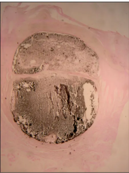
Vol. 23, Suppl. 1, 2011 S117 Fig. 3. Amorphous black to dark brown colored materials were confirmed as calcium by staining with von Kossa (von Kossa stain, ×40).
Fig. 2. Haematoxylin and eosin-stained sections showed a hyperkeratotic and acanthotic epidermis overlying a cystic structure in the upper dermis. The cystic space contained amorphous eosinophilic materials as multilobulated masses (H&E stain, ×40).
hood SCN on the sole without trauma.
CASE REPORT
A 21-month-old boy visited our clinic because of an asymptomatic, warty papule on the left sole that had de- veloped over the last 4 months. There was no history of preceding trauma. On physical examination, a painless, oval shaped, whitish papule measuring 5 mm on an eryth- ematous base was observed on the left sole (Fig. 1). It re- sembled a verruca. The boy was otherwise in good health.
Serum calcium and phosphorus levels were within normal limits.
The nodule was excised by punch biopsy under local anesthesia. Haematoxylin and eosin-stained sections showed a hyperkeratotic and acanthotic epidermis overlying a cystic structure of the upper dermis and narrow pointed rete ridges in the dermis. The cystic space contained amorphous eosinophilic materials as multilobulated mass- es (Fig. 2). The amorphous basophilic material described in this paper appeared to be eosinophilic, indicating oc- currence of degenerative and insufficient calcium on the amorphous basophilic material.
The materials were confirmed as calcium by staining with von Kossa (Fig. 3), which looks brown in color rather than black. It can be explained by two reasons: The first, the tis- sue sample itself showed faint staining. Early calcification, detected by von Kossa stain, could not be discovered by routine H&E staining12. The second, the high calcium area shows a black color in contrast to the brown colored cal- cium-lacking site. In another study of calcified lesions, 53
von Kossa-positive cases showed only 29 positive H&E (basophilic), while another 24 cases were negative on H&E13. In addition, PAS, Alcian blue (pH 2.5), and congo red stain were performed for determination of glycogen, mucopolysaccharides, and amyloid, which all showed a negative result. It suggested that the lesion contained de- posits of calcium, though it appeared to show poor stain- ing on H&E.
The lesion was completely removed by punch and there was good healing without recurrence.
DISCUSSION
In the human body, normal calcification appears only in bones and teeth. Cutaneous calcium deposits were de- scribed by Duhring as early as 1877; however, but they were first recognized by Winer14 in 1952 and named by Woods and Kellaway15 in 1963. SCN occurs more com- monly in children and can also occur at birth. Incidence among male and female children is approximately equal16. Clinically, SCN is presented as a single raised, well circumscribed, whitish and hard nodule with a smooth or verrucous surface9. Multiple lesions can also be seen, but less often17. The most common location of SCN is the face18.
Pathogenesis of this disease is still unknown; however, in 1980, Tezuka19 proposed that SCN is caused by de- granulation of mast cells with subsequent deposition of calcium and phosphates on the discharged mast cell contents. Histopathology of SCN consists of homogeneous basophilic masses and variable sized granules in the upper
IS Ahn, et al
S118 Ann Dermatol
dermis with hyperkeratosis, focal parakeratosis, papil- lomatosis, and epidermal hyperplasia. Calcium forms granules, globules, or large masses, which often contain nuclei. A lymphohistocytic infiltrate, macrophages, foreign body granulomatous giant cells, can be seen. Calcium- containing tissue is positive for von Kossa staining18,19. Surgical excision is the treatment of choice for patients with SCN20. However, intralesional therapy (corticoste- roids) has also been reported21.
In Korea, most cases of SCN occurred on the eyelid9-11 and cheek8, and, in some cases, lesions showed bilateral occurrence on thighs22, buttocks23, and fingertips24 in adulthood. Patients vary in age, from 3 to 55 years. There have been 4 cases reported as having less than a 1-year of duration of illness22,23, 1 case with 7 years8, and 2 cases with 20 years11,24. In this case, age of onset was 21 months, the youngest among domestic reports, after 4 months of spontaneous development without any trauma history. This is the age when toddling begins; therefore, we could consider the possibility that toddling itself might stimulate the onset of disease; however, there was no evi- dence of trauma. At the beginning, spontaneous tiny white papules without trauma were observed, and the lesion grew larger over a period of 4 months, suggesting that the lesion occurred naturally.
Our patient is asymptomatic; however, Rho et al.3 re- ported that this condition does not always remain asymp- tomatic and show spontaneous resolution. There have been several reports of calcinosis cutis classified as dystro- phic calcinosis cutis, due to chronic dermal damage by multiple heel sticks in children with a medical history of admission to the neonatal intensive care unit (NICU)5-7. Development of calcinosis cutis on the sole and without trauma is rare. For this reason, we report on this case, in- volving asymptomatic occurrence on the sole.
REFERENCES
1. Elder DE, Elenitsas R, Johnson BL Jr, Murphy GF, Xu G.
Lever’s histopathology of the skin. 10th ed. Philadelphia:
Lippincott Williams & Wilkins, 2009:435-438.
2. Baselga E, Fairley JA. Calcification and ossification in the skin. In: Harper J, Oranje A, Prose N, editors. Textbook of pediatric dermatology. Oxford: Blackwell Science, 2000:
788-789.
3. Rho NK, Youn SJ, Park HS, Kim WS, Lee ES. Calcified nodule on the heel of a child following a single heel stick in
the neonatal period. Clin Exp Dermatol 2003;28:502-503.
4. Hyun JS, Park CJ, Yi JY. A case of calcinosis cutis following heel sticks. Korean J Dermatol 2000;38:1270-1272.
5. Cambiaghi S, Restano L, Imondi D. Calcified nodule of the heel. Pediatr Dermatol 1997;14:494.
6. Williamson D, Holt PJ. Calcified cutaneous nodules on the heels of children: a complication of heel sticks as a neonate.
Pediatr Dermatol 2001;18:138-140.
7. Lemont H, Brady J. Infant heel nodules. Calcification of epidermal cysts. J Am Podiatr Med Assoc 2002;92:112-113.
8. Son SJ, Kim WS. A case of subepidermal calcified nodules.
Korean J Dermatol 1976;14:173-178.
9. Paek SH, Kim YH, Kim DW, Jun JB, Chung SL. Subepidermal calcified nodule. Ann Dermatol 1996;8:296-271.
10. Jung GD, Choi YH, Jeon YM, Song ES. Subepidermal calcified nodule arising in the lesion of clear cell syringoma.
Korean J Dermatol 2000;38:1660-1663.
11. Kim YK, Choi YH, Choi KC, Kim HK. A case of subepi- dermal calcified nodules showing an unusual clinical mani- festation. Korean J Dermatol 1983;21:595-599.
12. Neidner KH. Light microscopic findings. Clinics in Derma- tology 1988;6:93-99.
13. Barson AJ, Symonds J. Calcified pituitary concretions in the newborn. Arch Dis Child 1997;52:642-645.
14. Winer LH. Solitary congenital nodular calcification of the skin. AMA Arch Derm Syphilol 1952;66:204-211.
15. Woods B, Kellaway TD. Cutaneous calculi subepidermal calcified nodules. Br J Dermatol 1963;75:1-11.
16. Plott T, Wiss K, Raimer SS, Solomon AR. Recurrent subepi- dermal calcified nodule of nose. Pediatr Dermatol 1988;5:
107-111.
17. Shmunes E, Wood MG. Subepidermal calcified nodules.
Arch Dermatol 1972;105:593-597.
18. Evans MJ, Blessing K, Gray ES. Subepidermal calcified nodule in children: a clinicopathologic study of 21 cases.
Pediatr Dermatol 1995;12:307-310.
19. Tezuka T. Cutaneous calculus - its pathogenesis. Derma- tologica 1980;161:191-199.
20. Evans LA, Evans CM, Cobb MW. An asymptomatic papule on the face. Pediatr Dermatol 1996;13:253-254.
21. Lee SS, Felsenstein J, Tanzer FR. Calcinosis cutis circums- cripta. Treatment with an intralesional corticosteroid. Arch Dermatol 1978;114:1080-1081.
22. Jun JH, Lee JB, Kim SJ, Lee SC, Won YH. A case of subepi- dermal calcified nodule with transepidermal elimination.
Korean J Dermatol 2003;41:89-91.
23. Kim DH, Ham SH, Kang H, Cho SH, Park YM. Subepidermal calcified nodule of the buttock. Ann Dermatol 2000;12:
74-76.
24. Lee WC, Cha YC, Lee SJ, Na GY, Kim DW, Lee SK. Subepi- dermal calcified nodule of the finger. Korean J Dermatol 2003;41:1414-1416.