Received April 21, 2015, Revised May 22, 2015, Accepted for publication July 13, 2015
Corresponding author: Seung Ho Lee, Department of Dermatology, Dongguk University Ilsan Hospital, 27 Dongguk-ro, Ilsandong-gu, Goyang 10326, Korea. Tel: 82-31-961-7252, Fax: 82-31-961-7258, E-mail: heydoc74@
hanmail.net
This is an Open Access article distributed under the terms of the Creative Commons Attribution Non-Commercial License (http://creativecommons.
org/licenses/by-nc/4.0) which permits unrestricted non-commercial use, distribution, and reproduction in any medium, provided the original work is properly cited.
Copyright © The Korean Dermatological Association and The Korean Society for Investigative Dermatology
Ann Dermatol Vol. 28, No. 2, 2016 http://dx.doi.org/10.5021/ad.2016.28.2.210
ORIGINAL ARTICLE
The Importance of Collagen Tissue in Papular Elastorrhexis, Eruptive Collagenoma, and Nevus Anelasticus
Seung Ho Lee, Nam Hee Sung
Department of Dermatology, Dongguk University Ilsan Hospital, Dongguk University College of Medicine, Goyang, Korea
Background: Papular elastorrhexis (PE), eruptive collageno- ma (EC), and nevus anelasticus (NA) are described as multi- ple small papules with decrease, fragmentation, or lack of dermal elastic fibers. These diseases are suggested to be the same entity. The change of collagen fibers in the conditions has not been addressed to date. Objective: We compared the clinical features of the 3 diseases and investigated changes in the collagen fibers involved. Methods: Twenty-four cases of PE, 12 cases of EC, and 2 cases of NA found in PubMed and the Korean database were reviewed. Changes in dermal col- lagen fibers in 10 cases with histological figures were investigated. Results: There were significant similarities be- tween the 3 entities in terms of their clinical features. Four pa- tients with PE and 2 with EC with fine, dense collagen fibers were women who had multiple white to hypopigmented, slightly indurated to firm, millimeter-size papules on the trunk and/or extremities that progressed gradually after de- veloping in the patients’ first to third decades. Conclusion:
The 3 conditions are the same clinical entity in our opinion;
such cases with fine, dense collagen manifest typical features. (Ann Dermatol 28(2) 210∼215, 2016)
-Keywords-
Collagen fiber, Connective tissue nevus, Elastic fiber, Eruptive collagenoma, Nevus anelasticus, Papular elastor- rhexis
INTRODUCTION
Papular elastorrhexis (PE), eruptive collagenoma (EC) and nevus anelasticus (NA) have been described as multiple small papules with decrease, fragmentation, or lack of der- mal elastic fibers. The conditions were first described in 1987, 1966, and 1921, respectively1-3. The term nevus anelasticus was introduced by Staricco and Mehregan4 in 1961 as a more adequate expression for describing the en- tity named by Lewandowsky3 as naevus elasticus regionis mammariae in 1921.
It is suggested that PE, EC, and NA might be the same entity. Bordas et al.1 suggested that PE was a variant of NA in the first reported case of PE. In addition, it was sug- gested that EC is inseparable from PE5. Other authors have suggested that NA and EC should be considered variants of PE6.
Ryder and Antaya7 reviewed previous reports of the 3 enti- ties and found remarkable similarities between them. All 3 begin to appear before the age of 20 years, and the sites of the lesions are mainly the trunk and upper extremities. A lack of history of trauma, inflammation, family history, or extracutaneous manifestations is also a common feature.
All 3 showed a decreased amount of elastic tissue in biop- sy specimens. Therefore, the authors concluded that PE, EC, and NA are the same entity.
Although changes in elastic fibers have been well eval- uated in the 3 conditions, changes in collagen fibers have not attracted attention. There have been inconsistencies in
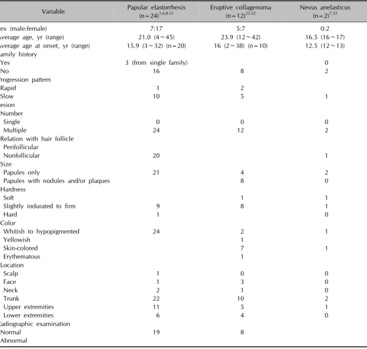
Table 1. Demographic information and clinical manifestation of reported cases
Variable Papular elastorrhexis
(n=24)1,6,8-21
Eruptive collagenoma (n=12)22-32
Nevus anelasticus (n=2)7,33
Sex (male:female) 7:17 5:7 0:2
Average age, yr (range) 21.0 (4∼45) 23.9 (12∼42) 16.5 (16∼17)
Average age at onset, yr (range) 15.9 (3∼32) (n=20) 16 (2∼38) (n=10) 12.5 (12∼13) Family history
Yes No
3 (from single family)
16 8
0 2 Progression pattern
Rapid Slow
1 10
2
5 1
Lesion Number Single Multiple
0 24
0 12
0 2 Relation with hair follicle
Perifollicular
Nonfollicular 20 1
Size
Papules only
Papules with nodules and/or plaques
21 4
8
2 0 Hardness
Soft
Slightly indurated to firm Hard
9 1
1 8
1 1 0 Color
Whitish to hypopigmented Yellowish
Skin-colored Erythematous
24 2
1 7 1
1 1 Location
Scalp Face Neck Trunk
Upper extremities Lower extremities
1 1 2 22 11 6
0 3 1 10 5 4
0 0 0 2 1 0 Radiographic examination
Normal Abnormal
19 8
previous reports’ descriptions of collagen fibers. Some au- thors reported homogenized, thick collagen fibers. On the other hand, others reported that the collagen tissue was normal. We experienced an interesting case in 2011 and reported it as PE8. The patient had fine, compacted colla- gen fibers in the upper dermis, where elastic fibers were reduced and fragmented, which prompted our interest in changes in collagen fibers as an important histological fea- ture in these diseases.
We reviewed previous reports of PE, EC, and NA, and changes in collagen and elastic fibers were investigated.
MATERIALS AND METHODS
We searched reports of PE, EC, and NA in PubMed and a Korean database. Demographic information and clinical manifestation of reported cases of the 3 diseases were summarized, and similarities and differences between the 3 conditions were analyzed.
We selected cases with histological figures from which we could evaluate the status of dermal collagen and elastic fibers. Changes in dermal collagen and elastic fibers of the select cases of the 3 diseases were summarized and compared.
According to the status of dermal collagen fibers, the se-
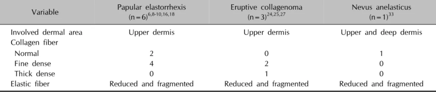
Table 2. Changes in dermal collagen and elastic fibers in cases with histological figures in which the status of dermal collagen and elastic fibers could be evaluated
Variable Papular elastorrhexis
(n=6)6,8-10,16,18 Eruptive collagenoma (n=3)24,25,27
Nevus anelasticus (n=1)33
Involved dermal area Upper dermis Upper dermis Upper and deep dermis
Collagen fiber
Normal 2 0 1
Fine dense 4 2 0
Thick dense 0 1 0
Elastic fiber Reduced and fragmented Reduced and fragmented Reduced and fragmented lected cases were reclassified into 3 groups: (i) normal col-
lagen group, (ii) fine, dense collagen group, and (iii) thick, dense collagen group. Demographic information and clin- ical manifestations for the 3 groups were summarized and compared.
RESULTS
Twenty-four cases of PE1,6,8-21, 12 cases of EC22-32, and 2 cases of NA7,33 were found. One case of EC24 was re- ported in Korean in the Korean literature, and the others were in English. Demographic information and clinical manifestations for the cases are summarized in Table 1.
There were significant similarities between the 3 entities.
All 3 diseases showed female predominance, and the average onset age was mid second decade. There was no family history except in 3 cases of PE from a single fam- ily20. The lesions progressed gradually in most cases. The lesional features of number, hardness, and location showed no significant difference between the 3 diseases. All cases had multiple lesions, and most lesions were slightly indu- rated to firm. The trunk and extremities were the major in- volved sites. All PE and NA cases in which relationships with hair follicles were described had nonfollicular lesions. Relationships with hair follicles were not men- tioned in the EC cases. All lesions were papules of several millimeters in size in PE and NA, but 8 cases of EC had nodules and/or plaques along with papules. All PE lesions were whitish to hypopigmented, whereas 7 cases of EC had skin-colored lesions. There were no abnormal radio- logic findings.
Among the reports, we selected only cases with histo- logical figures in which we could evaluate the status of dermal collagen and elastic fibers. Six cases of PE6,8-10,16,18, 3 cases of EC24,25,27, and 1 case of NA33 were selected.
Changes in dermal collagen and elastic fibers in those cas- es are summarized in Table 2. The dermal area in which elastic fiber was reduced and fragmented was the upper dermis in 9 cases except in 1 case of NA, in which elastic
fiber alteration was observed from the upper to deep dermis. Changes in collagen fibers in the elastic fiber-re- duced area could be classified into 3 patterns: (i) normal collagen, (ii) fine, dense collagen, and (iii) thick, dense collagen. Four cases of PE8,10,16,18 and 2 cases of EC24,27 had fine, dense collagen tissue. In 2 cases of PE6,9 and 1 case of NA33, collagen fibers did not show any change;
and only 1 case of EC25 had the thick, dense collagen fibers.
The 10 selected cases were reclassified into 3 groups ac- cording to the status of collagen fibers in the elastic fi- ber-reduced dermal area: (i) normal collagen group, (ii) fine, dense collagen group, and (iii) thick, dense collagen group. Demographic information and clinical manifes- tations for the 3 groups are summarized in Table 3. The fine, dense collagen group had all women, and the aver- age onset age was 13.3 years (range, 3∼25 years). Three cases progressed gradually, but 1 case progressed rapidly, with all lesions developing within 1 week. In all cases, multiple asymptomatic, less-than-1-cm-size, scattered pap- ules developed on the trunk and/or extremities. Exceptio- nally, a few lesions also developed on the face and neck in 1 patient. The lesions were whitish to hypopigmented and slightly indurated to firm in most patients.
Three cases in the normal collagen group differed from cases in the fine, dense collagen group in their clinical manifestations. The first case9 reported as “eruptive PE”
was the only male patient and had the latest onset age among the 10 selected cases. The involved sites of face and scalp also differed from the main involved areas. The lesions had developed rapidly, which was also unusual.
The second patient33 was a 17-year-old female who had papules that were grouped and localized on the right areola. Elastic fiber alteration was observed from the up- per to deep dermis in the biopsy specimen. Judging from the clinical figure, the last patient6 seemed to have numer- ous lesions.
One case with thick, dense collagen25 had a unique clin- ical manifestation: dozens of lesions were localized on the
Table 3. Demographic information and clinical manifestations of three groups classified according to the status of collagen fibers in the elastic fiber-reduced dermal area
Variable Normal collagen
(n=3)6,9,33
Fine dense collagen
(n=6)8,10,16,18,24,27 Thick dense collagen (n=1)25
Sex (male:female) 1:2 0:6 0:1
Average age, yr (range) 21.7 (17∼30) 15.5 (9∼26) 15
Average age at onset, yr (range) 18.3 (12∼29) 13.3 (3∼25) 15
Family history Yes
No
0 3
0 6
0 1 Progression pattern
Rapid Slow
1 2
1 3
0 1 Lesion
Number
Multiple but not numerous Numerous
1 2
6 0
0 1 Size
Papules only
Papules with nodules and/or plaques
3 0
6 0
0 1 Relation with hair follicle
Perifollicular
Nonfollicular 2 3
Hardness Soft
Slightly indurated to firm 1
1 4
0 1 Color
Whitish to hypopigmented Skin-colored
Erythematous
3 0 0
5 1 0
0 0 1 Location
Scalp Face Neck Trunk
Upper extremities Lower extremities
1 1 0 2 0 0
0 1 1 6 6 6
0 0 0 1 0 0 Radiographic examination
Normal Abnormal
2 4
left trunk in a zosteriform distribution. Peculiarly, the col- or of the lesions was erythematous, and some lesions were confluent.
DISCUSSION
We searched reports of PE, EC, and NA in PubMed and the Korean database and found 24 cases of PE, 12 cases of EC, and 2 cases of NA. Demographic information and clinical manifestations of the 3 diseases had significant similarities on comparative evaluation. EC has been known to present with an acute onset2. However, 5 of the 7 cases of EC whose progression pattern was described
progressed gradually, as in PE and NA. NA was first de- scribed as perifollicular lesions3. However, 1 of the 2 NA cases included had nonfollicular lesions, and a relation- ship with hair follicles was not described for the other patient.
We selected 10 cases with published histological figures in which we could evaluate the status of dermal collagen and elastic fibers. The cases were reclassified into 3 groups according to collagen tissue changes in the dermal area where elastic fibers were reduced and fragmented: (i) normal collagen group, (ii) fine, dense collagen group, and (iii) thick, dense collagen group.
Six cases in the fine, dense collagen group consisting of 4
cases of PE and 2 cases of EC showed similar clinical features. The condition developed in women between the 1st and 3rd decades, and the lesions progressed gradually.
There were multiple asymptomatic, white to hypo- pigmented, slightly indurated to firm, less-than-1-cm-size, scattered nonfollicular papules on the trunk and/or extremities. Electron microscopic examination was per- formed in 1 case of that group27. Scanning electron micro- scopy of the dermis showed that collagen fibers did not form bundles. Collagen fibers were individualized, form- ing waved, compact masses resembling noodles, and hence, empty spaces normally seen between dermal colla- gen bundles disappeared. On the other hand, normal col- lagen bundles were easily observed in the normal skin, and empty spaces were seen among the bundles.
Three cases in the normal collagen group differed from the fine, dense collagen group in clinical manifestations.
Each case had peculiar clinical features with few si- milarities. One case in the thick, dense collagen group al- so had clinical features that differed from other cases.
Ryder and Antaya7 suggested that PE, EC, and NA are the same entity. Our conclusions corroborated that suggestion.
Furthermore, the fine, dense collagen group showed typi- cal clinical and histological features of this condition.
Until now, decrease in elastic tissue has been empha- sized, but changes in collagen fibers also should be em- phasized because collagen tissue mainly contributes to the formation of the lesions. It is possible that the decrease in elastic tissue is secondary to the change in collagen bun- dles—such as a dilutional effect.
Connective tissue nevi are acquired dermal connective tis- sue hamartomas characterized predominantly by an im- balance in the relative amount and distribution of colla- gen, elastin, or proteoglycans34. The nevi are classified ac- cording to histological aspects. When collagen predom- inates, they are called collagenomas. Lesions in which elastic tissue predominates are called elastomas34,35. There- fore, we suggest papular collagenoma as a new name for the disease because it represents the disease’s clinical and histological features.
Four cases with normal collagen or thick, dense collagen showed different clinical features as well as histological features from the fine, dense collagen group. We consid- ered them to be conditions different from papular collage- noma or its variant.
REFERENCES
1. Bordas X, Ferrándiz C, Ribera M, Galofré E. Papular elastorrhexis: a variety of nevus anelasticus? Arch Dermatol 1987;123:433-434.
2. Cramer HJ. On the clinical aspects of the so-called
"eruptive collagenoma". Hautarzt 1966;17:437-440.
3. Lewandowsky F. Naevus elasticus regionis mammariae.
Arch Clin Exp Derm 1921;90:131.
4. Staricco RG, Mehregan AH. Nevus elasticus and nevus elasticus vascularis. Arch Dermatol 1961;84:943-947.
5. Lee MW, Choi JH, Sung KJ, Moon KC, Koh JK. A case of eruptive collagenoma. Pediatr Dermatol 2002;19:565-567.
6. Choonhakarn C, Jirarattanapochai K. Papular elastorrhexis:
a distinct variant of connective tissue nevi or an incomplete form of Buschke-Ollendorff syndrome? Clin Exp Dermatol 2002;27:454-457.
7. Ryder HF, Antaya RJ. Nevus anelasticus, papular elas- torrhexis, and eruptive collagenoma: clinically similar en- tities with focal absence of elastic fibers in childhood.
Pediatr Dermatol 2005;22:153-157.
8. Choi Y, Jin SY, Lee JH, Kwon HB, Lee AY, Lee SH. Papular elastorrhexis: a case and differential diagnosis. Ann Der- matol 2011;23 Suppl 1:S53-S56.
9. Sahin S, Durmaz EÖ, Sezer E, Cetin ED. Eruptive papular elastorrhexis of the face and scalp. J Am Acad Dermatol 2013;69:e251-e252.
10. Emre S, Metin A, Demir-Pektaş S, Kılıçarslan A. Papular elastorrhexis. Cutis 2013;92:E4-E5.
11. Thomé EP, Steglich RB, Meotti CD, Schwartz J, Boff AL.
Case for diagnosis. Papular elastorrhexis. An Bras Dermatol 2012;87:651-653.
12. Cañueto J, Román C, Santos-Briz Á, Ciria S, González R, Unamuno P. Papular elastorrhexis and Buschke-Ollendorff syndrome are different entities. J Am Acad Dermatol 2011;
65:e7-e9.
13. Flores PB, Cuevas J, Sánchez C, De Eusebio E, Vergara A.
Papular elastorrhexis: an acquired disorder of elastic tissue.
Eur J Dermatol 2010;20:525-526.
14. Tan C, Zhu WY, Min ZS. Papular elastorrhexis located on occipito-cervical and mandibular regions. Eur J Dermatol 2009;19:399-400.
15. Del Pozo J, Martínez W, Sacristán F, Fernández-Jorge B, Fonseca E. Papular elastorrhexis, a distinctive entity? Am J Dermatopathol 2008;30:188-190.
16. Kim YY, Lee JH, Lee JD, Cho SH. A case of papular elastorrhexis. Ann Dermatol 2007;19:185-188.
17. Do MO, Kim SH, Choi HY, Myung KB, Choi YW. Papular elastorrhexis. Ann Dermatol 2007;19:16-18.
18. Buechner SA, Itin P. Papular elastorrhexis. report of five cases. Dermatology 2002;205:198-200.
19. Lee SH, Park SH, Song KY, Yoon TJ, Kim TH. Papular elastorrhexis in childhood improved by intralesional injec- tions of triamcinolone. J Dermatol 2001;28:569-571.
20. Schirren H, Schirren CG, Stolz W, Kind P, Plewig G.
Papular elastorrhexis: a variant of dermatofibrosis lenti- cularis disseminata (Buschke-Ollendorff syndrome)? Der- matology 1994;189:368-372.
21. Sears JK, Stone MS, Argenyi Z. Papular elastorrhexis: a variant of connective tissue nevus. Case reports and review of the literature. J Am Acad Dermatol 1988;19:409-414.
22. Xiao M, Yang L, Dong L, Wang Y, Sun X, Tao J. Three cases
of eruptive collagenoma and a literature review, 1970- 2012. Eur J Dermatol 2014;24:384-385.
23. Sharma R, Verma P, Singal A, Sharma S. Eruptive colla- genoma. Indian J Dermatol Venereol Leprol 2013;79:256- 258.
24. Kim HR, Na CH, Shin BS, Choi KC, Kim MS. A case of eruptive collagenoma. Korean J Dermatol 2012;50:539- 543.
25. Zhao C, Ma W, Wang Y, Sun Q. Female with eruptive collagenoma clustered in the left lateral aspect of the abdomen. J Dermatol 2010;37:843-845.
26. Marque M, Gardie B, Bressac de Paillerets B, Rustin P, Guillot B, Richard S, et al. Novel FH mutation in a patient with cutaneous leiomyomatosis associated with cutis verticis gyrata, eruptive collagenoma and Charcot-Marie-Tooth disease. Br J Dermatol 2010;163:1337-1339.
27. de Almeida HL Jr, Breunig Jde A, Wolter M, de Castro LA, Rocha NM. Light and electron microscopy of eruptive collagenoma. J Cutan Pathol 2009;36 Suppl 1:35-38.
28. Kang MH, Kim HJ, Seo YJ, Park EJ, Kim CW, Cho HJ, et al.
A case of eruptive collagenoma on the left calf. Ann
Dermatol 2008;20:130-133.
29. Yahya H, Rafindadi AH. Eruptive collagenoma in a Nigerian girl. Int J Dermatol 2006;45:1344-1346.
30. Mukhi SV, Kumar P, Yuvarajkumar D, Raghuveer CV.
Eruptive collagenoma. Indian J Dermatol Venereol Leprol 2002;68:98-99.
31. Berberian BB, Wood C. Asymptomatic nodules on the back and abdomen. Connective tissue nevi, eruptive colla- genoma type. Arch Dermatol 1987;123:811-812, 815.
32. Smith LR, Bernstein BD. Eruptive collagenoma. Arch Der- matol 1978;114:1710-1711.
33. Wang AR, Kent K, Jagdeo J, Robinson-Bostom L, Bercovitch L. Nevus anelasticus: how should such lesions be classified?
J Cutan Pathol 2014;41:519-523.
34. Uitto J, Santa Cruz DJ, Eisen AZ. Connective tissue nevi of the skin. Clinical, genetic, and histopathologic classification of hamartomas of the collagen, elastin, and proteoglycan type. J Am Acad Dermatol 1980;3:441-461.
35. Pierard GE, Lapiere CM. Nevi of connective tissue. A reappraisal of their classification. Am J Dermatopathol 1985;7:325-333.