악성중피종 (malignant mesothelioma)은 흉막 (pleura)이나 복막 (peritoneum)의 중피세포 (mesothe- lial cell)에서 기원하는 악성 종양으로 미국의 경우 매년 대 략 3300명, 우리나라에서는 2010년의 경우 연 96건으로 전체 암 발생의 0.1% 미만에 속하는 매우 드문 종양이다 (1, 2). 대부분은 석면에 노출된 과거력이 있는 환자에게 발생 하며 이 중 80% 이상이 악성흉막중피종 (malignant pleural mesothelioma, MPM)으로 알려져 있다 (3, 4).
최근에는 새로운 항암제 및 표적 치료제 (targeted agents)등의 개발, 방사선치료, 수술법 및 다방법 병합치료 (multimodality therapy)등의 발달로 이환률 및 사망률이
감소하고 있기는 하지만 대부분 완화치료 (palliative treatment)에 불과하고 아직도 예후는 매우 좋지 않아 중 앙 생존 기간은 4에서 18개월 정도이다 (5-8). 최근에는 완 치를 위해서 완전한 수술적 절제를 포함한 다방법 병합치료 들이 제시되고 있는데 이를 위해서는 적합한 환자의 선택이 필수이다 (9-11). 이 환자 선택에 있어서 가장 중요한 요소 중 하나는 정확한 암 병기 (cancer stage)를 아는 것이다 (9, 11). 악성흉막중피종의 암 병기를 결정하는데 있어서 중 요한 것은 흉벽 (chest wall)이나 종격동 (mediastinum) 의 침범 여부 및 림프절 (lymph node)과 폐로의 전이 여부 인데 컴퓨터단층촬영 (computed tomography, CT)이 현 재로는 진단 및 병기, 그리고 치료 반응을 보는데 있어서 일 차적인 영상기법으로 널리 사용되고 있다 (6, 12-15). 그러 나 CT는 흉벽 침범을 과소평가하는 경향이 있고 또 림프절 침범 여부를 평가하는 데에도 제한이 있다 (14, 16). 자기공 명영상 (magnetic resonance imaging, MR)은 연조직에 대한 대조도가 매우 높아 종양의 흉벽이나 횡격막 침범 여 부를 보다 손쉽게 볼 수 있어 종양의 완전 절제 가능성을 평 가하는데 유리하다 (14, 15). 또 최근에는 T1-weighted image (WI)를 3-dimensional, gradient recalled echo (3D GRE) sequence의 기법을 통해 더욱 높은 해상도의 사 진을 얻는 것이 가능해지고 respiratory dynamic cine 서 론
�Received; August 20, 2013�Revised; September 9, 2013
�Accepted; September 17, 2013 Corresponding author : Eun Jin Chae, M.D.
Department of Radiology and Research Institute of Radiology, University of Ulsan College of Medicine, Asan Medical Center, 88 Olympic-ro 43-gil, Songpa-gu, Seoul 138-736, Korea.
Tel. 82-2-3010-4355, Fax. 82-2-476-0090 E-mail : [email protected]
악성흉막중피종의 병기판정에서 자기공명영상의 진단적 유용성 평가
이은솔∙채은진∙강선지∙염유경∙이현주∙박종천∙신소연∙최윤영∙최준호∙도경현 울산대학교 의과대학, 서울아산병원 영상의학과 영상의학연구소
목적 :악성흉막중피종 (이하 MPM)의 임상적 병기판정에 있어 자기공명영상 (이하 MR)의 진단적 유용성을 컴퓨터단층촬영 (이하 CT)과 비교하였다.
대상 및 방법 : 1997년부터 2012년까지 MPM으로 확인된 환자 중 진단 시 CT와 MR을 얻은 환자 20명 (남:여=14:6; 평
균연령= 53.5세)을 대상으로 하였다. CT 혹은 MR 단독을 이용하여 두 명의 영상의학과 의사가 의견 일치 하에 병기판정을 시행하였다. 수술적 병기판정을 받은 환자의 경우 이와 비교하여 CT 단독 및 CT와 MR을 함께 시행하였을 때의 진단적 정확 도를 평가하였다. 또 CT 단독과 CT와 MR을 함께 시행하였을 때의 임상병기의 일치도를 비교하였다.
결과 : CT를 이용한 T stage의 정확도는 23.1% (3/13)였고 MR과 함께 시행한 경우 38.5% (5/13)였다. CT 및 MR을
함께 시행한 경우 수술 후 수술적 병기에서 5명의 환자는 상위 진단을, 3명의 환자는 하위 진단을 보였다. CT 단독 및 CT와 MR을 함께 시행한 병기를 서로 비교하였을 때 85.0% (17/20)의 일치도를 보였고 불일치한 예(3명)에서는 함께 시행한 경우 CT 단독보다 상위 진단을 하였다.
결론 : MPM의 병기판정에 있어서 MR을 함께 시행한 경우 CT 단독에 비해 보다 높은 정확도의 병기판정을 제공하여 수술
대상이 되는 환자에서 수술 전 검사로서 중요한 역할을 수행할 것으로 기대된다.
Original Article
MR기법 역시 더욱 발전하였다 (17-19). 이전 연구들에서 몇 차례 악성흉막중피종의 병기 판정에 있어서 CT와 MR을 비교하긴 하였지만 과거 연구이거나 이 둘을 직접 비교한 연구는 드물다 (13, 14, 20, 21). 이는 악성흉막중피종의 낮 은 발생률에 기인한 것으로 판단된다. 최근에는 폐암 (lung cancer)에서도 MR기법의 발전에 따라 점점 더 정확한 진 단이 가능하다고 밝혀지고 있고 이 역시 악성흉막중피종에 서 마찬가지의 적용이 가능할 것으로 사료된다 (18, 19). 이 에 추가연구가 필요할 것으로 보여 본 연구에서는 악성흉막 중피종의 병기 설정에 있어서 MR의 정확도 (accuracy)를 알아보고, CT를 단독으로 시행하였을 때와의 비교를 해보 고자 한다.
환자군
1997년 1월부터 2012년 12월까지 본원에 내원하였던 환 자 중 수술 또는 생검을 통해 병리조직검사 결과 악성흉막 중피종으로 확인된 환자들을 대상으로 하였다. 총 71명의 환자가 병리조직검사 결과 악성흉막중피종으로 확인되었으 며 이 중 처음진단 당시 CT와 MR 중 한 가지 검사라도 시 행하지 않은 환자는 연구에서 제외하여 총 20명 (남:여 = 14:6)이 포함되었다. 연령 분포는 41세에서 73세까지였고, 평균연령은 53.5세였다.
연구에 포함된 환자 중 수술로 병기가 확인된 환자는 총 13명 (65.0%)이며 이 환자들을 대상으로 수술적 병기와 CT, MR 각각의 임상적 병기를 비교하였다. 또 전체 환자를 대상으로 CT를 단독으로 시행하였을 때와 CT와 MR을 함 께 시행하였을 때의 임상적 병기를 서로 비교하였다.
CT 영상기법
본원 및 여러 외부병원에서 다양한 기종으로 시행된 CT 영상이 연구에 포함되었다. 대부분 16 또는 64채널 CT (Sensation 16, 64, Definition AS+, FLASH; Siemens Medical Solutions, Forchheim, Germany; Light- Speed16, VCT; General Electric Medical Systems, Milwaukee, WI, USA; Mx8000 IDT 16; Philips Medical Systems, Best, Netherlands)가 사용되었으나 한 환자에서 4채널의 CT도 사용되었다 (LightSpeedQx/I;
General Electric Medical Systems). 모든 환자에서 전체 thoracic cage를 포함하여 조영증강후 CT가 얻어졌고 절 편 (slice)의 간격 및 두께는 3 mm에서 6 mm사이로 연속 으로 얻었다.
MR 영상기법
모든 MR은 1.5T MR기종이었으며 Magnetom Avanto
scanner (Siemens, n=5), Vision scanner (Siemens, n=4), Integra (Philips, n=10), Achieva scanner (Philips, n=1), Signa scanner (GE, n=1)등으로 검사가 시행되었다.
모든 환자에서 전체 thoracic cage를 포함하여 조영증강 없이 T1WI, T2WI, 그리고 조영증강후 T1WI를 축상면 (axial plane), 시상면 (sagittal plane), 그리고 관 상면 (coronal plane)의 3방향으로 사진을 얻었다. 절편의 간격은 6.6~10 mm, 그리고 절편의 두께는 6~8 mm로 절 편 사이에는 1.2~2 mm의 틈 (gap)을 두고 사진을 얻었다.
또 11명 (55%)의 환자에서는 역동적 호흡 자기공명영상 (dynamic breathing MR imaging)을 얻기 위해 real time true Fast Imaging with Steady-state Precession (FISP) sequence나 3D GRE sequence를 추 가로 얻었다. 3명 (15%)의 환자에서는 조영증강후 3D GRE sequence를 추가로 얻었다. 3D GRE sequence의 경우 전 체 volume data를 얻은 후 2~4 mm의 절편 두께로 절편 사이의 틈 없이 연속으로 사진을 재구성 (reconstruction) 하였다.
임상적 병기판정
2명의 영상의학과 의사 (각각 13년, 4년의 판독 경험 보 유)가 수술 소견 및 병리 소견을 모르는 상태로 합의를 통해 CT 단독, 그리고 MR 단독으로 임상병기판정을 시행하였 다. 임상병기판정은 Petavision (Asan Medical Center, Seoul)이라는 picture archiving and communicating system (PACS) 소프트웨어를 이용하여 이루어졌다. 병기 판정은 International Mesothelioma Interest Group이 제시한 New International Staging System for MPM을 따랐다 (22).
조직검사 및 수술
조직검사는 CT 및 투시 유도 아래 경피적 흉부 세침검사 및 흉강경 (thoracoscopy)을 통한 폐 조직검사를 통해 이 루어졌다. 영상 진단을 통해 수술을 할 수 없는 경우라고 판 단된 환자를 제외하고는 진단적 흉강경이나 탐색 개흉술 (exploratory thoracotomy)을 통해 수술적 병기 설정을 하거나 흉막외 폐절제술 (extrapleural pneumonectomy) 을 통해 수술적 치료가 이루어졌으며 이를 통해 수술적 병 기판정이 이루어졌다.
자료 및 통계 분석
본 연구에서 사용된 모든 통계는 SPSS (version 12.0 for Windows, Chicago, IL, USA)를 이용하여 구하였다.
수술을 시행한 환자에서 수술적 병기를 기준으로 CT 단독 및 CT와 MR을 함께 시행하였을 때의 병기의 정확도 (accuracy)를 평가하였다. 또 전체 환자에서 CT 단독 병기 대상 및 방법
와 MR 및 CT를 함께 시행하였을 때의 병기를 비교하였다.
전체 환자의 임상적 특성, 임상병기, 수술 병기를 표 1에 요약하였다 (Table 1). 악성흉막중피종의 조직학적 아형 중 상피성 중피종 (epithelial mesothelioma)이 11명 (52.0%)으로 가장 많았고 이상성 중피종 (biphasic mesothelioma)이 3명 (15.0%)으로 두 번째로 많았으며 섬 유조직형성 중피종 (desmoplastic mesothelioma)이 1명 (5.0%)로 가장 적었다. 7명의 환자에서는 조직학적 아형을 병리적으로 판단하기 어려웠다. 생존기간 (survival time) 은 70일에서 5568일까지로 중간값 (median)은 458일이었 다. 7명 (35.0%)의 환자에서 수술적 치료가 이루어졌고 10
명 (50.0%)의 환자에서 항암치료, 그리고 3명 (15.0%)의 환 자에서는 완화요법 (palliative therapy)만이 이루어졌다.
T staging에 있어서 수술 병기와 비교하였을 때 CT는 (3/13, 23.1%)의 정확도를 보였으며 CT 및 MR을 함께 시 행한 경우 (5/13, 38.5%)의 정확도를 보였다 (Fig. 1). 수술 병기와 비교하였을 때 MR은 5명에서 수술 병기보다 하위 진단 (downstaged)을 하였지만 3명에서는 수술 병기보다 상위 진단을 하였다 (Fig. 2). 전체 환자를 대상으로 CT 단 독 및 CT와 MR을 함께 시행한 병기를 서로 비교하였을 때 (Fig. 3), 20명중 17명 (85.0%)이 일치하였고 불일치한 경 우 (15.0%, 3/20) MR 및 CT를 함께 시행한 경우에서 CT 단독보다 상위 진단을 하였다 (Fig. 4). 3D GRE sequence 를 이용하여 검사한 3명의 환자에서는 CT와 MR이 동일한 T4 stage를 보였으며 이에 따라 수술적 병기 설정은 이루 어지지 않았다. N staging에 있어서는 수술 병기와 비교하 결 과
Table 1. Clinical Characteristics Including Clinical and Surgical Staging of Subjects
Patient Age Sex Histology Survival Procedure Treatment CT MR Surgical CT MR Surgical MR
No. day TNM TNM TNM stage stage stage technique
1 49 F U 144 Bx C T4N0M0 T4N0M0 IV IV 3D+DB+B
2 62 M E 659 Bx C T4N0M0 T4N0M0 IV IV 3D+DB+B
3 44 M U 180 ET C T2N0M0 T4N0M0 T4N0M0 II IV IV DB+B
4 60 F U 1511 EPP S T2N0M0 T2N0M0 T3N0M0 II II III DB+B
5 55 M E 248 Bx C T4N0M0 T4N0M0 IV IV DB+B
6 41 M B 128 Bx P T4N0M0 T4N0M0 IV IV Basic
7 60 M E 1085 EPP S T4N0M0 T4N0M0 T3N0M0 IV IV III DB+B
8 38 F B 458 ET C T2N0M0 T3N0M0 T4N0M0 II III IV Basic
9 64 F E 441 Bx C T4N0M0 T4N0M0 IV IV 3D+DB+B
10 62 M D 70 EPP S T2N0M0 T3N0M0 T3N0M0 II III III Basic
11 45 M B 517 VATS C T4N0M0 T4N0M0 T3N0M0 IV IV III Basic
12 42 M E 924 ET C T3N0M0 T3N0M0 T4N0M0 III III IV Basic
13 56 M E 137 EPP S T2N0M0 T2N0M0 T2N1M0 II II III Basic
14 52 M E 248 EPP S T4N0M0 T4N0M0 T2N2M0 IV IV III Basic
15 69 M U 571 Bx C T1N0M0 T1N0M0 I I DB+B
16 60 F E 301 EPP S T2N0M0 T2N0M0 T3N1M0 II II IV DB+B
17 50 M E 651 VATS P T2N0M0 T2N0M0 T4N0M0 II II IV Basic
18 58 M E 1223 EPP S T2N0M0 T2N0M0 T2N0M0 II II II DB+B
19 36 F U 813 Bx C T4N3M0 T4N3M0 IV IV DB+B
20 28 M E 191 ET P T4N0M0 T4N0M0 T4N0M0 IV IV IV Basic
Note.─ E, epithelial; B, biphasic, D, desmoplastic, U, unknown; Bx, image-guided biopsy; ET, exploratory thoracotomy; EPP, extrapleural pneumonectomy; VATS, video-assisted thoracoscopic surgery; C, chemotherapy; S, surgery; P, palliative therapy; 3D, contrast-enhanced, 3-dimensional, gradient recalled echo sequence; DB, dynamic breathing imaging; B, Basic, basic MR sequence (3 direction T1 weighted image, T2 weighted image, contrast-enhanced T1 weighted image)
였을 때 CT는 85.0%의 정확도를 보였고 MR은 80.0%의 정확도를 보였다. 역동적 호흡 자기공명영상은 11명의 환자 에서 이루어졌는데 이에 의한 추가적 병기 변화는 없었다.
악성흉막중피종의 치료에 있어 흉막 외 폐절제술이나 확 대 흉막절제술/피질제거술 (extended pleurectomy/
decortication)을 비롯한 근치적 수술이 생존률 향상에 도 움이 되는지는 논란이 있지만 육안적 완전 절제 (macro- scopic complete resection)가 가능하다면 수술적 절제술
을 시행하자는 쪽으로 의견이 모아지고 있다 (11, 23). 현재 는 유일한 무작위 시험인 MARS (Mesothelioma and Radical Surgery)에 이어 MARS-2가 진행 중으로 근치적 수술의 효과에 대한 결론을 내어줄 것으로 기대되고 있다 (24). 이러한 근치적 수술 여부를 결정하는 데에 있어서는 나이, 동반 질환 등의 임상상 외에도 임상적 병기판정이 매 우 중요하다 (9). 이러한 악성흉막중피종의 수술 전 영상 진 단 방법은 CT, MR, fluorodeoxyglucose positron emission tomography (FDG-PET), positron emission tomography (PET)-CT 등의 여러 가지 영상 진단 방법이 사용되고 있다 (6, 21, 25). 그러나 이들을 이용한 진단이나 임상병기판정 및 영상 진단 방법간의 비교 연구는 매우 부 족한 실정이다 (16, 21, 25). 현재 가장 널리 쓰이고 있는 International Mesothelioma Interest Group이 제시한 악성흉막중피종의 병기분류에서도 수술적 절제 가능성을 중요시하여 국소적으로 진행하였지만 절제 가능한 경우를 T3, 기술적으로 절제 불가능한 경우를 T4로 분류하였다 (22). 그러나 아직까지 CT나 MR을 이용한 악성흉막중피종 의 정확한 수술 전 병기판정은 매우 어려운 것으로 알려져 있다 (6, 14, 26). 본 연구에서도 악성흉막중피종의 수술 전 병기판정을 수술 후와 비교하였을 때 CT (23.1%), MR (42.9%) 모두에서 낮은 정확도를 보였다. 아직까지 병기판 정의 정확도를 비교한 연구는 없지만 병기 판정에 대해 연 구한 다른 논문들에서는 다양한 정확도를 간접적으로 시사 하는 결과를 제시하였다. Patz 등 (13)은 악성흉막중피종 고 찰
a b
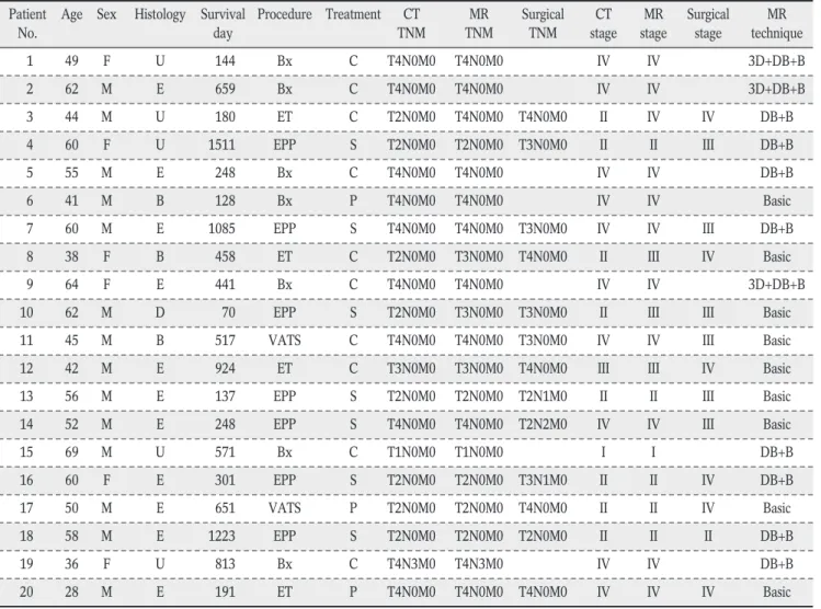
Fig. 2. A representative case of upstaging by MR compared to surgical stage.
a. A CT image shows circumferential pleural thickening involving visceral and parietal pleura. Areas of obliteration of extrapleural fat probably over the expected line of endothoracic fascia suggest chest wall invasion (T4) (arrows).
b. Gadolinium-enhanced T1-weighted MR image shows some indentation of chest wall by the pleural mass suggesting chest wall invasion (T4) (arrows). However, patient underwent pleuropneumonectomy and surgical and histologic findings revealed no evidence of chest wall invasion (T2).
Fig. 1. Differences between clinical T stage and corrected surgical T stage in patients with available surgical stage.
환자에서 CT의 횡격막 및 흉벽 침범의 민감도를 각각 94%
와 93%로 보고하였고 MR의 경우 횡격막 및 흉벽 침범의 민감도를 모두 100%로 보고 하였다. Heelan 등 (14)은 횡 격막 침범 정확도를 CT 및 MR 각각 52% 와 82%, 그리고 흉내근막 (endothoracic fascia) 침범 및 절제 가능한 흉벽 침범의 정확도를 CT 및 MR 각각 46%와 69%로 보고 하였 다.
본 연구에서 CT의 수술 전 병기판정의 정확도가 낮은 것 은 환자군 설정에 따른 문제 일 가능성이 있는데, 초기 진단 시 CT를 먼저 시행한 후 CT에서 흉곽 및 종격동 등의 침범 여부, 즉 병기가 불확실한 경우만 MR을 시행하였고 이러한 환자들이 주로 환자군에 포함되었기 때문으로 사료된다.
MR의 경우 CT보다는 정확도가 높았지만 기대만큼 정확도 가 높지는 않았다. 하지만 비록 낮은 정확도이긴 하지만 CT 에 비해 상대적으로 MR에서 높은 정확도를 보인 점은 CT 에서 병기를 판단하기 어려운 환자군 이었다는 점에서 의미 가 있다. 또 다른 이유로는 긴 환자포함기간에 걸쳐 여러 종 류의 MR 기종 및 프로토콜을 이용하여 다양한 MR 영상이 얻어진 것이 한 요인이 될 수 있겠다. 초기 프로토콜에는 스 핀 에코 계열의 T1WI, T2WI, 그리고 조영증강을 한 T1WI 만이 포함되었으며 절편의 두께도 두껍고 공간 해상도도 낮 았다. 가장 최근에 시행한 프로토콜에는 3D 영상을 쉽게 얻 을 수 있게 되어 조영증강 후 3D GRE sequence가 포함되 었다 (17, 27). 3명의 환자에서 이를 적용하였으며 모두 stage IV로 판정되어 수술을 하지 않았지만 흉벽이나 횡격 막 침범에 높은 민감도 및 특이도를 보일 것으로 기대된다 (17). 역동적 호흡 자기공명영상 역시 본 연구에서 일부 환 자에서 함께 시행을 하였으나 대부분의 종양이 넓게 퍼져 있어 흉벽과의 유착이 심해 병기 판정에 도움이 되지 않았 다.
병기판정에 있어서 MR을 시행하는 가장 큰 이유는 흉벽, 횡격막, 그리고 종격동의 침범 여부 등 병변의 범위를 보기 위한 것이다 (9). MR에서 흉벽여부 침범을 판단하는 중요 한 방법 중 하나는 흉막외 지방 (extrapleural fat)층이 침 범 당하지 않고 존재하는지 판단하는 것이다 (15). 또 연조 직 (soft tissue) 대조도가 높기 때문에 CT에서 연조직 음 영이 근육에 있는 경우 이것이 근육 자체에 의한 음영인지
a b
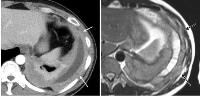
Fig. 4. A representative case of upstaging by MR compared to CT.
a. A CT image shows pleural effusion with diffuse pleural thickening. Soft tissue density lesions are suspicious in intercostal spaces in the lower portion of hemithorax, however chest wall invasion is not clear.
b. Gadolinium-enhanced, 3-dimensional, gradient recalled echo sequence MR image clearly shows enhancing soft tissue lesions in the intercostal spaces suggesting diffuse chest wall invasion (T4) (arrow).
Fig. 3. Differences between clinical stage by CT only and CT and MR.
종양의 침범에 의한 것인지 알기 어렵지만 MR에서는 쉽게 구별 가능하다 (14, 15). 본 연구에서도 MR을 CT와 함께 시행하였을 때 CT단독으로 병기설정을 한 경우보다 상위 진단을 한 경우는 모두 이런 연조직 대조도의 차이에 의해 보다 쉽게 종양의 침범 여부를 알 수 있었기 때문이다
본 연구에 포함되어 있는 일부 증례에서는 MR에서는 흉 벽이나 종격동을 침범한 것처럼 보였지만 실제 수술 및 병 리 소견에서는 침범하지 않은 경우도 있었다. 이는 주로는 부분 부피 인공물 (partial volume artifact)에 의한 것으 로 여겨지며 3D MR영상을 얻음으로써 해결 가능하다 (28). MR에서 나타나는 T1WI에서의 조영증강이나 T2WI 에서의 고신호는 종양의 침범뿐만 아니라 주변 조직의 염증 에 의해서도 매우 민감하게 나타난다 (29).
본 연구의 제한점으로는 후향적 연구이며 매우 드문 질환 이라는 점 때문에 환자군의 수가 작아 통계적 분석을 할 수 있는 표본수를 갖추지 못했다는 것이다. 또 다양한 프로토 콜로 CT 및 MR을 시행하였다는 것 역시 제한점이 되겠으 나 초기의 MR 프로토콜과 후기의 3D GRE sequence가 포함된 프로토콜과의 비교는 앞으로의 MR protocol에 대 한 제안이 될 수 있을 것이다.
악성흉막종피종의 병기판정에 있어서 MR을 CT와 함께 시행하였을 때 CT를 단독으로 시행한 경우보다 높은 정확 도의 병기판정을 제공하여 수술 대상이 되는 환자에서 수술 전 검사로서 중요한 부가적 역할을 수행할 것으로 기대된 다. 그러나 MR을 함께 시행하더라도 병기판정의 정확도가 충분히 높지 않아 fast imaging을 이용한 다양한 프로토콜 을 시도한 MR 영상의 병기판정의 정확도 등에 대한 추가적 인 연구가 필요할 것이다.
References
1. Jung KW, Won YJ, Kong HJ, Oh CM, Seo HG, Lee JS. Cancer statistics in Korea: incidence, mortality, survival and prevalence in 2010. Cancer Res Treat 2013;45:1-14
2. Teta MJ, Mink PJ, Lau E, Sceurman BK, Foster ED. US mesothelioma patterns 1973-2002: indicators of change and insights into background rates. Eur J Cancer Prev 2008;17:525- 534
3. Price B, Ware A. Mesothelioma trends in the United States: an update based on Surveillance, Epidemiology, and End Results Program data for 1973 through 2003. Am J Epidemiol 2004;
159:107-112
4. Delgermaa V, Takahashi K, Park EK, Le GV, Hara T, Sorahan T. Global mesothelioma deaths reported to the World Health Organization between 1994 and 2008. Bull World Health Organ 2011;89:716-724
5. Cao C, Tian DH, Pataky KA, Yan TD. Systematic review of pleurectomy in the treatment of malignant pleural mesothe- lioma. Lung Cancer 2013;81:319-327
6. Erasmus JJ, Truong MT, Smythe WR, et al. Integrated computed tomography-positron emission tomography in patients with potentially resectable malignant pleural mesothe- lioma: Staging implications. J Thorac Cardiovasc Surg 2005;
129:1364-1370
7. Scherpereel A, Astoul P, Baas P, et al. Guidelines of the European Respiratory Society and the European Society of Thoracic Surgeons for the management of malignant pleural mesothelioma. Eur Respir J 2010;35:479-495
8. Martino D, Pass HI. Integration of multimodality approaches in the management of malignant pleural mesothelioma. Clin Lung Cancer 2004;5:290-298
9. Neumann V, Loseke S, Nowak D, Herth FJ, Tannapfel A.
Malignant pleural mesothelioma: incidence, etiology, diagnosis, treatment, and occupational health. Dtsch Arztebl Int 2013;110:
319-326
10. Sugarbaker DJ, Flores RM, Jaklitsch MT, et al. Resection margins, extrapleural nodal status, and cell type determine postoperative long-term survival in trimodality therapy of malignant pleural mesothelioma: results in 183 patients. J Thorac Cardiovasc Surg 1999;117:54-63
11. Rusch V, Baldini EH, Bueno R, et al. The role of surgical cytore- duction in the treatment of malignant pleural mesothelioma:
meeting summary of the International Mesothelioma Interest Group Congress, September 11-14, 2012, Boston, Mass. J Thorac Cardiovasc Surg 2013;145:909-910
12. Nowak AK. CT, RECIST, and malignant pleural mesothelioma.
Lung Cancer 2005;49 Suppl 1:S37-40
13. Patz EF, Jr., Shaffer K, Piwnica-Worms DR, et al. Malignant pleural mesothelioma: value of CT and MR imaging in predict- ing resectability. AJR Am J Roentgenol 1992;159:961-966 14. Heelan RT, Rusch VW, Begg CB, Panicek DM, Caravelli JF,
Eisen C. Staging of malignant pleural mesothelioma: comparison of CT and MR imaging. AJR Am J Roentgenol 1999;172:1039- 1047
15. Wang ZJ, Reddy GP, Gotway MB, et al. Malignant pleural mesothelioma: evaluation with CT, MR imaging, and PET.
Radiographics 2004;24:105-119
16. Boiselle PM, Patz EF, Jr., Vining DJ, Weissleder R, Shepard JA, McLoud TC. Imaging of mediastinal lymph nodes: CT, MR, and FDG PET. Radiographics 1998;18:1061-1069
17. Karabulut N, Martin DR, Yang M, Tallaksen RJ. MR imaging of the chest using a contrast-enhanced breath-hold modified three- dimensional gradient-echo technique: comparison with two- dimensional gradient-echo technique and multidetector CT. AJR Am J Roentgenol 2002;179:1225-1233
18. Hintze C, Dinkel J, Biederer J, Heussel CP, Puderbach M. New procedures. Comprehensive staging of lung cancer by MRI.
Radiologe 2010;50:699-705
19. Kajiwara N, Akata S, Uchida O, et al. Cine MRI enables better therapeutic planning than CT in cases of possible lung cancer chest wall invasion. Lung Cancer 2010;69:203-208
20. Plathow C, Staab A, Schmaehl A, et al. Computed tomography, positron emission tomography, positron emission tomography/
computed tomography, and magnetic resonance imaging for staging of limited pleural mesothelioma: initial results. Invest Radiol 2008;43:737-744
결 론
21. Zahid I, Sharif S, Routledge T, Scarci M. What is the best way to diagnose and stage malignant pleural mesothelioma? Interact Cardiovasc Thorac Surg 2011;12:254-259
22. Rusch VW. A proposed new international TNM staging system for malignant pleural mesothelioma. From the International Mesothelioma Interest Group Chest 1995;108:1122-1128 23. Treasure T, Lang-Lazdunski L, Waller D, et al. Extra-pleural
pneumonectomy versus no extra-pleural pneumonectomy for patients with malignant pleural mesothelioma: clinical outcomes of the Mesothelioma and Radical Surgery (MARS) randomised feasibility study. Lancet Oncol 2011;12:763-772
24. Datta A, Smith R, Fiorentino F, Treasure T. Surgery in the treatment of malignant pleural mesothelioma: recruitment into trials should be the default position. Thorax. 2013 Epub 25. Gill RR. Imaging of mesothelioma. Recent Results Cancer Res
2011;189:27-43
26. Gill RR, Gerbaudo VH, Jacobson FL, et al. MR imaging of benign and malignant pleural disease. Magn Reson Imaging Clin N Am 2008;16:319-339
27. Donmez FY, Yekeler E, Saeidi V, Tunaci A, Tunaci M, Acunas G. Dynamic contrast enhancement patterns of solitary pulmonary nodules on 3D gradient-recalled echo MRI. AJR Am J Roentgenol 2007;189:1380-1386
28. Lee VS, Lavelle MT, Krinsky GA, Rofsky NM. Volumetric MR imaging of the liver and applications. Magn Reson Imaging Clin N Am 2001;9:697-716
29. Weber U, Lambert RG, Rufibach K, et al. Anterior chest wall inflammation by whole-body magnetic resonance imaging in patients with spondyloarthritis: lack of association between clinical and imaging findings in a cross-sectional study. Arthritis Res Ther 2012;14:R3
Address reprint requests to: Eun Jin Chae, M.D., Department of Radiology and Research Institute of Radiology, University of Ulsan College of Medicine, Asan Medical Center,
88 Olympic-ro 43-gil, Songpa-gu, Seoul 138-736, Korea.
Tel. 82-2-3010-4355 Fax. 82-2-476-0090 E-mail: [email protected]
Added Value of Magnetic Resonance Imaging in Staging of Malignant Pleural Mesothelioma
Eunsol Lee, Eun Jin Chae, Sunji Kang, Yoo Kyeong Yeom, Hyun Joo Lee, Jong Chun Park, So Youn Shin, Yoon Young Choi, Joon Ho Choi, Kyung-Hyun Do
Department of Radiology and Research Institute of Radiology, University of Ulsan College of Medicine, Asan Medical Center
Purpose : We investigated the possible added value of magnetic resonance imaging (MR) in staging of malignant
pleural mesothelioma (MPM) compared to computed tomography (CT).Materials and Methods: We retrospectively enrolled 20 patients (M;F = 14:6; mean age, 53.5 yrs) who diagnosed
as MPM by histology and underwent CT and MR at initial evaluation from Jan 1997 to Dec 2012. Two radiologists performed clinical staging by using CT alone or MR alone in consensus. In patients underwent surgery (n = 13), we evaluated the diagnostic accuracy of CT and MR in terms of staging compared to surgical staging. In all patients, we compared clinical staging of CT only and CT with MR.Results: The diagnostic accuracy for T staging of CT only was 23.1% (3/13) and that of combined CT and MR was
38.5% (5/13), respectively. Among 13 patients underwent surgery, surgical stage was higher than combined CT and MR stage in 5 patients, but lower in 3 patients. CT only and combined CT and MR agreed in 85.0% (17/ 20).In cases of disagree (15.0%, 3/20), combined CT and MR showed higher stage than CT only.
Conclusion: Combined CT and MR increases the diagnostic accuracy in staging of MPM compared to CT only and
is important in determining the appropriate treatment in patients being considered for surgery.Index words : Malignant pleural mesothelioma∙Magnetic resonance imaging (MR)∙TNM staging
JKSMRM 17(3) : 232-238, 2013