Introduction
Since Sakaguchi and colleagues identified a sub- population of CD4+ T cells constitutively expressing the IL-2 receptor α-chain (CD25) in 1995 (1), CD4+ CD25+ regulatory T cells have been a main theme of immunological studies over the past few years.
Due to the arduous efforts of many researchers to unravel the underlying mechanisms of development and function of Tregs, there have been tremendous advances in our knowledge about biology of the Tregs (2). In this review, we will briefly discuss the current understanding of developmental charac- teristics of Tregs, especially in the context of peri- pheral generation of Tregs.
Role of CD4+CD25+ regulatory T cells for maintaining the peripheral tolerance
One of the major roles of the immune system is to distinguish between self and non-self (2). In the thymus, potentially self-reactive T cells are almost completely deleted and thus T cells that egress to the periphery are mostly self-tolerant (2,3). This highly sophisticated process to maintain self-tolerance is so-called central tolerance. However, because not all peripheral tissue antigens are expressed in the thymus
and thymic selection of self-reactive T cells is not perfect, some self-reactive repertoires of T cells are still present in the periphery (2). Therefore, to prevent the possible deleterious effects of remaining peripheral self-reactive T cells, another tolerance mechanism that can effectively control self-reactivity is also required. Fortunately, our immune system has evolved to develop another means of control, namely peripheral tolerance (2,4-7).
Peripheral tolerance consists of several mechanisms including deletion (4,5), anergy (6,7) and immuno- logical ignorance (8). In addition to these passive mechanisms, an active process mediated by cells with regulatory activity is also well-known (9,10). So far, a number of T cell populations with regulatory function have been reported (1,11-13) and among them CD4+CD25+ regulatory T cells have been most intensively studied (14,15). Although the existence of suppressor T cells was first described by Sakakura and colleagues more than 30 years ago (16), it was not until Sakaguchi and colleagues identified CD4+CD25+ regulatory T cells that regulatory T cells made their way into the main stream of immunological research (1).
CD4+CD25+ regulatory T cells comprise 5∼10%
of peripheral CD4+ T cells in na ve mice and 1∼
3% of peripheral CD4+ T cells in normal humans (17). CD4+CD25+ regulatory T cells constitutively express several cell surface markers such as CD25, CTLA-4 and GITR (17). These cell surface molecules, however, can also be up-regulated by activated T cells
Peripheral Generation of CD4 CD25 Foxp3 Regulatory T Cells
Byung-Seok Kim, Young-Jun Park and Chang-Yuil Kang
Laboratory of Immunology, Institute of Pharmaceutical Sciences, College of Pharmacy, Seoul National University, Seoul, Korea
ABSTRACT
CD4+CD25+ regulatory T cells (Tregs) expressing the lineage-specific marker Foxp3 represent an important regulatory T cell that is essential for maintaining peripheral tolerance. Although it was believed that Treg development is solely dependent on the thymus, accumulating evidence demonstrates that Tregs can also be induced in the periphery. Considering the various origins of peripherally developed CD4+CD25+Foxp3+ regulatory T cells, it seems likely that multiple factors are involved in the peripheral generation of Tregs. (Immune Network 2007;7(1):1-9)
Key Words: CD4+CD25+Foxp3+ regulatory T cell, development, generation
Correspondence to: Chang-Yuil Kang, Laboratory of Immunology, College of Pharmacy, Seoul National University, Sillim-9-dong, Gwanak-gu, Seoul 151-742, Korea (Tel) 82-2-880-7860, (Fax) 82-2-872-1795, (E-mail) [email protected]
1
Immune Network
so it is impossible to discriminate between regulatory T cells and activated T cells using these molecules (2,17). However, the advent of transcription factor Foxp3, which is the most Treg-specific marker at present and does not induced to express in activated T cells at least in mice, makes it possible to distinguish CD4+CD25+ regulatory T cells (which are Foxp3+) from activated CD4+CD25+ T cells (which are Foxp3-) (18,19). In general, Tregs are anergic to T cell receptor (TCR) stimulation in vitro and upon TCR stimulation they can suppress the proliferation of CD4+ and CD8+ T cell (20). Al- though Tregs were originally thought to primarily control autoimmune diseases mediated by self- reactive immune cells, recent studies suggest that they also regulate other immune responses to non-self antigens such as microbial antigens (21), alloantigens (22) and tumor antigens (13).
Thymic development of CD4+CD25+ regula- tory T cells
Several findings indicated that CD4+CD25+ regulatory T cells as well as other conventional T cells can develop in the thymus (23,24). As thymic development of Tregs is not the main focus of this review, we will briefly summarize the evidence that demonstrates the critical role of the thymus in the generation of Tregs.
In the mid 1990s, Sakaguchi and colleagues reported that neonatal thymectomy 3 days after birth in mice can cause spontaneous organ-specific auto- immunity which correlates with reduced numbers of naturally occurring CD4+CD25+ regulatory T cells (nTregs) in the peripheral lymphoid pool (25). On the other hand, mice thymectomized prior to day 3 showed a marginal autoimmune phenotype most likely due to the decreased development of self- reactive T cells. Furthermore, mice thymectomized even later (day 7 and day 14) also manifested an alleviated autoimmune phenotype presumably because at these time points a sufficient number of functional nTregs have already emerged to the periphery.
Collectively, these results suggest that functional CD4+ CD25+ regulatory T cells begin to develop in the thymus about 3 days after birth and egress to the periphery repeatedly until complete thymic involution occurs.
In line with this speculation, Sakaguchi and col- leagues also reported that CD4+CD8- thymocytes depleted of CD25+cells induce autoimmune diseases when transferred into athymic nu/nu mice (26). In addition, a number of studies demonstrate that both human and mouse CD4+CD8-CD25+ thymocytes are phenotypically and functionally similar to peripheral CD4+CD25+ regulatory T cells in that
they express Foxp3 as well as Treg-specific cell surface molecules and suppress both mitogen- and antigen-induced T cell proliferation in vitro (19,27,28).
Therefore, it seems likely that the thymus plays a critical role not only in the development of conventional T cells but also in the development of naturally occurring CD4+CD25+ regulatory T cells.
Evidence for peripheral generation of CD4+ CD25+ regulatory T cells
Although CD4+CD25+ regulatory T cells have been proved to develop in the thymus (24,29), accumulating evidence, mostly obtained from animal in vivo studies, suggest that these cells may also develop in the periphery (30-46). Most of them took advantage of either the precursor T cell adoptive transfer system (35-38,40-44) or the Ab-mediated in vivo depletion of CD4+CD25+ regulatory T cells following thymectomy (39,45) or both (46). Further- more, several in vitro studies demonstrating the de novo generation of CD4+CD25+ regulatory T cells using various experimental protocols strengthen the relevance of the in vivo studies and explain the possible mechanisms of peripheral generation of Tregs (18,47-58).
Soluble foreign antigen-induced Tregs. To our knowledge, Thorstenson and Khoruts reported indisputable evidence for the peripheral de novo generation of CD4+CD25+ regulatory T cells for the first time (35). They adoptively transferred ovalbumin (OVA)- specific CD4+CD25-T cells from DO11.10-RAG2-/- mice into syngenic BALB/c mice and induced peripheral tolerance with soluble intravenous or oral OVA. Within 1 week after Ag exposure, significant numbers of OVA-specific CD4+CD25+ T cells with immunoregulatory properties emerged in the periphery regardless of antigen delivery routes, although the researchers didn’t check the Foxp3 expression level of these induced-Tregs (iTregs). As TCR-transgenic mice on a RAG-/- background actually have no CD4+CD25+ Foxp3+ regulatory T cells due to the lack of endogenous TCR (26) it was not the result of expansion of contaminant CD4+CD25+ regulatory T cells but the consequence of de novo generation of CD4+CD25+regulatory T cells from CD4+CD25- precursors. Lafaille and colleagues reached the same conclusion using a similar but a little different system (59). They used T/B monoclonal mice which have monoclonal naïve T (OVA-specific) and B (Influenza HA-specific) cells and are devoid of Tregs. Oral administration of soluble cognate antigen into T/B monoclonal mice induced antigen specific CD4+ CD25+ Foxp3+ T cells with anergic and suppressive properties.
Boehmer and colleagues also generated antigen- specific Tregs in the periphery with noble techniques (36,38). In an earlier study, they described that prolonged subcutaneous infusion of low dose antigenic peptides by means of osmotic pumps generated antigen-specific CD4+CD25+regulatory T cells from naïve CD4+CD25-precursors. Moreover, in a more recent study, they also generated antigen-specific CD4+CD25+ regulatory T cells in the periphery by targeting antigens to the immature dendritic cells.
Alloantigen-induced Tregs. Antigen-specific Tregs against alloantigen can also be induced in the periphery (45).
According to the report by Wood and colleagues, alloantigen-specific CD4+CD25+ regulatory T cells can develop in vivo from CD4+CD25- precursors in a thymus independent process (45). They intravenously administered alloantigen into mice that had previously been thymectomized and depleted of CD4+CD25+ regulatory T cells, and then confirmed the emergence of CD4+CD25+T cells that have the ability to suppress skin allograft rejection mediated by CD4+CD45RBhigh effector T cells.
Tumor microenvironment-induced Tregs. Performing thy- mectomy and anti-CD25 depleting Ab treatment followed by tumor inoculation, Colombo and colleagues have shown that a tumor microen- vironment can induce conversion of CD4+CD25- T cells into CD4+CD25+ regulatory T cells (46). They also conducted the experiment adoptively transferring precursor CD4+CD25- T cells into tumor bearing mice and came to the same conclusion. In addition, a recent study by Levitsky and colleagues de- monstrated that the tumor microenvironment is necessary but not sufficient to generate Tregs and nominal tumor antigen encounter is also essential in inducing tumor antigen-specific Tregs (44).
Peripheral generation of Tregs under homeostatic proliferation and steady state. In addition to antigen-induced peripheral generation of Tregs, lymphopenia induced homeostatic proliferation of polyclonal CD4+CD25- T cells can also lead to the generation of polyclonal CD4+CD25+ regulatory T cells. Indeed, there are more than five reports that have shown the peripheral generation of polyclonal CD4+CD25+ regulatory T cells under lymphopenic conditions (37,39,41-43). Most of them adoptively transferred polyclonal CD4+CD25- T cells into lymphopenic mice such as CD3ε-/-, RAG-/- and T/B monoclonal mice (37,42,43), whereas one of them used sublethally irradiated mice as a lymphopenic host (41). In these studies, the CD4+CD25+ regulatory T cells generated through homeostatic proliferation of CD4+CD25- T cells are almost completely identical to naturally occurring CD4+
CD25+ regulatory T cells in their phenotypic and functional characteristics. However, it is noteworthy that these studies used polyclonal CD4+CD25- T cells derived from RAG+/+ mice as adoptively transferred precursor cells. Although some of the studies proved that peripheral CD4+CD25+ regulatory T cells are induced by homeostatic proliferation of CD4+CD25- T cells rather than by expansion of pre-existing CD4+CD25+ contaminant using splendid numerical methods (37,41), it seems unlikely that it was the sole result of de novo generation of CD4+CD25+Foxp3+Tregs from CD4+ CD25-Foxp3- T cells as these polyclonal CD4+ CD25- precursors also contain few CD4+CD25- Foxp3+ T cells which is reported to constitute a reservoir of committed Tregs (42).
One of the studies also reported that conversion of CD4+CD25- T cells into CD4+CD25+ regula- tory T cells can occur spontaneously under natural conditions in a thymus-independent manner (41).
These Tregs converted in steady state may be induced by natural endogenous antigen and also exhibit characteristics of the nTregs.
Experimental generation of Tregs in vitro. It has been shown that in vitro treatment of TGF-β can generate CD4+CD25+regulatory T cells from CD4+ CD25- T cells in the presence of TCR stimulation in both mouse and human systems (47,49,50,55,56, 58). Furthermore, cytokines other than TGF-β, such as IL-4 and IFN-γ, can also induce the de novo generation of Tregs (52,54). It is important to note that activated human CD4+CD25- T cells can develop into CD4+CD25+Foxp3+regulatory T cells even in the absence of exogenous regulatory cytokines (48,53), although stable Foxp3 expression and suppressive activity of these cells have some discrepancies between experimental systems (48,53, 60,61).
Factors involved in the peripheral generation of CD4+CD25+ regulatory T cells
At present, it is unclear which factors are involved in the peripheral generation of Tregs. Considering the combined outcomes from several in vitro and in vivo studies, there may be multiple conditions that can induce peripheral generation of Tregs (33,34). We will discuss in detail about the factors involved in the peripheral development of Tregs.
TGF-β. As mentioned earlier, several lines of evidence suggests that TGF-β is important in generating CD4+CD25+ regulatory T cells from CD4+CD25- T cells in the periphery (33,38,47,49, 51,56-59). However, it is unlikely that TGF-β is indispensable for the peripheral generation of Tregs.
When Lafaille and colleagues investigated the role of
TGF-β in the peripheral generation of CD4+CD25+ regulatory T cells using oral tolerance model, TGF-β neutralization has a marginal effect on the generation of CD4+CD25+ regulatory T cells although early Foxp3 expression levels of MLN CD4+ T cells are somewhat decreased (59). Moreover, several in vitro studies demonstrated that CD4+CD25- T cells can develop into CD4+CD25+ regulatory T cells in the absence of exogenous TGF-β (48,52-54) and most of them ruled out the possibility of endogenous TGF-β involvement as well (52-54). Therefore, it appears that TGF-β is critical but not necessary for the peripheral generation of Tregs.
IL-4/IL-13 (IL-4 receptor α-chain (IL-4Rα) cytokine).
Th2 cytokines such as IL-4 and IL-13 have been related to amelioration of several autoimmune diseases but precise mechanisms of action remains elusive (62-64). A recent study by Skapenko and colleagues proposed induction of Tregs as one of the mechanisms of autoimmune regulation mediated by IL-4 and IL-13 (52). They cocultured human PBMC CD4+CD25-T cells with irradiated autologous APC
in the presence of IL-4 and confirmed the development of CD4+CD25+ regulatory T cells. In contrast to other Th2-type cytokines including IL-5 and IL-9, IL-13 also have the ability to induce CD4+ CD25+ regulatory T cells. In that experimental system, neutralization of endogenous IL-10 or TGF- β has little effect on the Treg generation. Because IL-4Rα is a high affinity receptor for IL-4 and also involved in IL-13 signaling, it seems likely that signaling through the IL-4Rα may play a critical role in the generation of Tregs in this condition.
IFN-γ. IFN-γ is an important Th1 proinflam- matory cytokine but also has a paradoxical regulatory effect on Th1-mediated autoimmune disease in that EAE is unexpectedly exacerbated in IFN-γ-deficient mice (65-67). A recent study by Zhang and colleagues suggests one explanation for this enigma by showing that IFN-γ is critical for the conversion of CD4+ CD25- T cells into CD4+CD25+ regulatory T cells (54). In this study, IFN-γ was sufficient to convert CD4+CD25-T cells into CD4+CD25+regulatory T cells in the absence of TCR stimulation although
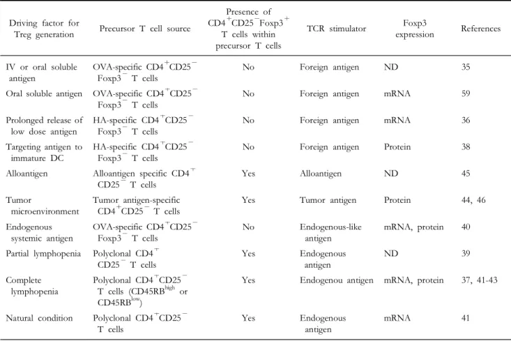
Table I. Evidence for peripheral generation of CD4+CD25+ regulatory T cells (mouse in vivo studies)
Driving factor for
Treg generation Precursor T cell source
Presence of CD4+CD25-Foxp3+
T cells within precursor T cells
TCR stimulator Foxp3
expression References
IV or oral soluble antigen
OVA-specific CD4+CD25- Foxp3- T cells
No Foreign antigen ND 35
Oral soluble antigen OVA-specific CD4+CD25- Foxp3- T cells
No Foreign antigen mRNA 59
Prolonged release of low dose antigen
HA-specific CD4+CD25- Foxp3- T cells
No Foreign antigen mRNA 36
Targeting antigen to immature DC
HA-specific CD4+CD25- Foxp3- T cells
No Foreign antigen Protein 38
Alloantigen Alloantigen specific CD4+ CD25- T cells
Yes Alloantigen ND 45
Tumor
microenvironment
Tumor antigen-specific CD4+CD25- T cells
Yes Tumor antigen Protein 44, 46
Endogenous systemic antigen
OVA-specific CD4+CD25- Foxp3- T cells
No Endogenous-like
antigen
mRNA, protein 40
Partial lymphopenia Polyclonal CD4+ CD25- T cells
Yes Endogenous
antigen
ND 39
Complete lymphopenia
Polyclonal CD4+CD25- T cells (CD45RBhigh or CD45RBlow)
Yes Endogenou antigen mRNA, protein 37, 41-43
Natural condition Polyclonal CD4+CD25- T cells
Yes Endogenous
antigen
mRNA 41
Treg: CD4+CD25+ regulatory T cell, Foxp3: Forkhead box P3, TCR: T cell receptor, IV: Intravenous, OVA: Ovalbumin, HA:
Hemagglutinin, ND: Not determined, DC: Dendritic cell.
Foxp3 expression level and suppressive activity of converted Tregs was inferior to those of naturally occurring Tregs. And the rate of conversion induced by IFN-γ was enhanced by TCR stimulation. These findings indicated that IFN-γ may contribute to the peripheral generation of Tregs in some conditions such as inflammation.
IL-2. Because CD4+CD25+ regulatory T cells constitutively express high affinity IL-2 receptor CD25, many researchers have investigated the role of IL-2 in the development and function of Tregs (40,68-71). According to the recent reports, IL-2 is not required for the thymic development of CD4+ CD25+ regulatory T cells but plays a critical role in the peripheral maintenance and function of Tregs (68-71). However, Abbas and colleagues announced that IL-2 is required for the peripheral generation of Tregs (40). Furthermore, two recent reports extended the conclusion deduced by Abbas and colleagues to
the point that IL-2 has a non-redundant role for TGF-β∼mediated induction of CD4+CD25+Foxp3
+ regulatory T cells and also enables induced-Tregs to expand (56,58). The essential role of IL-2 in the generation and expansion of iTregs cannot be compensated by other common γ-chain (γc) cyto- kines such as IL-4, IL-7 and IL-15 (56,58).
TCR stimulation and co-stimulatory molecules. Most of the above mentioned studies indicated that TCR sti- mulation is required for the peripheral generation of Tregs, except when IFN-γ was sufficient for the de novo generation of Tregs without TCR stimulation (54). TCR stimulators, in this context, include mi- togen (47-50,53,55,56,58), soluble foreign antigen (35,36,38,59), autologous antigen (37,41-43,52), alloantigen (45,51), tumor antigen (44,46,57) and so on.
Whereas CD28/B7 co-stimulation but not CTLA4/
B7 co-stimulation is essential for the generation of
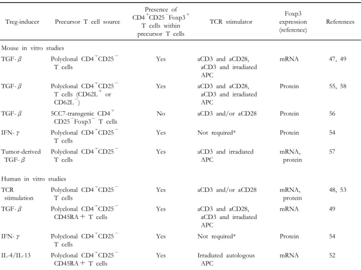
Table II. Experimental generation of CD4+CD25+ regulatory T cells in vitro
Treg-inducer Precursor T cell source
Presence of CD4+CD25-Foxp3+
T cells within precursor T cells
TCR stimulator
Foxp3 expression (reference)
References
Mouse in vitro studies
TGF-β Polyclonal CD4+CD25- T cells
Yes aCD3 and aCD28,
aCD3 and irradiated APC
mRNA 47, 49
TGF-β Polyclonal CD4+CD25- T cells (CD62L+ or CD62L-)
Yes aCD3 and aCD28,
aCD3 and irradiated APC
Protein 55, 58
TGF-β 5CC7-transgenic CD4+ CD25-Foxp3- T cells
No aCD3 and/or aCD28 Protein 56
IFN-γ Polyclonal CD4+CD25- T cells
Yes Not required* Protein 54
Tumor-derived TGF-β
Polyclonal CD4+CD25- T cells
Yes aCD3 and irradiated APC
mRNA, protein
57
Human in vitro studies TCR
stimulation
Polyclonal CD4+CD25- T cells
Yes aCD3 and/or aCD28 mRNA,
protein
48, 53
TGF-β Polyclonal CD4+CD25- CD45RA+ T cells
Yes aCD3 and aCD28,
aCD3 and irradiated APC
mRNA 49
IFN-γ Polyclonal CD4+CD25- T cells
Yes Not required* Protein 54
IL-4/IL-13 Polyclonal CD4+CD25- CD45RA+ T cells
Yes Irradiated autologous APC
mRNA 52
Treg: CD4+CD25+ regulatory T cell, Foxp3: Forkhead box P3, TCR: T cell receptor, APC: Antigen-presenting cell. *The rate of conversion was enhanced by the addition of anti-CD3 Ab.
nTregs (23), whether B7-mediated co- stimulation is required for the peripheral generation of Tregs remains unclear. There have been a few studies exploring the role of B7 co-stimulation in the de novo generation of CD4+CD25+ regulatory T cells (41,52,55). For example, Kosiewicz and colleagues reported that B7 co-stimulation is required for the conversion of CD4+CD25-T cells into CD4+CD25+ regulatory T cells in steady state (41). Schulze-Koops and colleagues have also shown that IL-4-induced generation of CD4+CD25+regulatory T cells in vitro is dependent upon B7 co-stimulation (52). Moreover, a recent study by Horwitz and colleagues described that CTLA-4 ligation of CD80 (B7-1) is required for TGF-β-mediated generation of Tregs and CD28/B7 co-stimulation, which is critical for the development of nTregs, is dispensable for generating TGF-β -induced Tregs (55). Distinct developmental require- ments for B7 co-stimulation of nTregs and iTregs may have implications for defining the intrinsic differences between the two cell populations.
Thymus. As mentioned earlier, the thymus is a site of nTreg development and a few mature peripheral T cells can re-circulate into the thymus (72). So, there is a possibility that conversion of CD4+CD25- T cells into CD4+CD25+ regulatory T cells occurs in the thymus. However, several studies have shown that thymectomized mice can efficiently generate Tregs in the periphery (36,39,41,45,46). Therefore, it looks like peripheral generation of Tregs is independent of the thymus.
Relative contribution of nTregs and iTregs to the total peripheral Treg pool
There is much debate about whether induced- Tregs substantially contribute to the total peripheral Treg pool. It has been suggested that a small number of nTregs may maintain peripheral tolerance by increasing total Treg number through converting CD4+CD25- T cells into CD4+CD25+ Tregs (51,73). This phenomenon is so-called infectious tolerance. However, recent reports using Foxp3 knock-in mice contradicted this possibility by showing that contribution of iTregs to the peripheral Treg pool is little, if any, during either immune response (29) or lymphopenia-induced proliferation (74). And a recent report by Freitas and colleagues consolidated this view using lymphopenia model (43).
Nevertheless, considering the differences in life span between mice and humans, iTregs may play a pivotal role for maintaining total peripheral Treg pool as the thymus begin to involute at least in humans (34). In line with this perspective, Akbar and colleagues proved that human CD4+CD25hiFoxp3+ regulatory T cells are derived by rapid turnover of
memory CD4+CD25- T cells especially in older subjects (75). Therefore, although nTregs make up a major part of the peripheral Treg pool, iTregs may also substantially contribute to the peripheral Treg pool throughout the life time.
Conclusion
Due to their potent suppressive activity and convenience of manipulation, CD4+CD25+ regula- tory T cells have been proved useful for the treatment of various diseases such as autoimmune disease (76,77), graft rejection (22) and allergy (78-80). However, it is difficult to obtain a sufficient number of CD4+CD25+ regulatory T cells from human blood as CD4+CD25+ regulatory T cell is a minor population in human PBMC (53). Therefore, generation and expansion of CD4+CD25+ regulatory T cells from relatively abundant CD4+CD25- T cells, using above-mentioned Treg-inducing stimulus, would be an alternative candidate for clinical application. To this end, it is important to establish standard methods for efficient generation and expansion of CD4+CD25+ regulatory T cells from CD4+CD25- T cells in vitro.
There has been remarkable progress in the peripherally induced CD4+CD25+Foxp3+regulatory T cell research field, but complexity has also increased as different researchers used different experimental systems. So, it is necessary to uncover the intrinsic differences between the systems and form a more precise definition of induced-Treg to resolve this problem. Furthermore, the physiological role of peripherally generated CD4+CD25+Foxp3+ regulatory T cells for maintaining peripheral tolerance would be an important open question in the future.
Acknowledgements
This work was supported by a Rheumatism Research Center grant by the Korean Science and Engineering Foundation (R11-2002-098-03002-0).
References
1. Sakaguchi S, Sakaguchi N, Asano M, Itoh M, Toda M:
Immunologic self-tolerance maintained by activated T cells expressing IL-2 receptor alpha-chains (CD25). Breakdown of a single mechanism of self-tolerance causes various auto- immune diseases. J Immunol 155;1151-1164, 1995 2. Sakaguchi S: Naturally arising CD4+ regulatory t cells for
immunologic self-tolerance and negative control of immune responses. Annu Rev Immunol 22;531-562, 2004
3. Kappler JW, Roehm N, Marrack P: T cell tolerance by clonal elimination in the thymus. Cell 49;273-280, 1987
4. Jones LA, Chin LT, Longo DL, Kruisbeek AM: Peripheral clonal elimination of functional T cells. Science 250;1726-
1729, 1990
5. Webb S, Morris C, Sprent J: Extrathymic tolerance of mature T cells: clonal elimination as a consequence of immunity. Cell 63;1249-1256, 1990
6. van Parijs L, Perez VL, Abbas AK: Mechanisms of peripheral T cell tolerance. Novartis Found Symp 215;5-14, 1998 7. Lechler R, Chai JG, Marelli-Berg F, Lombardi G: The
contributions of T-cell anergy to peripheral T-cell tolerance.
Immunology 103;262-269, 2001
8. Miller JFAP, Heath WR: Self-ignorance in the peripheral T-cell pool. Immunol Rev 133;131-150, 1993
9. Oliveira V, Agua-Doce A, Duarte J, Soares MP, Graca L:
Regulatory T cell maintenance of dominant tolerance:
induction of tissue self-defense? Transpl Immunol 17;7-10, 2006
10. Coutinho A, Hori S, Carvalho T, Caramalho I, Demengeot J: Regulatory T cells: the physiology of autoreactivity in dominant tolerance and "quality control" of immune responses. Immunol Rev 182;89-98, 2001
11. Groux H, O'Garra A, Bigler M, Rouleau M, Antonenko S, de Vries JE, Roncarolo MG: A CD4+T-cell subset inhibits antigen-specific T-cell responses and prevents colitis. Nature 389;737-742, 1997
12. Chen Y, Kuchroo VK, Inobe J, Hafler DA, Weiner HL:
Regulatory T cell clones induced by oral tolerance:
suppression of autoimmune encephalomyelitis. Science 265;
1237-1240, 1994
13. Zou W: Regulatory T cells, tumour immunity and im- munotherapy. Nat Rev Immunol 6;295-307, 2006
14. Piccirillo CA, Thornton AM: Cornerstone of peripheral tolerance: naturally occurring CD4+CD25+ regulatory T cells. Trends Immunol 25;374-380, 2004
15. Piccirillo CA, Shevach EM: Naturally-occurring CD4+CD25+
immunoregulatory T cells: central players in the arena of peripheral tolerance. Semin Immunol 16;81-88, 2004 16. Nishizuka Y, Sakakura T: Thymus and reproduction:
sex-linked dysgenesia of the gonad after neonatal thymec- tomy in mice. Science 166;753-755, 1969
17. Wing K, Suri-Payer E, Rudin A: CD4+CD25+-regulatory T cells from mouse to man. Scand J Immunol 62;1-15, 2005 18. Hori S, Nomura T, Sakaguchi S: Control of regulatory T cell development by the transcription factor Foxp3. Science 299;1057-1061, 2003
19. Fontenot JD, Gavin MA, Rudensky AY: Foxp3 programs the development and function of CD4+CD25+ regulatory T cells. Nat Immunol 4;330-336, 2003
20. Shevach EM: CD4+ CD25+ suppressor T cells: more questions than answers. Nat Rev Immunol 2;389-400, 2002 21. Mills KH: Regulatory T cells: friend or foe in immunity to
infection? Nat Rev Immunol 4;841-855, 2004
22. Wood KJ, Sakaguchi S: Regulatory T cells in transplantation tolerance. Nature Reviews Immunology 3;199-210, 2003
23. Liston A, Rudensky AY: Thymic development and peripheral homeostasis of regulatory T cells. Curr Opin Immunol 19;176-185, 2007
24. Pacholczyk R, Ignatowicz H, Kraj P, Ignatowicz L: Origin and T cell receptor diversity of Foxp3+CD4+CD25+ T cells. Immunity 25;249-259, 2006
25. Asano M, Toda M, Sakaguchi N, Sakaguchi S: Autoimmune disease as a consequence of developmental abnormality of a T cell subpopulation. J Exp Med 184;387-396, 1996 26. Itoh M, Takahashi T, Sakaguchi N, Kuniyasu Y, Shimizu J,
Otsuka F, Sakaguchi S: Thymus and autoimmunity: pro- duction of CD25+CD4+ naturally anergic and suppressive T cells as a key function of the thymus in maintaining immunologic self-tolerance. J Immunol 162;5317-5326, 1999 27. Cosmi L, Liotta F, Lazzeri E, Francalanci M, Angeli R, Mazzinghi B, Santarlasci V, Manetti R, Vanini V, Romagnani P, Maggi E, Romagnani S, Annunziato F: Human CD8+
CD25+ thymocytes share phenotypic and functional features with CD4+CD25+ regulatory thymocytes. Blood 102;
4107-4114, 2003
28. Wing K, Ekmark A, Karlsson H, Rudin A, Suri-Payer E:
Characterization of human CD25+CD4+T cells in thymus, cord and adult blood. Immunology 106;190-199, 2002 29. Fontenot JD, Rasmussen JP, Williams LM, Dooley JL, Farr
AG, Rudensky AY: Regulatory T cell lineage specification by the forkhead transcription factor foxp3. Immunity 22;329- 341, 2005
30. Akbar AN, Taams LS, Salmon M, Vukmanovic-Stejic M: The peripheral generation of CD4+CD25+ regulatory T cells.
Immunology 109;319-325, 2003
31. Bluestone JA, Abbas AK: Natural versus adaptive regulatory T cells. Nat Rev Immunol 3;253-257, 2003
32. Taams LS, Akbar AN: Peripheral generation and function of CD4+CD25+ regulatory T cells. Curr Top Microbiol Immunol 293;115-131, 2005
33. Lohr J, Knoechel B, Abbas AK: Regulatory T cells in the periphery. Immunol Rev 212;149-162, 2006
34. Akbar AN, Vukmanovic-Stejic M, Taams LS, Macallan DC:
The dynamic co-evolution of memory and regulatory CD4+
T cells in the periphery. Nat Rev Immunol 7;231-237, 2007 35. Thorstenson KM, Khoruts A: Generation of anergic and potentially immunoregulatory CD25+CD4 T cells in vivo after induction of peripheral tolerance with intravenous or oral antigen. J Immunol 167;188-195, 2001
36. Apostolou I, von Boehmer H: In vivo instruction of suppressor commitment in naive T cells. J Exp Med 199;1401-1408, 2004
37. Curotto de Lafaille MA, Lino AC, Kutchukhidze N, Lafaille JJ: CD25- T cells generate CD25+Foxp3+ regulatory T cells by peripheral expansion. J Immunol 173;7259-7268, 2004
38. Kretschmer K, Apostolou I, Hawiger D, Khazaie K,
Nussenzweig MC, von Boehmer H: Inducing and expanding regulatory T cell populations by foreign antigen. Nat Immunol 6;1219-1227, 2005
39. Laurie KL, Van Driel IR, Gleeson PA: The role of CD4+
CD25+ immunoregulatory T cells in the induction of autoimmune gastritis. Immunol Cell Biol 80;567-573, 2002 40. Knoechel B, Lohr J, Kahn E, Bluestone JA, Abbas AK:
Sequential development of interleukin 2-dependent effector and regulatory T cells in response to endogenous systemic antigen. J Exp Med 202;1375-1386, 2005
41. Liang S, Alard P, Zhao Y, Parnell S, Clark SL, Kosiewicz MM: Conversion of CD4+ CD25- cells into CD4+ CD25+
regulatory T cells in vivo requires B7 costimulation, but not the thymus. J Exp Med 201;127-137, 2005
42. Zelenay S, Lopes-Carvalho T, Caramalho I, Moraes-Fontes MF, Rebelo M, Demengeot J: Foxp3+ CD25- CD4 T cells constitute a reservoir of committed regulatory cells that regain CD25 expression upon homeostatic expansion. Proc Natl Acad Sci USA 102;4091-4096, 2005
43. Almeida ARM, Zaragoza B, Freitas AA: Competition con- trols the rate of transition between the peripheral pools of CD4+CD25- and CD4+CD25+ T cells. Int Immunol 18;1607-1613, 2006
44. Zhou G, Levitsky HI: Natural regulatory T cells and de novo-induced regulatory T cells contribute independently to tumor-specific tolerance. J Immunol 178;2155-2162, 2007 45. Karim M, Kingsley CI, Bushell AR, Sawitzki BS, Wood KJ:
Alloantigen-induced CD25+CD4+ regulatory T cells can develop in vivo from CD25-CD4+ precursors in a thymus-independent process. J Immunol 172;923-928, 2004 46. Valzasina B, Piconese S, Guiducci C, Colombo MP: Tumor- induced expansion of regulatory T cells by conversion of CD4+CD25- lymphocytes is thymus and proliferation independent. Cancer Res 66;4488-4495, 2006
47. Chen W, Jin W, Hardegen N, Lei KJ, Li L, Marinos N, McGrady G, Wahl SM: Conversion of peripheral CD4+
CD25- naive T cells to CD4+CD25+ regulatory T cells by TGF-beta induction of transcription factor Foxp3. J Exp Med 198;1875-1886, 2003
48. Walker MR, Kasprowicz DJ, Gersuk VH, Benard A, Van Landeghen M, Buckner JH, Ziegler SF: Induction of FoxP3 and acquisition of T regulatory activity by stimulated human CD4+CD25- T cells. J Clin Invest 112;1437-1443, 2003 49. Fantini MC, Becker C, Monteleone G, Pallone F, Galle PR, Neurath MF: Cutting edge: TGF-{beta} induces a regulatory phenotype in CD4+CD25- T cells through Foxp3 induction and down-regulation of Smad7. J Immunol 172;5149-5153, 2004
50. Park HB, Paik DJ, Jang E, Hong S, Youn J: Acquisition of anergic and suppressive activities in transforming growth factor-beta-costimulated CD4+CD25- T cells. Int Immu- nol 16;1203-1213, 2004
51. Zheng SG, Wang JH, Gray JD, Soucier H, Horwitz DA:
Natural and induced CD4+CD25+ cells educate CD4+
CD25- cells to develop suppressive activity: the role of IL-2, TGF-beta, and IL-10. J Immunol 172;5213-5221, 2004 52. Skapenko A, Kalden JR, Lipsky PE, Schulze-Koops H: The IL-4 receptor alpha-chain-binding cytokines, IL-4 and IL-13, induce forkhead box P3-expressing CD25+CD4+ regulato- ry T cells from CD25-CD4+ precursors. J Immunol 175;6107-6116, 2005
53. Walker MR, Carson BD, Nepom GT, Ziegler SF, Buckner JH: De novo generation of antigen-specific CD4+CD25+
regulatory T cells from human CD4+CD25- cells. Proc Natl Acad Sci USA 102;4103-4108, 2005
54. Wang Z, Hong J, Sun W, Xu G, Li N, Chen X, Liu A, Xu L, Sun B, Zhang JZ: Role of IFN-{gamma} in induction of Foxp3 and conversion of CD4+ CD25- T cells to CD4+
Tregs. J Clin Invest 116;2434-2441, 2006
55. Zheng SG, Wang JH, Stohl W, Kim KS, Gray JD, Horwitz DA: TGF-beta requires CTLA-4 early after T cell activation to induce FoxP3 and generate adaptive CD4+CD25+
regulatory cells. J Immunol 176;3321-3329, 2006
56. Davidson TS, Dipaolo RJ, Andersson J, Shevach EM:
Cutting edge: IL-2 is essential for TGF-beta-mediated induction of Foxp3+ T regulatory cells. J Immunol 178;4022-4026, 2007
57. Liu VC, Wong LY, Jang T, Shah AH, Park I, Yang X, Zhang Q, Lonning S, Teicher BA, Lee C: Tumor evasion of the immune system by converting CD4+CD25- T cells into CD4+CD25+ T regulatory cells: role of tumor-derived TGF-beta. J Immunol 178;2883-2892, 2007
58. Zheng SG, Wang J, Wang P, Gray JD, Horwitz DA: IL-2 is essential for TGF-beta to convert naive CD4+CD25- Cells to CD25+Foxp3+ Regulatory T Cells and for Expansion of These Cells. J Immunol 178;2018-2027, 2007 59. Mucida D, Kutchukhidze N, Erazo A, Russo M, Lafaille JJ, Curotto de Lafaille MA: Oral tolerance in the absence of naturally occurring Tregs. J Clin Invest 115;1923-1933, 2005 60. Ziegler SF: FOXP3: not just for regulatory T cells anymore.
Eur J Immunol 37;21-23, 2007
61. Wang J, Ioan-Facsinay A, van der Voort EI, Huizinga TW, Toes RE: Transient expression of FOXP3 in human activated nonregulatory CD4+ T cells. Eur J Immunol 37;
129-138, 2007
62. Youssef AR, Shen CR, Lin CL, Barker RN, Elson CJ: IL-4 and IL-10 modulate autoimmune haemolytic anaemia in NZB mice. Clin Exp Immunol 139;84-89, 2005
63. Ostlie N, Milani M, Wang W, Okita D, Conti-Fine BM:
Absence of IL-4 facilitates the development of chronic autoimmune myasthenia gravis in C57BL/6 mice. J Immunol 170;604-612, 2003
64. Young DA, Lowe LD, Booth SS, Whitters MJ, Nicholson L, Kuchroo VK, Collins M: IL-4, IL-10, IL-13, and
TGF-beta from an altered peptide ligand-specific Th2 cell clone down-regulate adoptive transfer of experimental auto- immune encephalomyelitis. J Immunol 164;3563-3572, 2000 65. Willenborg DO, Fordham S, Bernard CC, Cowden WB, Ramshaw IA: IFN-gamma plays a critical down-regulatory role in the induction and effector phase of myelin oligo- dendrocyte glycoprotein-induced autoimmune encephalo- myelitis. J Immunol 157;3223-3227, 1996
66. Krakowski M, Owens T: Interferon-gamma confers re- sistance to experimental allergic encephalomyelitis. Eur J Immunol 26;1641-1646, 1996
67. Ferber IA, Brocke S, Taylor-Edwards C, Ridgway W, Dinisco C, Steinman L, Dalton D, Fathman CG: Mice with a disrupted IFN-gamma gene are susceptible to the induction of experimental autoimmune encephalomyelitis (EAE). J Immunol 156;5-7, 1996
68. Setoguchi R, Hori S, Takahashi T, Sakaguchi S: Homeostatic maintenance of natural Foxp3(+) CD25(+) CD4(+) regulatory T cells by interleukin (IL)-2 and induction of autoimmune disease by IL-2 neutralization. J Exp Med 201;
723-735, 2005
69. Fontenot JD, Rasmussen JP, Gavin MA, Rudensky AY: A function for interleukin 2 in Foxp3-expressing regulatory T cells. Nat Immunol 6;1142-1151, 2005
70. Zorn E, Nelson EA, Mohseni M, Porcheray F, Kim H, Litsa D, Bellucci R, Raderschall E, Canning C, Soiffer RJ, Frank DA, Ritz J: IL-2 regulates FOXP3 expression in human CD4+
CD25+ regulatory T cells through a STAT-dependent mechanism and induces the expansion of these cells in vivo.
Blood 108;1571-1579, 2006
71. Fehervari Z, Yamaguchi T, Sakaguchi S: The dichotomous role of IL-2: tolerance versus immunity. Trends Immunol 27;109-111, 2006
72. Surh CD, Sprent J, Webb SR: Exclusion of circulating T cells from the thymus does not apply in the neonatal period. J Exp Med 177;379-385, 1993
73. Jonuleit H, Schmitt E, Kakirman H, Stassen M, Knop J, Enk AH: Infectious tolerance: human CD25+ regulatory T cells convey suppressor activity to conventional CD4+ T helper cells. J Exp Med 196;255-260, 2002
74. Wan YY, Flavell RA: Identifying Foxp3-expressing suppre- ssor T cells with a bicistronic reporter. Proc Natl Acad Sci USA 102;5126-5131, 2005
75. Vukmanovic-Stejic M, Zhang Y, Cook JE, Fletcher JM, McQuaid A, Masters JE, Rustin MH, Taams LS, Beverley PC, Macallan DC, Akbar AN: Human CD4+ CD25hi Foxp3+ regulatory T cells are derived by rapid turnover of memory populations in vivo. J Clin Invest 116;2423-2433, 2006
76. Baecher-Allan C, Hafler DA: Human regulatory T cells and their role in autoimmune disease. Immunol Rev 212;203-216, 2006
77. Sakaguchi S, Ono M, Setoguchi R, Yagi H, Hori S, Fehervari Z, Shimizu J, Takahashi T, Nomura T: Foxp3+ CD25+
CD4+ natural regulatory T cells in dominant self-tolerance and autoimmune disease. Immunol Rev 212;8-27, 2006 78. Wing K, Sakaguchi S: Regulatory T cells as potential immu-
notherapy in allergy. Curr Opin Allergy Clin Immunol 6;
482-488, 2006
79. Ahern DJ, Robinson DS: Regulatory T cells as a target for induction of immune tolerance in allergy. Curr Opin Allergy Clin Immunol 5;531-536, 2005
80. Akbari O, Stock P, DeKruyff RH, Umetsu DT: Role of regulatory T cells in allergy and asthma. Curr Opin Immunol 15;627-633, 2003