Endocrinol Metab 2015;30:584-592
http://dx.doi.org/10.3803/EnM.2015.30.4.584 pISSN 2093-596X · eISSN 2093-5978
Original Article
Thyroid Hormone Regulates the mRNA Expression of Small Heterodimer Partner through Liver Receptor Homolog-1
Hwa Young Ahn1,2, Hwan Hee Kim1, Ye An Kim1, Min Kim1, Jung Hun Ohn1, Sung Soo Chung1, Yoon-Kwang Lee3, Do Joon Park1, Kyong Soo Park1, David D. Moore3, Young Joo Park1
1Department of Internal Medicine, Seoul National University College of Medicine; 2Department of Internal Medicine, Chung-Ang University College of Medicine, Seoul, Korea; 3Department of Molecular and Cellular Biology, Baylor College of Medicine, Houston, TX, USA
Background: Expression of hepatic cholesterol 7α-hydroxylase (CYP7A1) is negatively regulated by orphan nuclear receptor small heterodimer partner (SHP). In this study, we aimed to find whether thyroid hormone regulates SHP expression by modulat- ing the transcriptional activities of liver receptor homolog-1 (LRH-1).
Methods: We injected thyroid hormone (triiodothyronine, T3) to C57BL/6J wild type. RNA was isolated from mouse liver and used for microarray analysis and quantitative real-time polymerase chain reaction (PCR). Human hepatoma cell and primary he- patocytes from mouse liver were used to confirm the effect of T3 in vitro. Promoter assay and electrophoretic mobility-shift assay (EMSA) were also performed using human hepatoma cell line.
Results: Initial microarray results indicated that SHP expression is markedly decreased in livers of T3 treated mice. We con- firmed that T3 repressed SHP expression in the liver of mice as well as in mouse primary hepatocytes and human hepatoma cells by real-time PCR analysis. LRH-1 increased the promoter activity of SHP; however, this increased activity was markedly de- creased after thyroid hormone receptor β/retinoid X receptor α/T3 administration. EMSA revealed that T3 inhibits specific LRH- 1 DNA binding.
Conclusion: We found that thyroid hormone regulates the expression of SHP mRNA through interference with the transcription factor, LRH-1.
Keywords: Small heterodimer partner; Thyroid hormones; Cholesterol; Liver receptor homolog-1; Bile acids and salts
Received: 3 April 2015, Revised: 24 August 2015, Accepted: 24 September 2015
Corresponding authors: Young Joo Park
Department of Internal Medicine, Seoul National University College of Medicine, 101 Daehak-ro, Jongno-gu, Seoul 03080, Korea
Tel: +82-2-2072-4183, Fax: +82-2-764-2199, E-mail: [email protected] David D. Moore
Department of Molecular and Cellular Biology, Baylor College of Medicine, One Baylor Plaza, Houston, Texas 77030, USA
Tel: +1-713-798-3313, Fax: +1-713-798-3017, E-mail: [email protected]
Copyright © 2015 Korean Endocrine Society
This is an Open Access article distributed under the terms of the Creative Com- mons Attribution Non-Commercial License (http://creativecommons.org/
licenses/by-nc/3.0/) which permits unrestricted non-commercial use, distribu- tion, and reproduction in any medium, provided the original work is properly cited.
pISSN 2093-596X · eISSN 2093-5978
INTRODUCTION
Small heterodimer partner (SHP) is an orphan nuclear receptor that, unlike other nuclear receptors, contains a putative ligand binding domain but lacks a classical DNA binding domain [1].
SHP mRNA is predominantly expressed in the liver, but it is also expressed in the adrenal gland, spleen, small intestine [2], and pancreas [3]. SHP has also been shown to interact with other nuclear receptors and transcription factors [4].
The expression of SHP is regulated by several other nuclear receptors. The orphan nuclear receptor, steroidogenic factor 1 (SF-1) and its liver homologue, liver receptor homolog-1 (LRH-1) transactivate the SHP promoter [5]. To date, at least 5 SF-1 binding sites have been identified in the promoter region of SHP [5], and LRH-1 is essential for basal SHP expression.
When SHP protein expression is elevated by LRH-1, SHP forms a heterodimeric SHP/LRH-1 complex and this complex inactivates LRH-1 thus reducing SHP expression, which is an established auto-regulatory negative feedback loop for SHP [6].
Farnesoid X receptor (FXR) is another well-known inducer of SHP gene expression; an FXR binding site is located in the SHP promoter region [7]. In addition, liver X receptor α (LXRα) and hepatocyte nuclear factor 4α (HNF4 α) directly regulate SHP promoter activity, and their respective binding sites have been previously identified [8,9]. Other nuclear re- ceptors and inducers, such as estrogen receptor-related receptor γ and sterol regulatory element binding protein-1 (SREBP-1), have also been known to regulate SHP expression [4].
In the liver, SHP is known to perform several metabolic functions. It regulates bile acid synthesis and cholesterol me- tabolism by modulating cholesterol 7α-hydroxylase (CYP7A1) expression through inhibition of LRH-1 and HNF4α activity [10]. In addition, SHP regulates lipogenesis by inhibiting SREBP-1c expression via LXR [11], and serum triglyceride levels by repressing microsomal triglyceride transfer protein (MTP) expression by inhibiting LRH-1 binding to the MTP promoter [12].
Thyroid hormone is known to regulate cholesterol and bile acid metabolism, mainly through regulation of 3-hydroxy- 3-methyl-glutaryl-CoA (HMG-CoA) reductase [13], low densi- ty lipoprotein (LDL) receptor [14], and CYP7A1 gene expres- sion [15]. SHP has also been shown to play an important role in cholesterol metabolism [6] in the liver, mainly through tran- scriptional regulation of the CYP7A1 gene, which suggests a possible relationship or interaction between SHP and thyroid hormone. In our preliminary data using microarray analysis, we
found that SHP expression was decreased by thyroid hormone.
Thyroid hormone acts mainly through its nuclear receptors, thyroid hormone receptor (TR) α and β. TRβ1 is putatively ex- pressed in the liver [16]. The functional TR complex consists of a heterodimer with retinoid X receptor (RXR). This complex then binds to a thyroid hormone response element (TRE) and regulates gene expression [17]. Although we observed that thy- roid hormone inhibited SHP expression, we were unable to identify TR binding to a TRE in the SHP promoter. However, TR could function through interactions with other nuclear re- ceptors, like peroxisome proliferator-activated receptor (PPAR) and LXR [16,18] at the level of transcription as well as by di- rect binding to TREs in the promoter region. Therefore, we speculated that TRβ might regulate SHP expression by inter- acting with other transcription factors that regulate SHP pro- moter. To confirm this hypothesis, we performed this study to find whether thyroid hormone regulates SHP expression by modulating the transcriptional activities of LRH-1, which is re- quired for basal SHP promoter activity in the liver.
METHODS
Animals
C57BL/6J wild type mice were used. Mice were housed in groups of 4 or 5 in plastic microisolator cages at 22°C with a 12-hour light/dark cycle. All animals were provided laboratory chow diet (Purina irradiated laboratory chow 38057, Purina Korea, Seoul, Korea) and water ad libitum. Animals were di- vided into 3 groups according to the duration of thyroid hor- mone treatment (n=4 or n=5 in each group). All experiments were performed in triplicate to confirm the results. Thyroid hormone, 3,5,3ʹ-triiodothyronine (T3; Sigma Chemical Co., St.
Louis, MO, USA) was prepared at a concentration of 1 mg/kg body weight in 20% DMSO and was administered via intra- peritoneal injection. Six hours group: T3 treated group was ad- ministered T3 6 hours before sacrifice. Five days group: the T3 treated group was administered T3 once daily for 5 days. Ani- mals were sacrificed 24 hours after the last T3 injection. All animals were sacrificed after fasting for 6 hours beginning at 6:00 AM. Mice were anesthetized by an intraperitoneal injec- tion of Zoletil (Virvac, Carros, France). Blood was drawn by inferior vena cava puncture. The liver was quickly removed, frozen in liquid nitrogen, and then used for RNA extraction. All procedures were performed in accordance with the guidelines of the Seoul National University Bundang Hospital Animal Policy and Welfare Committee.
Cholesterol measurement
Serum was prepared from whole blood by centrifugation at 1,200 ×g for 15 minutes. Serum total cholesterol was mea- sured with an enzymatic assay kit (Thermo DMA Inc., St. Lou- isville, CO, USA) following the manufacturer’s protocol.
Cell culture and transient transfection
Hepatocytes were isolated from wild type mice. Animals were anesthetized with Zoletil. The inferior vena cava was cannulat- ed, and the portal vein was severed. Collagenase dissolved in Hanks’ balanced salt solution (GIBCO Laboratories, Grand Is- land, NY, USA) was perfused through the liver at 37°C to dis- perse hepatocytes, and then the hepatocytes were further puri- fied by Percoll (Sigma). The cells were seeded into collagen- coated 6-well plates at density of 1 to 2×10⁶ cells/well in 2 mL of William’s E medium supplemented with 10% fetal calf se- rum, penicillin (100 U/mL), streptomycin (0.1 mg/mL), insulin (10 mg/mL), and triamcinolone (1 mmol/L). We also used HepG2 and HEK293 cells. Cells were grown at 37°C in a hu- midified atmosphere of 5% CO2/95% air. HepG2 cells were maintained in modified Eagle’s medium plus 10% fetal bovine serum (FBS). T3 (100 nmol/L) was incubated with primary he- patocytes and HepG2 cells according to the response time (control 2, and 6 hours). After T3 treatment, RNA was isolated from the cells.
One day before transfection, confluent cells were trypsinized and plated into 6-well plates at a 1:4 ratio to allow the cells to reach 50% to 60% confluency at the time of transfection. Six hours before transfection, fresh medium containing 10% char- coal-stripped FBS was added to the cells. All transfections were performed using the calcium phosphate method. Lucifer- ase values shown are the averages of triplicate samples. All plasmids used in this study were previously reported. Various 5ʹ deletions of the human SHP promoter [5], the human SHP promoter [19], LRH-1 [19], TRβ [20], and RXRα [21] were used.
RNA isolation and quantitative real-time polymerase chain reaction
Total RNA was isolated from frozen liver samples and cells us- ing TRIzol Reagent (Invitrogen, Carlsbad, CA, USA) accord- ing the manufacturer’s instruction. First-strand cDNA was syn- thesized from 1 µg of RNA using superscript II reverse tran- scriptase (Invitrogen). Quantitative real-time polymerase chain reaction (PCR) was performed using SYBR Green PCR master mix and an ABI prism 7500 Sequence Detection System (Ap-
plied Biosystems, Foster City, CA, USA). The PCR primers used for each gene are listed below (5ʹ to 3ʹ): mouse SHP-F (forward), TTG CAC CTG CAT CTC ACA GC; SHP-R (re- verse), TCT TGG CTA GGA CAT CCA AG; glyceraldehyde 3-phosphate dehydrogenase (GAPDH)-F, TGT GTC CGT CGT GGATCT GA; GAPDH-R, CCT GCT TCA CCA CCT TCT TGA; human SHP-F, GGA CTT CCT TGG TTT GGA CA; SHP-R, CTC ATC CCA AGA AGG GAC AG; GAPDH-F, GAT CAT CAG CAA TGC CTC CT; GAPDH-R, TGT GGT CAT GAG TCC TTC CA. Relative mRNA expression was quantified using the comparative cycle threshold (Ct) method and expressed as 2–ΔΔCt.
Identification of differentially expressed genes
RNA was hybridized to Affymetrix GeneChip Human Gene 1.0 ST Arrays (Affymetrix, Santa Clara, CA, USA). Robust mul- tichip analysis was used to normalize the expression data in the Expression Console software (Affymetrix) [22]. We calculated the log fold change of each probe in different conditions and independent samples. Student t test was performed to deter- mine the significance of differential expression using GenePat- tern software [23].
Electrophoretic mobility-shift assay
Double-stranded oligonucleotides of the following sequences were used as probes: LRH-1 response element (LRE) in the hu- man SHP promoter (LRE WT (–518 to –489), 5ʹ-GAAGCA- GGGCCCAAGGTTAGGCAACAAG-3ʹ, LRE is underlined);
LRE mutant competitor (LRE MT, 5ʹ-GAAGCAGGGCCCttc- caaAGCAACAAG-3ʹ); consensus LRE (5ʹ-GACTTCTGGAGT CAAGGTTGTTGGGCCATTC-3ʹ). The probes were labeled with [α-32P] dATP using Klenow polymerase (Roche, Mannheim, Germany), and the labeled probe (35,000 cpm) was incubated with nuclear extracts of HepG2 cells treated with 100 nmol/L T3 in 10 mmol/L Hepes (pH 7.9) containing 50 mmol/L KCl, 0.1 mmol/L ethylenediaminetetraacetic acid, 0.25 mmol/L DTT, 0.1 mg/mL poly (dIdC), 0.01% nonidet P-40, and 10% glycerol at room temperature for 10 minutes. Competitors were added in 50- fold molar excess to the labeled probe.
Statistical analysis
Values are expressed as the mean±SD. The significance of dif- ferences between mean values was evaluated using Student t test. A P<0.05 was considered statistically significant.
RESULTS
Thyroid hormone regulates the expression of genes involved in bile acid synthesis and cholesterol metabolism Before conducting the microarray analyses, to confirm the ef- fect of thyroid hormone on serum cholesterol, we measured se- rum cholesterol levels after intraperitoneal administration of T3 (1 mg/kg) to mice for 5 days. Serum total cholesterol levels de- creased significantly after T3 injection (101.7±12.0 mg/dL vs.
47.0±9.7 mg/dL, P<0.05). We profiled the transcriptional changes in mouse liver before and after 5 days of T3 treatment and examined the expression of genes related to the regulation of cholesterol and bile acid metabolism.
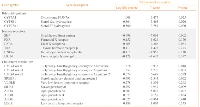
In microarray analyses (Table 1), genes related to cholesterol metabolism, HMG-CoA synthase and reductase was up-regu- lated by thyroid hormone. Among genes associated with high density lipoprotein cholesterol, apolipoprotein A-1 was up-reg- ulated and scavenger receptor was down-regulated by thyroid hormone. However, the LDL receptor, the main receptor re- sponsible for clearing cholesterol-laden LDL particles from the blood, was not significantly up-regulated by thyroid hormone, which is different from the results of previous studies [14,24].
Among the genes related to bile acid synthesis, CYP7A1 and mitochondrial sterol 27 hydroxylase (Cyp27a1) were up-regu- lated after T3 treatment, although the fold change of Cyp27a1 was small. In a previous study, thyroxine enhanced CYP7A1 expression but not Cyp27a1 expression in rat hepatocytes when added to dexamethasone-containing medium [25].
The expression of CYP7A1 and SHP was regulated by sev- eral transcription factors, such as FXR, LRH-1, HNF-4α, and LXRα [4,26]. However, expression of these transcription fac- tors did not significantly change in response to thyroid hor- mone in microarray analysis (Table 1). In contrast, the expres- sion of SHP was markedly decreased by thyroid hormone;
therefore, we assumed that thyroid hormone regulates SHP ex- pression independent of other transcription factors, and might regulate A expression indirectly through changes in SHP ex- pression.
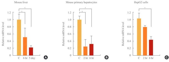
To confirm this, we examined the changes in SHP mRNA level after T3 in mouse liver by real-time PCR. Level of serum T3 was significantly increased in 6-hour and 5-day group than control (231.4±42.4 and 310.2±65.8 ng/dL vs. 70.1±20.7 ng/
dL). As expected, the expression of SHP decreased after treat- ment with T3 for 5 days (Fig. 1A).
Table 1. Changes in Gene Expression after 5 Days of T3 Treatment in Mouse Liver
Gene symbol Gene description T3 treatment vs. control
Log fold changea T-score P value
Bile acid synthesis CYP7A1 CYP8B1 CYP27A1
Cytochrome P450 7A Sterol 12α-hydroxylase Sterol 27-hydroxylase
1.008 –0.364 0.500
3.477 –3.465 3.784
0.025 0.026 0.020 Nuclear receptors
SHPFXR LXRαTRβ HNF4α LRH-1
Small heterodimer partner Farnesoid X receptor Liver X receptor α Thyroid hormone receptor β Hepatocyte nuclear receptor 4α Liver receptor homolog-1
–0.698 0.152 0.465 0.119 –0.115 –0.128
–7.061 1.624 2.266 1.423 –1.955 –1.623
0.002 0.176 0.083 0.225 0.118 0.177 Cholesterol metabolism
HMG-CoA R HMG-CoA S1 HMG-CoA S2 SREBP1 VLDLR SR-B1 APOA1 APOBAPOE LDLR
3-Hydroxy-3-methylglutaryl-coenzyme A reductase 3-Hydroxy-3-methylglutaryl-coenzyme A synthase 1 3-Hydroxy-3-methylglutaryl-coenzyme A synthase 2 Sterol regulatory element binding protein 1 Very low density lipoprotein receptor Scavenger receptor
Apolipoprotein A1 Apolipoprotein B Apolipoprotein E
Low density lipoprotein receptor
1.516 1.252 0.074 –0.593 –0.278 –0.792 0.201 0.077 –0.025 0.386
3.935 5.776 0.699 –2.543 –1.301 –4.942 5.047 0.158 –0.864 1.007
0.018 0.005 0.529 0.062 0.262 0.009 0.007 0.884 0.440 0.373
aThe fold changes are shown in log scale.
1.2 1.0 0.8 0.6 0.4 0.2 0
1.2 1.0 0.8 0.6 0.4 0.2 0
1.2 1.0 0.8 0.6 0.4 0.2 0
a a
a a a
Relative mRNA level Relative mRNA level Relative mRNA level
Mouse liver Mouse primary hepatocytes HepG2 cells
C 6 hr 5 day A C 2 hr 6 hr B C 2 hr 6 hr C
Fig. 1. Effect of thyroid hormone (T3) treatment on small heterodimer partner (SHP) expression. Comparison of SHP expression in mouse liver (A, n=4 or n=5 in each group) after 6 hours or 5 days of thyroid hormone treatment (1 mg/g). Comparison of SHP expres- sion in mouse primary hepatocytes (B) and human hepatoma cells (C) after 2 and 6 hours of T3 treatment (100 nmol/L). aSignificant dif- ferences compared to the control are P<0.05.
Fig. 2. Effect of thyroid hormone (T3) on small heterodimer partner (SHP) promoter activity with or without thyroid hormone receptor (TR)/retinoid X receptor (RXR), and liver receptor homolog-1 (LRH-1). In each HepG2 cell sample, 300 ng of mouse SHP (A) or hu- man SHP (B, C) promoter DNA was co-transfected with or without 75 ng of TRβ and 75 ng of RXRα or 75 ng of LRH-1. Vehicle or T3 (100 nmol/L) was administered for 24 hours to determine the effect of thyroid hormone on SHP promoter expression. Luciferase activity was measured and normalized to β-galactosidase activity. Significant differences compared to the control are aP<0.05 and bP<0.01.
6 5 4 3 2 1 0
a
b b
a
a
Relative luciferase activity
Human SHP Luc
TRβ/RXRα LRH-1
T3 100 nmol/L – + – + – + – +
– – – – – + – +
– – + + – – + + C
2.5 2.0 1.5 1.0 0.5 0
3.0 2.5 2.0 1.5 1.0 0.5 0
a a a a
Relative luciferase activity Relative luciferase activity
Mouse SHP Human SHP
Control TRβ/RXRα Control TRβ/RXRα
Vehicle T3 100 nmol/L
Vehicle T3 100 nmol/L
A B
To confirm the specific regulatory effects of T3 on the ex- pression of SHP, an in vitro study was performed using freshly isolated primary hepatocytes and human hepatoma HepG2 cells. Hepatocytes from C57BL/6L mice were isolated, and T3 (100 nmol/L) or vehicle was added to the culture media. As shown Fig. 1B, the expression of SHP decreased at 2 and 6 hours after T3 treatment. In HepG2 cells, T3 also decreased SHP expression after 6 hours of T3 treatment (Fig. 1C). Al- though the time sequence differed, T3 decreased the expression of SHP in both human and mouse; therefore, we evaluated the mechanism underlying the regulation of SHP by T3.
TRβ/RXRα plus T3 decreases SHP promoter activity by interfering with LRH-1
We used full-length (~2 Kb) mouse and human SHP promoter constructs [5] to determine whether TRβ affects the activity of the SHP promoter. We cotransfected HepG2 cells with SHP promoters along with an empty or TRβ/RXRα expression vec- tor and evaluated the effect of vehicle or T3 (100 nmol/L) ad- ministered over 24 hours on promoter activity. T3 treatment alone did not have a significant effect on mouse or human SHP promoter activity (Fig. 2A, B). When the SHP promoter was cotransfected with TRβ/RXRα in the absence of T3, SHP pro- moter expression increased. However, this increased activity decreased significantly after administration of T3 (Fig. 2A, B).
This result suggests that unliganded TRβ/RXRα increases the activity of the SHP promoter and T3/TRβ/RXRα repressed ac- tivity of SHP.
Chromatin immunoprecipitation and high throughput se- quencing (ChIP-Seq) was used to define genome wide TRβ binding sites in the liver [27]. In accord with the negative re- sults of bioinformatics searches for conserved TREs in the hu- man and mouse SHP promoters, no such sites were observed.
Therefore, we presumed that the T3/TRβ/RXRα complex might interact with other SHP promoter transcription factors.
When we transfected the SHP promoter along with FXR, HNF4, or LRH-1, SHP promoter activity was increased (data not shown). Among these transcription factors, LRH-1 induced SHP promoter activity most strongly. LRH-1 is an abundant transcription factor in the liver; therefore, we checked for the interaction between LRH-1 and the T3/TRβ/RXRα complex using the human SHP promoter construct (Fig. 2C). Interest- ingly, LRH-1 and unliganded TRβ/RXRα markedly increased SHP promoter activity; however, when we added T3, promoter activity decreased significantly (Fig. 2C).
We then tested the functional interaction of TR and LRH-1
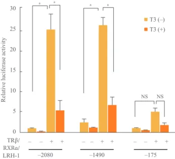
using SHP promoter deletion constructs affecting LRH-1 bind- ing sites [5]. To check the interaction, we used 3 fragments (–2080, –1490, and –175) of human SHP promoter in our study. As LRH-1 response element (LRE) in the human SHP promoter was located from –518 to –489, the activation of pro- moter by unliganded TRβ/RXRα/LRH-1 was decreased in –175 fragments and when we added T3, overall activity of hu- man SHP promoter by TRβ/RXRα/LRH-1 was significantly reduced (Fig. 3).
Electrophoretic mobility-shift assays were performed with a double-stranded oligonucleotide containing the –518 human SHP promoter LRE to explore the mechanism of the T3 depen- dent inhibition of LRH-1 transactivation (Fig. 4). The affinity of LRH-1 binding decreased in nuclear extracts of human hep- atoma cells treated with T3 for increasing times up to 24 hours (lanes 2 to 5). To verify the specific binding of LRH-1 to the probe, competition assays were also performed. An oligomer containing a mutated LRE sequence did not compete with the probe (lane 9), whereas an oligomer with the consensus LRE competed with the SHP LRE probe (lane 10).
30 25 20 15 10 5 0
a a a a
Relative luciferase activity
RXRα/TRβ/
LRH-1
– – + + – – + + – – + +
T3 (–) T3 (+)
NS NS
–2080 –1490 –175
Fig. 3. Repression of expression from the 5ʹ deletion small het- erodimer partner (SHP) promoter by the thyroid hormone. The 200 ng of 5ʹ human SHP promoter (full, –1490 and –175) was co- transfected with or without 100 ng of thyroid hormone receptor β (TRβ) and 30 ng of retinoid X receptor α (RXRα) or 100 ng of liver receptor homolog-1 (LRH-1) in 293 cells. Thyroid hormone (T3; 100 nmol/L) was added. Luciferase activity was measured and normalized against renilla activity. NS, not significant. Sig- nificant differences compared to the control aP<0.01.
DISCUSSION
In this study, we demonstrated that liganded TRβ suppressed the expression of SHP. Because there is no known TRE in SHP promoter, liganded TRβ might decrease the expression of SHP by disturbing the transcriptional activity of LRH-1 to SHP.
Therefore, our result could be one mechanism of regulation of CYP7A1 expression by thyroid hormone and TRβ.
In fact, role of thyroid hormone to regulation of CYP7A1 was already studied in several previous studies. Murine CY- P7A1 is highly inducible by thyroid hormone because there are TREs in the murine CYP7A1 promoter [28]. Therefore, thyroid hormone directly increases the expression of murine CYP7A1 and induces bile acid synthesis. In human, there are inconsis- tent results about regulation of CYP7A1 expression by thyroid hormone. Previously, human CYP7A1 expression was reduced by thyroid hormone in several studies [29,30]. Drover et al.
[30] found that, unlike the CYP7A1 promoter site II TRE, the
sequence of the site III TRE is not conserved among different species, which might explain the divergent responses of human and murine CYP7A1 to thyroid hormone. In contrast, recent study showed that thyroid hormone increased expression of CYP7A1 similarly in human and mouse primary liver culture, human hepatocytes and mouse liver [31]. However, it is diffi- cult to explain until now why several researchers announced different results about thyroid hormone and induction of CY- P7A1. For this reason, the relationship between CYP7A1 and thyroid hormone it is not sufficient to explain the regulation of bile acid metabolism by thyroid hormone. However, in our study, SHP expression in the mouse (both in vivo and in vitro) and human hepatoma cells was identically decreased by thy- roid hormone. Therefore, our results are important to determine the mechanism underlying bile acid metabolism regulation, as it is the first study to report repression of SHP expression by thyroid hormone.
Many potential transcription factors for SHP, such as LRH- 1, HNF-4, FXR, LXR, and SREPB-1 [4] were identified in previous studies. Among these, LRH-1 induces strong tran- scriptional activity because at least 5 LRH-1 binding sites were identified in the SHP promoter [5].
The results of ChIP-Seq, TRE bioinformatics and direct co- transfection all indicate that TRβ/RXRα does not regulate the SHP promoter directly. Among a number of transcription fac- tors that are known to have such direct effects, LRH-1 has most binding sites and is essential for basal SHP promoter activity.
In cotransfections of LRH-1 and TRβ/RXRα, we observed a T3-dependent suppression of promoter activity. This effect was diminished in –175 fragments of human SHP promoter. We also observed a time-dependent decrease in specific LRH-1 DNA binding in T3 treated livers, but the basis for this effect remains to be determined.
Thyroid hormone can act as both a positive and negative regulator of gene expression. In positive regulation, TR binds to TREs located in the promoters of target genes. In the ab- sence of T3, the unliganded TR/RXR complex with corepres- sors inhibits the transcriptional activity of target genes. Binding of T3 to TR induces structural changes and recruits coregulato- ry proteins, which enables transcriptional activation [17]. TRs also negatively regulate transcription with and without DNA binding. In contrast to positive regulation, when TR binds to specific negative TREs, liganded TR recruits a corepressor complex to repress transcriptional activity. Another model sug- gests that TR does not bind to DNA directly, but instead binds to another transcription factor via protein-protein interactions.
1 2 3 4 5 6 7 8 9 10 – 0 6 12 24 (hr) – Self MT LRE
Lane T3 (100 nmol/L) Nuclear extract
Fig. 4. Interaction between thyroid hormone (T3) and liver recep- tor homolog-1 (LRH-1). Electrophoretic mobility-shift assay was performed using an oligomer containing the LRH-1 responsive element (LRE) in the human SHP promoter (–518) as a probe and nuclear extracts of human hepatoma cells treated with 100 nmol/L of T3 for increasing times as indicated. For competition assays, unlabeled oligomers containing the hLRH-1(–518) LRE (self;
lane 8), an LRE mutant (lane 9), or a consensus LRE (lane 10) were used in a 50-fold molar excess. MT, mutant.
Then, liganded TR recruits a corepressor complex to inhibit transcriptional activity [32]. In contrast, unliganded TR binds to specific transcription factor and recruits a coactivator com- plex (CoA) leading to gene activation. The increased activity of SHP promoter by adding LRH-1 to unliganded TRβ/RXRα as shown Fig. 2C could be explained by this mechanism, al- though it is not clearly known yet from our results.
In our study, we assumed that TRβ did not bind to DNA di- rectly, but interacted with another transcription factor, LRH-1.
This interaction of TR with to other transcription factors has been demonstrated in previous studies; both TRβ and LXRα heterodimerize with RXRα and compete for binding to DR4 elements in the CYP7A1 promoter [16]. In addition, PPAR se- lectively inhibits the transcriptional activity of TRs by compet- ing for RXR and possibly non-RXR TR-auxiliary proteins [18].
Therefore, transcriptional regulation by TR through interaction with other transcriptional factors might be as important as reg- ulation through classic TREs.
In conclusion, we found that thyroid hormone indirectly reg- ulates the expression of SHP mRNA by interfering with the transcriptional activity of another transcription factor, LRH-1.
This provides an additional mechanism to better understanding of the regulation of bile acid and cholesterol metabolism regu- lation by thyroid hormone.
CONFLICTS OF INTEREST
No potential conflict of interest relevant to this article was re- ported.
ACKNOWLEDGMENTS
This study was supported by a grant from the Korean Health Technology R&D Project, the Ministry of Health and Welfare, Republic of Korea (A100589) and a grant from Seoul National University Bundang Hospital (03-2010-015). We thank Ste- phen D. Ayers from Methodist Hospital Research Institute for his assisstance with data interpretation.
REFERENCES
1. Seol W, Choi HS, Moore DD. An orphan nuclear hormone receptor that lacks a DNA binding domain and heterodi- merizes with other receptors. Science 1996;272:1336-9.
2. Lee HK, Lee YK, Park SH, Kim YS, Park SH, Lee JW, et al. Structure and expression of the orphan nuclear receptor
SHP gene. J Biol Chem 1998;273:14398-402.
3. Sanyal S, Kim JY, Kim HJ, Takeda J, Lee YK, Moore DD, et al. Differential regulation of the orphan nuclear receptor small heterodimer partner (SHP) gene promoter by orphan nuclear receptor ERR isoforms. J Biol Chem 2002;277:1739- 48.
4. Lee YS, Chanda D, Sim J, Park YY, Choi HS. Structure and function of the atypical orphan nuclear receptor small het- erodimer partner. Int Rev Cytol 2007;261:117-58.
5. Lee YK, Parker KL, Choi HS, Moore DD. Activation of the promoter of the orphan receptor SHP by orphan receptors that bind DNA as monomers. J Biol Chem 1999;274:20869- 73.
6. Lu TT, Makishima M, Repa JJ, Schoonjans K, Kerr TA, Au- werx J, et al. Molecular basis for feedback regulation of bile acid synthesis by nuclear receptors. Mol Cell 2000;6:507- 15.
7. Goodwin B, Jones SA, Price RR, Watson MA, McKee DD, Moore LB, et al. A regulatory cascade of the nuclear recep- tors FXR, SHP-1, and LRH-1 represses bile acid biosynthe- sis. Mol Cell 2000;6:517-26.
8. Goodwin B, Watson MA, Kim H, Miao J, Kemper JK, Kliewer SA. Differential regulation of rat and human CY- P7A1 by the nuclear oxysterol receptor liver X receptor-al- pha. Mol Endocrinol 2003;17:386-94.
9. Shih DQ, Screenan S, Munoz KN, Philipson L, Pontoglio M, Yaniv M, et al. Loss of HNF-1alpha function in mice leads to abnormal expression of genes involved in pancreatic islet development and metabolism. Diabetes 2001;50:2472-80.
10. Kir S, Zhang Y, Gerard RD, Kliewer SA, Mangelsdorf DJ.
Nuclear receptors HNF4alpha and LRH-1 cooperate in reg- ulating CYP7A1 in vivo. J Biol Chem 2012;287:41334-41.
11. Watanabe M, Houten SM, Wang L, Moschetta A, Mangels- dorf DJ, Heyman RA, et al. Bile acids lower triglyceride levels via a pathway involving FXR, SHP, and SREBP-1c.
J Clin Invest 2004;113:1408-18.
12. Huang J, Iqbal J, Saha PK, Liu J, Chan L, Hussain MM, et al. Molecular characterization of the role of orphan receptor small heterodimer partner in development of fatty liver.
Hepatology 2007;46:147-57.
13. Ness GC, Chambers CM. Feedback and hormonal regula- tion of hepatic 3-hydroxy-3-methylglutaryl coenzyme A re- ductase: the concept of cholesterol buffering capacity. Proc Soc Exp Biol Med 2000;224:8-19.
14. Ness GC, Zhao Z. Thyroid hormone rapidly induces hepat- ic LDL receptor mRNA levels in hypophysectomized rats.
Arch Biochem Biophys 1994;315:199-202.
15. Ness GC, Pendelton LC, Zhao Z. Thyroid hormone rapidly increases cholesterol 7 alpha-hydroxylase mRNA levels in hy- pophysectomized rats. Biochim Biophys Acta 1994;1214:229- 33.
16. Hashimoto K, Cohen RN, Yamada M, Markan KR, Mon- den T, Satoh T, et al. Cross-talk between thyroid hormone receptor and liver X receptor regulatory pathways is re- vealed in a thyroid hormone resistance mouse model. J Biol Chem 2006;281:295-302.
17. Cheng SY, Leonard JL, Davis PJ. Molecular aspects of thy- roid hormone actions. Endocr Rev 2010;31:139-70.
18. Juge-Aubry CE, Gorla-Bajszczak A, Pernin A, Lemberger T, Wahli W, Burger AG, et al. Peroxisome proliferator-acti- vated receptor mediates cross-talk with thyroid hormone receptor by competition for retinoid X receptor. Possible role of a leucine zipper-like heptad repeat. J Biol Chem 1995;270:18117-22.
19. Lee YK, Moore DD. Dual mechanisms for repression of the monomeric orphan receptor liver receptor homologous protein-1 by the orphan small heterodimer partner. J Biol Chem 2002;277:2463-7.
20. Park YJ, Lee EK, Lee YK, Park do J, Jang HC, Moore DD.
Opposing regulation of cytochrome P450 expression by CAR and PXR in hypothyroid mice. Toxicol Appl Pharma- col 2012;263:131-7.
21. Lee YK, Dell H, Dowhan DH, Hadzopoulou-Cladaras M, Moore DD. The orphan nuclear receptor SHP inhibits hepato- cyte nuclear factor 4 and retinoid X receptor transactivation:
two mechanisms for repression. Mol Cell Biol 2000;20:187- 95.
22. Irizarry RA, Bolstad BM, Collin F, Cope LM, Hobbs B, Speed TP. Summaries of Affymetrix GeneChip probe level data. Nucleic Acids Res 2003;31:e15.
23. Reich M, Liefeld T, Gould J, Lerner J, Tamayo P, Mesirov JP. GenePattern 2.0. Nat Genet 2006;38:500-1.
24. Lopez D, Abisambra Socarras JF, Bedi M, Ness GC. Acti- vation of the hepatic LDL receptor promoter by thyroid hormone. Biochim Biophys Acta 2007;1771:1216-25.
25. Stravitz RT, Vlahcevic ZR, Russell TL, Heizer ML, Avad- hani NG, Hylemon PB. Regulation of sterol 27-hydroxy- lase and an alternative pathway of bile acid biosynthesis in primary cultures of rat hepatocytes. J Steroid Biochem Mol Biol 1996;57:337-47.
26. Gilardi F, Mitro N, Godio C, Scotti E, Caruso D, Crestani M, et al. The pharmacological exploitation of cholesterol 7alpha-hydroxylase, the key enzyme in bile acid synthesis:
from binding resins to chromatin remodelling to reduce plasma cholesterol. Pharmacol Ther 2007;116:449-72.
27. Ayers S, Switnicki MP, Angajala A, Lammel J, Arumanaya- gam AS, Webb P. Genome-wide binding patterns of thyroid hormone receptor beta. PLoS One 2014;9:e81186.
28. Shin DJ, Plateroti M, Samarut J, Osborne TF. Two uniquely arranged thyroid hormone response elements in the far up- stream 5’ flanking region confer direct thyroid hormone regulation to the murine cholesterol 7alpha hydroxylase gene. Nucleic Acids Res 2006;34:3853-61.
29. Wang DP, Stroup D, Marrapodi M, Crestani M, Galli G, Chiang JY. Transcriptional regulation of the human choles- terol 7 alpha-hydroxylase gene (CYP7A) in HepG2 cells. J Lipid Res 1996;37:1831-41.
30. Drover VA, Wong NC, Agellon LB. A distinct thyroid hor- mone response element mediates repression of the human cholesterol 7alpha-hydroxylase (CYP7A1) gene promoter.
Mol Endocrinol 2002;16:14-23.
31. Lammel Lindemann JA, Angajala A, Engler DA, Webb P, Ayers SD. Thyroid hormone induction of human cholester- ol 7 alpha-hydroxylase (CYP7A1) in vitro. Mol Cell Endo- crinol 2014;388:32-40.
32. Weitzel JM. To bind or not to bind: how to down-regulate target genes by liganded thyroid hormone receptor? Thy- roid Res 2008;1:4.