ABSTRACT
Tumor localization is challenging in the context of ductal carcinoma in situ (DCIS) treated with breast-conserving surgery. Conventional localization methods are generally performed under the guidance of ultrasonography or mammography and are rarely performed with magnetic resonance imaging (MRI), which is more sensitive than the aforementioned modalities in detecting DCIS. Here, we report the application of MRI-based individualized 3-dimensional (3D)-printed breast surgical guides (BSGs) for patients with breast cancer.
We successfully resected indeterminate and suspicious lesions that were only detected using preoperative MRI, and the final histopathologic results confirmed DCIS with clear resection margins. MRI guidance combined with 3D-printed BSGs can be used for DCIS localization, especially for lesions easily detectable using MRI only.
Keywords: Breast neoplasms; Carcinoma, ductal; Magnetic resonance imaging;
Printing, three-dimensional
INTRODUCTION
In recent decades, with the widespread use of mammography for screening, the incidence of breast ductal carcinoma in situ (DCIS) has dramatically increased, and DCIS currently accounts for approximately 20% of all new breast cancer diagnoses [1]. Breast-conserving surgery combined with adjuvant radiotherapy is the standard treatment for small DCIS. The goal of breast-conserving surgery is to completely remove the tumor with clear margins while preserving normal tissue as much as possible for optimal cosmesis. However, most DCIS lesions are clinically impalpable, and they frequently involve a larger area than the extent visible on preoperative imaging. These features make it difficult to accurately localize the exact tumor extent before surgery. Magnetic resonance imaging (MRI) is more sensitive than mammography or ultrasonography in detecting DCIS [2,3], and lesions are often not
Case Report
Received: Oct 23, 2020 Revised: Jan 20, 2021 Accepted: Jan 25, 2021 Correspondence to BeomSeok Ko
Division of Breast Surgery, Department of Surgery, Asan Medical Center, University of Ulsan College of Medicine, 88 Olympic-ro 43-gil, Songpa-gu, Seoul 05505, Korea.
E-mail: [email protected]
© 2021 Korean Breast Cancer Society This is an Open Access article distributed under the terms of the Creative Commons Attribution Non-Commercial License (https://
creativecommons.org/licenses/by-nc/4.0/) which permits unrestricted non-commercial use, distribution, and reproduction in any medium, provided the original work is properly cited.
ORCID iDs Zhen-Yu Wu
https://orcid.org/0000-0003-1731-6370 Young Joo Lee
https://orcid.org/0000-0001-5352-8093 Yungil Shin
https://orcid.org/0000-0001-8635-4835 Soojeong Choi
https://orcid.org/0000-0003-3094-3651 Soo Yeon Baek
https://orcid.org/0000-0003-2026-0603 Jung Whan Chun
https://orcid.org/0000-0001-6438-5526
Zhen-Yu Wu 1,2,3, Young Joo Lee 2, Yungil Shin 2, Soojeong Choi 2, Soo Yeon Baek 2, Jung Whan Chun 2, Loai Saleh Albinsaad 2,4, Woo Jung Choi 5, Namkug Kim 5, BeomSeok Ko 5
1Department of Breast Surgery, Shanghai East Hospital, Tongji University School of Medicine, Shanghai, China
2 Division of Breast Surgery, Department of Surgery, Asan Medical Center, University of Ulsan College of Medicine, Seoul, Korea
3Biomedical Engineering Research Center, Asan Institute for Life Sciences, Asan Medical Center, Seoul, Korea
4Department of Surgery, College of Medicine, King Faisal University, Al-Ahsa, Saudi Arabia
5Department of Radiology, Asan Medical Center, University of Ulsan College of Medicine, Seoul, Korea
Usefulness of 3-Dimensional-Printed Breast Surgical Guides for Undetectable Ductal Carcinoma In Situ on
Ultrasonography: A Report of 2 Cases
Loai Saleh Albinsaad
https://orcid.org/0000-0003-1249-0919 Woo Jung Choi
https://orcid.org/0000-0003-1341-6470 Namkug Kim
https://orcid.org/0000-0002-3438-2217 BeomSeok Ko
https://orcid.org/0000-0001-7831-7874 Funding
This work was supported by a grant (2017- 672, 2018-672) from Asan Institute for Life Sciences, Asan Medical Center, Seoul, Korea.
Conflict of Interest
BeomSeok Ko and Namkug Kim hold a patent for the 3D-printed breast surgical guide and are the founding members of ANYMEDi, Inc.
Zhen-Yu Wu received consulting fees from ANYMEDi, Inc.
Author Contributions
Conceptualization: Wu ZY, Kim N, Ko B; Data curation: Wu ZY, Lee YJ, Shin Y, Choi S, Baek SY, Chun JW, Albinsaad LS, Choi WJ, Ko B; Formal analysis: Wu ZY, Ko B; Funding acquisition:
Ko B; Investigation: Wu ZY, Lee YJ, Shin Y, Choi S, Baek SY, Chun JW, Albinsaad LS, Choi WJ, Kim N, Ko B; Methodology: Wu ZY, Kim N, Ko B; Project administration: Kim N, Ko B;
Resources: Wu ZY, Kim N, Ko B; Software: Wu ZY, Ko B; Supervision: Wu ZY, Ko B; Validation:
Wu ZY, Kim N, Ko B; Visualization: Kim N, Ko B;
Writing - original draft: Wu ZY; Writing - review
& editing: Wu ZY, Ko B
detectable using imaging modalities other than MRI. However, conventional localization methods, such as hook wire or radioactive seeding techniques, are generally performed under ultrasonographic or mammography guidance and are rarely performed with MRI guidance in current clinical practice [4,5].
Under such circumstances, an MRI-based patient-specific surgical guide for localizing the tumor extent using 3-dimensional (3D) printing technology can be an effective alternative.
Here, we report the application of an MRI-based 3D-printed breast surgical guide (BSG) for breast-conserving surgery in patients with biopsy-proven invasive cancer concurrent with DCIS lesions not detected using preoperative ultrasonography.
CASE REPORT
This study was approved by the Institutional Review Board of Asan Medical Center, Seoul, Korea (No. 2016-1237, 2018-0690). Informed consent was obtained from all patients.
Case 1
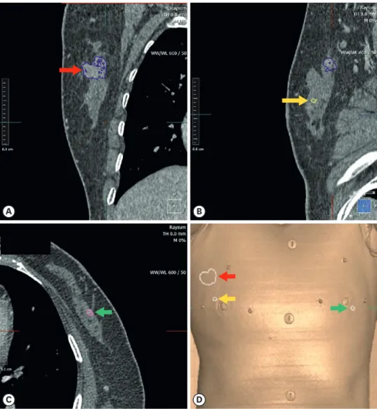
A 55-year-old woman diagnosed at her local hospital with invasive ductal carcinoma in the 10–11 o'clock position of her right breast by core needle biopsy was referred to our department in December 2018 for further examination and surgical management. Physical examination revealed a well-defined palpable mass in the upper outer quadrant of the right breast. Enhanced breast MRI revealed an irregular enhancing mass in the 11 o'clock position, 6.1 cm from the nipple, with a diameter of 2.8 cm, in the right breast, which was concordant with a biopsy-confirmed malignancy (Figure 1A). In addition, MRI revealed an indeterminate mass in the central portion of the right breast, 3.0 cm from the nipple, with a diameter of 0.6 cm (Figure 1B), and another indeterminate mass at 6 o'clock of the left breast, 3.6 cm from the nipple, with a diameter of 0.6 cm (Figure 1C). A prominent lymph node in the right axillary level I area was observed.
Magnification mammography revealed an isodense mass with calcifications measuring 2.3 cm in the upper outer quadrant of the right breast and another isodense mass measuring 0.8 cm deep in the outer portion of the right breast. No obvious abnormalities were observed in the left breast tissue. Ultrasonography did not reveal MRI-detected indeterminate masses in either breast (Figure 2). We proposed breast-conserving surgery using prone/supine MRI-based 3D-printed BSGs to excise the cancer and bilateral indeterminate masses not detected using ultrasonography. The patient approved of the treatment plan.
Breast imaging was performed using a 3.0-T MRI system (Ingenia; Philips Healthcare, Best, the Netherlands) with a dedicated bilateral 4-element breast coil. Additional MRI was performed with the patient in the supine position to replicate the patient's position during the surgical procedure. The patient provided written informed consent and agreed to undergo supine imaging in addition to the standard baseline MRI protocol. Data obtained from the prone/supine MRI were analyzed, and the tumors (the 2 indeterminate masses in both breasts and normal tissues) were divided using the image segmentation program Mimics Medical 17 (Materialise Inc., Leuven, Belgium) (Figure 3). The patient-specific hybrid-type BSGs (for each breast) containing blue dye injection columns, which can indicate the tumor extent requiring removal, were modeled at distances of 0.5 cm from the tumor boundaries to guarantee a margin of safety (Figure 4). The following specifications were used to model the BSGs to ensure an accurate display of the tumor resection boundaries: 1) they were made to precisely fit the breast skin surface, 2) a hole was provided to fit the nipple, and
3) guidelines were used to prevent rotation of the BSGs and indicate their placement relative to the opposite nipple and suprasternal notch. BSGs were made to fit the patient's skin surface and were manufactured as a hybrid type, with a groove for marking the breast surface and a column for precisely marking the underlying tissue.
R, UO, 11 o'clock, posterior
Diameters: 2.8 × 1.6 × 1.9 cm Angio volume: 4.8 cc
Cor
R
Ax-CC Sag-ML
Lat Med
A
R, central, middle
Diameters: 0.64 × 0.64 cm Cor
R
Ax-CC Sag-ML
Lat Med
B
L, LO, 6 o'clock, middle
Diameters: 0.62 × 0.37 cm Cor
L
Ax-CC Sag-ML
Med Lat
C
Figure 1. Enhanced breast magnetic resonance imaging (A) revealing an irregular enhancing mass in the 11 o'clock position, 6.1 cm from the nipple, with a diameter of 2.8 cm, in the right breast which was concordant with biopsy- confirmed malignancy. (B) an indeterminate mass in the central portion of the right breast, 3.0 cm from the nipple, with a diameter of 0.6 cm (C) and another indeterminate mass at 6 o'clock of the left breast, 3.6 cm from the nipple, with a diameter of 0.6 cm were detected.
A B
Figure 2. Preoperative ultrasonography did not reveal the magnetic resonance imaging–detected indeterminate masses (A) right 10 o'clock, (B) left 4 o'clock in either breast.
Surgery was performed in January 2019. BSGs were then printed and sterilized preoperatively.
The tumor resection boundaries were drawn on the breast skin surface along the lines designed to match the tumor shapes. Blue dye was injected through the columns to indicate the extent that needs to be removed around the tumors. Tumors in the 11 o'clock position and central portion of the right breast and those at the 6 o'clock position of the left breast were separately removed using the blue borders as excision boundaries. A sentinel lymph node biopsy sample was obtained from the right axilla, and frozen biopsy was negative for malignancy (0/4).
No sentinel lymph node biopsy sample was obtained from the left axilla. The intraoperative resection margin frozen biopsy yielded negative results (3, 6, 9, and 12 o'clock margins of the right breast biopsy-confirmed cancer). The operation was completed in 82 min.
The final histopathologic diagnosis of the right 11 o'clock mass was invasive ductal
carcinoma: Grade 2, T2 (2.7 cm) N0 (0/4) M0, Stage IIA, estrogen receptor (+), progesterone
A B
C D
Figure 3. Data obtained from supine magnetic resonance imaging were analyzed, and the tumors, including the right breast biopsy-confirmed cancer (A, D: red arrow) and the indeterminate tumors in the right breast (B, D: yellow arrow), left breast (C, D: green arrow), and normal tissues, were divided. The final histopathologic diagnosis of the 11 o'clock specimen from the right breast was invasive ductal carcinoma. Furthermore, the tumor in the central portion of the right breast was a DCIS, and the tumor in the left breast was a DCIS with lobular carcinoma in situ.
DCIS = ductal carcinoma in situ.
receptor (+), HER2 (−), and Ki67 index of 70%–80%. Moreover, the tumor in the central portion of the right breast was a multifocal DCIS measuring 0.3 cm. The tumors in the 6 o'clock position of the left breast were DCIS measuring 0.9 cm and multifocal lobular carcinoma in situ measuring 0.4 cm. All resection margins were free of tumor. The patient received adjuvant chemotherapy, radiotherapy, and hormonal therapy. At the last follow-up, before the composition of this report, the patient remained disease free for 18 months.
Case 2
A 61-year-old woman diagnosed with invasive ductal carcinoma in the 1 o'clock position of her right breast by core needle biopsy at her local hospital was referred to our center in January 2019 for surgical treatment. Physical examination showed a palpable mass in the upper inner quadrant of the right breast. Enhanced breast MRI revealed an irregular enhancing mass at 2 o'clock, 7 cm from the nipple, with a diameter of 2.0 cm, in the right breast, concordant with biopsy-confirmed malignancy. Furthermore, MRI revealed another suspicious mass in the 12 o'clock position of the right breast, 6.5 cm from the nipple, with a diameter of 0.7 cm. Magnification mammography revealed an irregular mass measuring 2.3 cm in the upper inner quadrant of the right breast. Ultrasonography did not reveal the corresponding suspicious mass in the 12 o'clock position of the right breast that was detected using MRI. After careful evaluation, we suggested breast-conserving surgery using an MRI- based 3D-printed BSG to excise the biopsy-confirmed cancer and suspicious mass in the right breast after obtaining patient's approval.
A B
D C
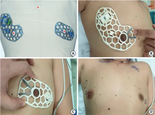
Figure 4. Patient-specific hybrid-type breast surgical guides (BSGs) containing the blue dye injection columns, which can indicate the tumor extent requiring removal, are modeled (A). The following specifications were used for modeling the BSGs to ensure an accurate display of the tumor resection boundaries: 1) they were made to precisely fit the breast skin surface, 2) a hole was provided to fit the nipple, and 3) guidelines to prevent rotation of the BSGs and indicate their placement relative to the opposite nipple and the suprasternal notch were included (B, C). The tumor resection boundaries were drawn onto the breast skin surface designed to match tumor shape (D).
BSG = breast surgical guide.
Breast-conserving surgery guided by a 3D-printed BSG was performed in January 2019 in the same manner as in case 1. A right sentinel lymph node biopsy was performed, and frozen biopsy revealed metastatic carcinoma in 2 of three lymph nodes. An axillary lymph node sampling was also performed. Intraoperative resection margin frozen biopsy yielded negative results. The operation was completed in 73 minutes.
The final histopathologic diagnosis of the right 2 o'clock specimen was invasive ductal carcinoma: Grade 2, T1 (2 cm) N1a (2/4) M0, stage IIA, estrogen receptor (+), progesterone receptor (+), HER2 (+), and Ki67 index of 20%−30%. In addition, the tumor in the 12 o'clock position of the right breast was a multifocal DCIS measuring 0.3 cm. All resection margins were tumor free. The patient received chemotherapy and radiotherapy along with trastuzumab and hormonal therapy as adjuvant treatments. At the last follow-up, the patient remained disease free for 18 months.
DISCUSSION
We used MRI-based 3D-printed BSGs for patients with DCIS who underwent breast- conserving surgery, and this allowed us to successfully resect DCIS lesions with clear resection margins only detected using preoperative MRI.
An appropriate preoperative tumor localization strategy is critical for achieving clear resection margins and optimal cosmetic results after breast-conserving surgery for DCIS.
However, as the vast majority of DCIS lesions are clinically undetectable or frequently have a diffuse growth pattern, accurate DCIS localization becomes extremely challenging. There are two generally used localization methods for DCIS: wire-guided and radioactive seeding localization. Although previous studies have reported the utility of these localization methods [4,5], the mean rate of re-excision associated with positive resection margins is 30% [6], and re-excision may lead to potential complications, as well as stress and additional costs, for patients. The sensitivity of MRI in detecting DCIS is higher than that of mammography or ultrasonography [2,3], and MRI is often performed for patients with DCIS. However, it is difficult to directly mark the tumor region on MRI using conventional localization methods. Moreover, patients screened using mammography or ultrasonography may not undergo biopsy or excision if they have undetected lesions. Lesions that were not identified using ultrasonography could be identified and confirmed via MRI-guided biopsy.
However, MRI-guided biopsy is time consuming and complicated, possibly leading to long waiting times for patients scheduled for breast cancer surgery.
In the cases reported herein, DCIS lesions were not detected on ultrasonography, but they were observed as indeterminate or suspicious tumors only using MRI. Thus, preoperative biopsies were not performed for these tumors. We used individualized MRI-based 3D-printed BSGs to excise these indeterminate or suspicious tumors, and the tumors were histopathologically confirmed as DCIS. If we had used conventional ultrasonography-guided localization for managing these patients instead of MRI-based BSG, these lesions would have likely been missed and not removed, leading to subsequent recurrence(s). In addition, the removal of suspicious lesions and histopathologically confirmed tumors may improve the efficacy of planned operations.
Three-dimensional printing is a versatile technology with expanding applications in various medical fields [7,8]. In a prospective pilot study conducted at our institution, Ko et al. [9]
employed MRI-based 3D-printed BSGs for breast-conserving surgery in patients receiving neoadjuvant chemotherapy. The authors found that this technique was useful for accurately marking the tumor region based on pretreatment MRI data, and they successfully used 3D-printed BSGs for patients with DCIS using a similar technique [9]. A patient-specific 3D-printed BSG can use MRI data to directly mark the tumor extent on the affected breast skin. Through the columns that are modeled in three dimensions, the blue dye injection into the breast tissue can provide additional quantitative representation of the extent of tumor requiring removal. Besides potentially improving patient care and surgical outcomes, this technique is less invasive than conventional localization methods (hook wire-guided or radioactive seeding localizations) and has advantages such as the absence of pain and radiation exposure, as well as a shorter procedure duration.
In conclusion, MRI guidance combined with 3D-printed BSGs can be useful for DCIS localization, especially for lesions that can only be detected on MRI but cannot be easily visualized using other imaging modalities. Large-scale prospective studies are required to establish the safety and effectiveness of this technique in patients with DCIS.
REFERENCES
1. Siegel RL, Miller KD, Jemal A. Cancer statistics, 2019. CA Cancer J Clin 2019;69:7-34.
PUBMED | CROSSREF
2. Berg WA, Gutierrez L, NessAiver MS, Carter WB, Bhargavan M, Lewis RS, et al. Diagnostic accuracy of mammography, clinical examination, US, and MR imaging in preoperative assessment of breast cancer.
Radiology 2004;233:830-49.
PUBMED | CROSSREF
3. Kuhl CK, Schrading S, Bieling HB, Wardelmann E, Leutner CC, Koenig R, et al. MRI for diagnosis of pure ductal carcinoma in situ: a prospective observational study. Lancet 2007;370:485-92.
PUBMED | CROSSREF
4. Agahozo MC, Berghuis SAM, van den Broek E, Koppert LB, Obdeijn IM, van Deurzen CHM. Radioactive seed versus wire-guided localization for ductal carcinoma in situ of the breast: comparable resection margins. Ann Surg Oncol 2020;27:5296-302.
PUBMED | CROSSREF
5. Gray RJ, Salud C, Nguyen K, Dauway E, Friedland J, Berman C, et al. Randomized prospective evaluation of a novel technique for biopsy or lumpectomy of nonpalpable breast lesions: radioactive seed versus wire localization. Ann Surg Oncol 2001;8:711-5.
PUBMED | CROSSREF
6. Toss MS, Pinder SE, Green AR, Thomas J, Morgan DA, Robertson JF, et al. Breast conservation in ductal carcinoma in situ (DCIS): what defines optimal margins? Histopathology 2017;70:681-92.
PUBMED | CROSSREF
7. Rybicki FJ. Medical 3D printing and the physician-artist. Lancet 2018;391:651-2.
PUBMED | CROSSREF
8. Michalski MH, Ross JS. The shape of things to come: 3D printing in medicine. JAMA 2014;312:2213-4.
PUBMED | CROSSREF
9. Ko BS, Kim N, Lee JW, Kim HJ, Chung IY, Kim J, et al. MRI-based 3D-printed surgical guides for breast cancer patients who received neoadjuvant chemotherapy. Sci Rep 2019;9:11991.
PUBMED | CROSSREF