Bee Venom이 세포자멸사를 통해 DU-145 세포의 증식에 미치는 영향
허근영ᆞ송호섭
경원대학교 한의과대학 침구학교실
목적 : 이 연구는 봉독이 세포자멸사 관련 단백질의 발현 조절을 통하여 세포자멸사를 유도하고 전립선 암세포주인 DU-145 세포의 성장을 억제하는지를 확인하고 해당 기전을 살펴보고자 하였다.
방법 : 봉독을 처리한 후 DU-145의 세포자멸사를 관찰하기 위해 TUNEL staining assay를 시행하였으 며, 세포자멸사 조절단백질의 변동 관찰에는 western blot analysis를 시행하였다.
결과 : DU-145 세포에 봉독을 처리한 후, 세포자멸사의 유발, 세포자멸사 관련 단백질의 발현에 미치는 영향을 관찰하여 다음과 같은 결과를 얻었다.
1. DU-145 세포에서 봉독을 처리한 후 세포자멸사가 유도되어 세포성장이 억제되었다.
2. 세포자멸사 관련 단백질 중 분리된 pro-apoptotic proteins인 PARP, caspase-3, caspase-9은 유의한 증 가를 나타내었다.
3. 세포자멸사 관련 단백질 중 분리된 anti-apoptotic proteins인 Bcl-2, p-AKT, XIAP, cIAP2는 유의한 감소를, MMP2, MMP13은 유의한 증가를 나타내었다.
결론 : 이상의 결과는 봉독이 인간 전립선 암세포주인 DU-145의 세포자멸사를 유발함으로써 전립선암세 포 증식억제 효과가 있음을 입증한 것으로 전립선암의 예방과 치료에 대한 효과적인 치료제 개발에 도움이 될 것으로 기대된다.
핵심 단어 : 봉독, 전립선암, DU-145, 세포자멸사
1)
Bee Venom Inhibits DU-145 Cell Proliferation Through Induction of Apoptosis
Hur Keun-young and Song Ho-sueb
Dept. of Acupuncture & Moxibustion, College of Oriental Medicine, Kyungwon University
* This research was supported by the Kyungwon University Research Fund in 2011
․Acceptance : 2011. 6. 1. ․Adjustment : 2011. 6. 10. ․Adoption : 2011. 6. 15.
․Corresponding author : Song Ho-sueb, Dept. of Acupuncture & Moxibustion, Kyungwon Gil Oriental Medical Hospital, 1200-1 Guwal-dong Namdong-gu Incheon 405-760 Republic of Korea
․Corresponding author : Tel. 82-70-7120-5012 E-mail : [email protected]
Original Article
국문초록
Ⅰ. Introduction
Prostate cancer is a significant health problem among men in most western countries1,2). The growth of prostate cancer has demonstrated multistage process, involving the onset as small latent carcinoma with androgen sensitivity to large metastatic lesion with androgen independency3). Androgen-resistant prostate cancer is a debilitating and lethal condition. Although androgen deprivation therapy is an established treatment for advanced prostate cancer and radical prostatectomy or radiation therapy is also available for organ confined one, local recurrence and disseminated, metastasis generally follow them due to androgen independency possibly induced by amplification, loss or changes in the specificity of the androgen receptor4).
Though related studies5-8) elucidated the molecular mechanisms responsible for androgen independency, it has not yet been clear.
Increasing studies have demonstrated that Bee Venom (BV) inhibits inflammation9) or cancer growth through induction of apoptotic cell death10,11). Park et al. reported that BV inhibited human prostate cancer PC-3 cell growth through induction of apoptotic cell death via down regulation of NF-κ B and alteration of expression of apoptosis regulatory proteins11). DU-145 cells have been well characterized and commonly used as a androgen refractory prostate cancer cell line, which are resistant to most of anti-cancer drugs, because the proliferation rate in DU-145 cells is much higher than in androgen sensitive prostate cancer cells such as LNCaP cells12).
Therefore, in order to gain better insight into the action mechanism of tumorigenesis and chemoresistance, the author conducted this study.
In the present study, to determine the inhibitory effect of BV on human androgen refractory prostate cancer, the author investigated the apoptosis and the expression of pro-apoptotic proteins as well as anti-apoptotic proteins in DU-145 cells.
Ⅱ. Materials and Methods
A. Materials
Dried BV was purchased from You-Miel Bee Venom Ltd (Hwasoon, Jeonnam, Korea). The com- position of the BV was as follow: 45~50% melittin, 2.5~3% apamin, 2~3% MCD peptide, 12% PLA2, 1% lyso-PLA, 1~1.5% histidine, 4~5% 6pp lipids, 0.5% , 0.1% tertiapin, 0.1% procamine, 1.5~2%
hyaluronidase, 2~3% amine, 4~5% carbohydrate, and 19~27% other, including protease inhibitor, glucosidase, invertase, acid phosphomonoesterase, dopamine, norepinephrine, and unknown amino acids, with >99.5% purity. All of the secondary antibodies such as AKT, phosphorylated AKT (p-AKT), Bax (Bcl-2 associated X protein), Bcl-2 (B-cell lymphoma 2), PARP (procyclic acidic repetitive protein), caspase-3, -9, cleaved caspase-3, -9, cIAP2 (celluar IAP2), XIAP (X-linked inhibitor of apoptosis protein), MMP2 (matrix metalloproteinase-2), MMP13 (matrix metalloproteinase-13), used in Western blot analysis were purchased from Santa Cruz Biotechnology (Santa Cruz, CA). All other reagents were purchased from Sigma unless otherwise stated.
B. Cell culture
The DU-145 human prostate cancer cell was obtained from ATCC (American Type Culture Collection, Rockville, MD). Prostate cells were cultured in RPMI-1640 medium (Life Technologies Inc, Gaithersberg, MD) supplement with 10% fetal calf serum (FCS; Collaborative Biomedical Products, Bedford, MA) and antibiotics, penicillin/streptomycin (100 unit/㎖, Bioproducts, Walkersville, MD). Cell cultures were then maintained at 37℃ in a humidified atmosphere of 5% CO2.
C. Apoptosis evaluation
Apoptosis was evaluated by TUNEL staining assay. In short, cells were cultured on 8-chamber
slides. After treatment with bee venom (1-10 ㎍/㎖) for 24 hr, the cells were washed twice with PBS and fixed by incubation in 4% paraformaldehyde in PBS for 1 hr at room temperature. TUNEL assays were performed by using the in situ Cell Death Detection Kit (Roche Diagonostics GmbH, Mannheim, Germany) according to manufacturer’s instructions.
Total number of cells in a given area was determined by using DAPI nuclear staining. The apoptotic rate was determined as a percentage calculated from the number of cells with TUNEL- positive stained cells divided by the total cell number of cell counted 100.
D. Western blot analysis
Cells were homogenized with lysis buffer [50 mM Tris pH 8.0, 150 mM NaCl, 0.02% sodium azide, 0.2% SDS, 1 mM PMFS, 10 ㎍/㎖ aprotinin, 1% igapel 630 (Sigma-Aldrich, St. Louis, MO, USA), 10 mM NaF, 0.5 mM EDTA, 0.1 mM EGTA and 0.5% sodium deoxycholate], and centrifuged at 23,000 g for 1 hr. Equal amount of proteins (80 ㎍) were separated on a SDS/12%-polyacrylamide gel, and then transferred to a nitrocellulose membrane (Hybond ECL, Amersham Pharmacia Biotech Inc, Piscataway, NJ). Blots were blocked for 2 hr at room temperature with 5% (w/v) non-fat dried milk in Tris-buffered saline [10 mM Tris (pH 8.0) and 150 mM NaCl] solution containing 0.05%
tween-20. The membrane was incubated for 5 hr at room temperature with specific antibodies AKT, p-AKT, Bax (Bcl-2 associated X protein), Bcl-2 (B-cell lymphoma 2), PARP (procyclic acidic repetitive protein), caspase-3, -9, cleaved caspase-3, -9, cIAP2 (celluar IAP2), XIAP (X-linked inhibitor of apoptosis protein), MMP2 (matrix metalloproteinase -2), MMP13 (matrix metalloproteinase-13). The blot was incubated with the corresponding conjugated anti-rabbit immuno- globulin G-horseradish peroxidase (Santa Cruz Biotechnology Inc). Immunoreactive proteins were detected with the ECL western blotting detection system. The relative density of the protein bands was scanned by densitometry using MyImage
(SLB, Seoul, Korea), and quantified by Labworks 4.0 software (UVP Inc, Upland, California).
E. Statistical analysis
Data were analyzed using one-way analysis of variance followed by Tuckey test as a post hoc test.
Differences were considered significant at p<0.05.
Ⅲ. Results
A. Induction of apoptosis
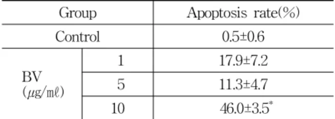
To delineate whether the inhibition of cell growth by the BV was due to increase of the induction of apoptosis, the author evaluated change of the chromatin morphology of human prostate cancer cells using DAPI staining. Apoptosis rate determined after 24 hr treatment was significantly increased by BV 10 ㎍/㎖ was 46.0±3.5% (Table 1, Fig. 1). As shown in Fig. 1, TUNEL-positive cells (stained green) were dose-dependently increased in BV-treated DU-145 cells, and the nuclei (stained blue) were found to be condensed (Fig. 1).
B. Expression of apoptosis regulatory proteins
Bax (Bcl-2 associated X protein) is a pro- apoptotic protein and its predominance over Bcl- 2 (B-cell lymphoma 2) promotes apoptosis. In the
Group Apoptosis rate(%)
Control 0.5±0.6
BV (㎍/㎖)
1 17.9±7.2
5 11.3±4.7
10 46.0±3.5*
Values are the mean ± SEM of three independent experiments performed in triplicate.
* : represents p<0.05, significant difference compared with control respectively.
BV : bee venom.
Table 1. Effect of BV on Apoptosis in DU-145 Cells
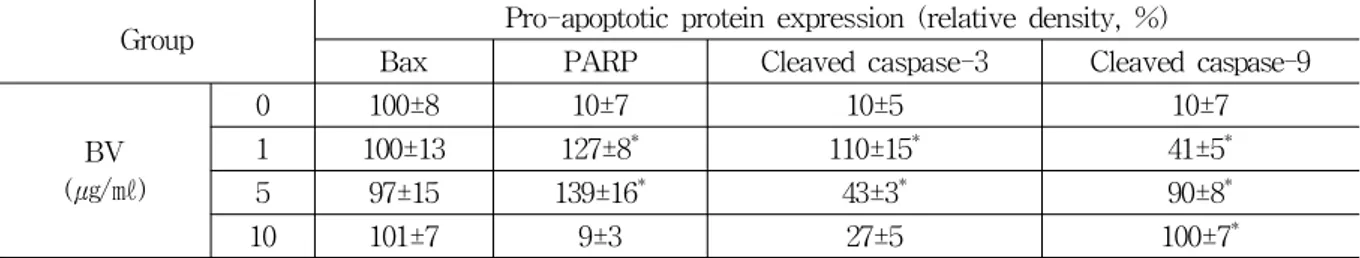
Group Pro-apoptotic protein expression (relative density, %)
Bax PARP Cleaved caspase-3 Cleaved caspase-9
BV (㎍/㎖)
0 100±8 10±7 10±5 10±7
1 100±13 127±8* 110±15* 41±5*
5 97±15 139±16* 43±3* 90±8*
10 101±7 9±3 27±5 100±7*
Values are the mean ± SEM of three independent experiments performed in triplicate.
* : represents p<0.05, significant difference compared with control respectively.
BV : bee venom.
Table 2. Effect of BV on Expression of Bax, PARP, Cleaved Caspase-3 and -9 in DU-145 Cells Fig. 1. Effect of BV on induction of apoptosis of DU-145 cells
The apoptotic cells were examined by fluorescence microscopy after DAPI staining, which were estimated by direct counting of fragmented nuclei. The apoptotic ratio was determined as a percentage calculated from the number of cells with TUNEL-positive stained cells divided by the total cell number of cell counted 100.
BV : bee venom
Fig. 2. Effect of BV on expression of Bax and Bcl-2 in DU-145 cells
Equal amounts (50 ㎍) of whole cell lysates were subjected to electrophoresis and analyzed by main apoptosis regulatory molecules such as Bax and Bcl-2. The relative density was analyzed by densitometry. Similar patterns of protein expression were obtained from three experiments.
BV : bee venom.
present study, Bax (Bcl-2 associated X protein) represents a tendency to overwhelm Bcl-2 (B-cell lymphoma 2), although it did not show significant increase (Fig. 2). Fig. 3 reveals a western blot analysis of Bax (Bcl-2 associated X protein), cleaved caspase-3, cleavaged PARP (procyclic acidic repetitive
protein), cleaved caspase-9, in which the increase of apoptotic action was confirmed by the ability of BV to induce Bax(Bcl-2 associated X protein), caspase-3, - 9, cleaved caspase-3, cleavaged PARP (procyclic acidic repetitive protein), cleaved caspase-9 up-regulation.
Contrast, to gain insight into mechanisms controlling apoptosis in DU-145 cells, the author also observed the effect of BV on anti-apoptotic proteins such as Bcl-2 (B-cell lymphoma 2), XIAP (X-linked inhibitor of apoptosis protein), cIAP2 (celluar IAP2), AKT, p-AKT, MMP2 (matrix metallo- proteinase-2), MMP13(matrix metalloproteinase-13).
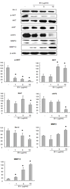
Similar to the above findings inducing apoptosis by up-regulation of pro-apoptotic proteins, compared with control, the anti-apoptotic proteins except for MMP2 (matrix metalloproteinase-2), MMP13 (matrix metalloproteinase-13) were significantly decreased by a different dose of BV in DU-145 cells, promoting apoptosis (Fig. 4).
MMPs (matrix metalloproteinases) play a central
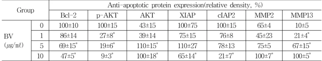
Group Anti-apoptotic protein expression(relative density, %)
Bcl-2 p-AKT AKT XIAP cIAP2 MMP2 MMP13
BV (㎍/㎖)
0 100±10 100±15 43±15 100±75 100±15 65±4 10±5
1 86±14 27±8* 39±14 75±15 76±8 45±23 21±4*
5 69±15* 19±6* 110±15* 110±27 78±13 75±5 67±15* 10 47±5* 9±3* 100±18* 65±14* 21±7* 100±7* 100±5* Values are the mean ± SEM of three independent experiments performed in triplicate.
* : represents p<0.05, significant difference compared with control respectively.
BV : bee venom.
Table 3. Effect of BV on Expression of Bcl-2, p-AKT, AKT, XIAP, cIAP2, MMP2 and MMP13 in DU-145 Cells
Fig. 3. Effect of BV on expression of pro-apoptotic proteins in DU-145 cells Equal amounts (50 ㎍) of whole cell lysates were subjected to electrophoresis and analyzed by pro-apoptosis regulatory molecules such as Bax, PARP, cleaved caspase-3, cleaved caspase-9. The relative density was analyzed by densitometry. Similar patterns of protein expression were obtained from three experiments. Values are mean ± SD of two experiments, with triplicate of each experiment.
* : p <0.05, significant difference compared with control respectively.
BV : bee venom.
role in cell proliferation and apoptosis, disregulation of which generally characterized by elevated expressions has been implicated in primary tumors or metastasis enhancing cancer cell invasion. In this study, MMP2 (matrix metalloproteinase-2), MMP13 (matrix metalloproteinase-13) were significantly increased, compared with control (Fig. 4).
PARP (procyclic acidic repetitive protein) and cleavaged caspase-3 significantly increased by 1 and 5 ㎍/㎖ of BV was 127±8 and 139±16%, and 110±15 and 43±3% (Table 2). Cleaved caspase-9 significantly increased by 1, 5 and 10 ㎍/㎖ of BV was 41±5, 90±8 and 100±7% (Table 2). p-AKT significantly increased by 1, 5 and 10 ㎍/㎖ of BV was 27±8, 19±6 and 9±3% (Table 3). Bcl-2 (B-cell lymphoma 2), XIAP (X-linked inhibitor of apoptosis protein) and cIAP2 (celluar IAP2) significantly decreased by 10 ㎍/㎖ of BV was 47±5, 65±14 and 21±7% respectively (Table 3). MMP2 (matrix metalloproteinase-2) significantly increased by 10
㎍/㎖ of BV was 100±7% and MMP13 (matrix metalloproteinase-13) significantly increased by 1, 5 and 10 ㎍/㎖ of BV was 21±4, 67±15 and 100±5%
(Table 3).
Ⅳ. Discussion
The crucial findings in the present study is the identification of anti-cancer efficacy of BV against human androgen independent prostate carcinoma DU-145 cells. Most of the current available cytotoxic anti-cancer agents exert their effect by promoting
Fig. 4 Effect of BV on expression of anti- apoptotic proteins in DU-145 cells
Equal amounts (50 ㎍) of whole cell lysates were subjected to electrophoresis and analyzed by anti-apoptosis regulatory molecules such as p-AKT, AKT, XIAP, cIAP2, MMP2 and MMP13. The relative density was analyzed by densitometry. Similar patterns of protein expression were obtained from three experiments. Values are mean ± SD of two experiments, with triplicate of each experiment.
* : p<0.05, significant difference compared with control respectively.
BV : bee venom.
apoptosis in cancer cells13,14), which is one of the major mechanisms for the targeted therapy of various cancers including prostate cancer13-16).
Advanced prostate cancer cells generally become resistant to apoptosis due to their androgen independency. Hence, there are limited treatment options available for this disease. Chemotherapy and radiation therapy are largely ineffective, leading to relapse, metastasis and fatal death15). The agents that potentially induce apoptotic cell death could be useful in controlling this advanced prostate cancer at the hormone refractory stage16). The author’s data demonstrated comparative low concentration (below 10 ㎍/㎖) of BV induced apoptotic cell death in a androgen independent DU-145 prostate cancer control, suggesting BV could be useful as a anti-cancer agent at hormone refractory stage consistent with the above assumption.
Apoptosis may be regarded as a protective mechanism against hyper-proliferation and metastasis of cancer due to its role in preventing the mutated cancer cells from growing in the system. Induction of apoptosis by various kinds of factors in cancer cells demonstrates a series of characteristic changes17), including increase in ROS level18), the release of cytochrome c from mitochondria to cytosol following loss of mitochondrial transmembrane potential (MTP)19), activation of caspase -9 and -3, subsequent cleaving PARP, an important marker of apoptosis during downstream caspase-3 activation20,21) in biochemistry, and cell shrinkage, chromatin conden- sation and nucleosomal degradation19) in microscopic morphology. Activation of caspase -3 and -9 activity22-24) has been known to be significant in the cancer cell death and considered as a major new strategic target for the cancer prevention and treatment24-26). Studies have also shown that Bax/Bcl-2 ratio increases during apoptosis24-27), which means the pro-apoptotic protein Bax has substantial influence upon MTP changes and subsequent cytochrome c release, promoting apoptosis, whereas Bcl-2 inhibits apoptotic cell death as an anti-apoptotic protein20,27-31). Reportedly, over-expression of Bcl-2 in numerous malignant tissues including prostate cancer28,29) can
be related with growth or metastasis of the malignancy, suggesting Bcl-2 could be also a major target of cancer drug and the agent down-regulating Bcl-2 be utilized to sensitize apoptosis-resistant prostate cancer cells30).
AKT phosphorylation acts as an anti-apoptotic factors through participating in control of apoptosis via regulating apoptosis related proteins such as anti-apoptotic protein Bcl-2 and pro-apoptotic factors caspase-932,33). The human inhibitor of apoptosis (IAP) family including cellular IAP1 (cIAP1), cIAP2, X-linked IAP (XIAP), neuronal apoptosis inhibitor protein, and survivin was also known as an anti- apoptotic protein, down regulating apoptosis in the systems34,35). Matrix metalloproteinase including a gelatinase MMP2 and a collagenase MMP13 is one of the attractive targets for the development of cancer drugs, because it is generally found up-regulated in the various cancers, contributing to the local prolif- erations and metastasis36,37).
From the above, to confirm BV exerts effects on apoptosis going in androgen independent prostate cancer, It needed to elucidate how BV involve the balance between the pro and anti- apoptotic proteins deciding apoptotic cell death in DU-145 cells.
Similar to Lee et al.’s38) and Oh et al.’s39) report, the present study demonstrated that BV consequently up-regulated pro-apoptotic proteins such as Bax, PARP, caspase -3 and -9 and down-regulated anti- apoptotic proteins such as Bcl-2, p-AKT, XIAP, cIAP2 in hormone refractory DU-145 cells, suggesting BV substantially promote apoptosis in DU-145 cells.
However, increased expression of MMP2 and MMP13 implies the possibility of existing hyper-prolifer- ation or metastasis in the DU-145 cells in spite of BV action.
Therefore, these data suggest that BV exerts inhibitory effect on androgen refractory DU-145 cell proliferation via induction of apoptotic cell death, and it could be used as a novel agent for human prostate cancer prevention and/or intervention if the drawbacks was compensated for in the further studies.
Ⅴ. References
1. Jemal A, Tiwari RC, Murray T et al. Cancer Statistics. CA Cancer J Clin 2005 ; 55 : 10-30.
2. Schally AV, Comaru-Schally AM, Plonowski A et al. Peptide analogs in the therapy of prostate cancer. Prostate. 2000 ; 45 : 158-66.
3. Sun M, Wang G, Paciga JE, Feldman RI, Yuan ZQ, Ma XL et al. AKT1/PKB alpha kinase is frequently elevated in human cancers and its constitutive activation is required for oncogenic transformation in NIH3T3 cells. Am J Pathol.
2001 ; 159(2) : 431-7.
4. Cai RZ, Qin Y, Ertl T et al. New pseudo- nonapeptide bombesin antagonist with C-terminal LeuW (CH2N)Tac-NH2 showing high binding affinity to bombesin/GRP receptors on CFPAC-1 human pancreatic cancer cell line. Int J Oncol.
1995 ; 6 : 1165-72.
5. Y Akao, S Kusakabe, Y Banno, M Kito, Y Nakagawa, K Tamiya-Koizumi, M Hattori, M Sawada, Y Hirabayasi, N Ohishi, Y Nozawa.
Ceramide accumulation is independent of camptothecin- induced apoptosis in prostate cancer LNCaP cells. Biochem Biophys Res Commun. 2002 ; 294 : 363-70.
6. Y Akao, Y Banno, Y Nakagawa, N Hasegawa, TJ Kim, T Murate, Y Igarashi, Y Nozawa.
High expression of sphingosine kinase 1 and S1P receptors in chemotherapy-resistant prostate cancer PC3 cells and their camptothecininduced up-regulation. Biochem Biophys Res Commun.
2006 ; 342 : 1284-90.
7. K Mizutani, K Matsumoto, N Hasegawa, T Deguchi, Y Nozawa. Expression of clusterin, XIAP and survivin, and their changes by camptothecin (CPT) treatment in CPT-resistant PC-3 and CPT-sensitive LNCaP cells. Exp Oncol. 2006 ; 28 : 209-15.
8. K Abdelmohsen, R Pullmann Jr, A Lal, HH Kim, S Galban, X Yang, JD Blethrow, M Walker, J Shubert, DA Gillespie, H Furneaux, M Gorospe. Phosphorylation of HuR by Chk2
regulates SIRT1 expression. Mol Cell. 2007 ; 25 : 543-57.
9. YB Kwon, HJ Lee, HJ Han, WC Mar, SK Kang, OB Yoon, Beitz AJ, JH Lee. The water-soluble fraction of bee venom produces antinociceptive and anti-inflammatory effects on rheumatoid arthritis in rats. Life Sci. 2002 ; 71 : 191-204.
10. Holle L, Song W, Holle E, Wei Y, Wagner T, Yu X. A matrix metalloproteinase 2 cleavable melittin/avidin conjugate specifically targets tumor cells in vitro and in vivo. Int J Oncol.
2003 ; 22 : 93-8.
11. HJ Park, YK Lee, HS Song, KJ Kim, DJ Son, JW Lee and JT Hong. Melittin inhibits human prostate cancer cell growth through induction of apoptotic cell death. J Toxicol Pub Health. 2006 ; 22(1) : 31-7.
12. Keitaro K, Riyako O, Yasunori F, Nanako H, Yukihiro A, Yoshinori N, Takashi D, Masafumi I. A role for SIRT1 in cell growth and chemo- resistance in prostate cancer PC3 and DU145 cells. Biochemical and Biophysical Research Communications. 2008 ; 373 : 423-8.
13. Lowe SW, Lin AW. Apoptosis in cancer. Carci- nogenesis. 2000 ; 21 : 48595.
14. Guseva NV, Taghiyev AF, Rokhlin OW, Cohen MB. Death receptor-induced cell death in prostate cancer. J Cell Biochem. 2004 ; 91 : 7099.
15. Gurumurthy S, Vasudevan KM, Rangnekar VM.
Regulation of apoptosis in prostate cancer.
Cancer Metastasis Rev. 2001 ; 20 : 22543.
16. Kantoff PW. New agents in the therapy of hormone-refractory patients with prostate cancer. Semin Oncol. 1995 ; 22(1) : 324.
17. Simon HU, Haj-Heyia A, Levi Schaffer F. Role of reactive oxygen species (ROS) in apoptosis induction. Apoptosis. 2000 ; 5 : 415-8.
18. Wyllie AH. The genetic regulation of apoptosis.
Curr. Opin. Genet.Dev. 1995 ; 5(1) : 97-104.
19. Green DR, Reed JC. Mitochondria and Apoptosis.
Science. 1998 ; 281 : 1309-12.
20. Thornberry NA, Lazbrik Y. Caspases : enemies within. Science. 1998 ; 281 : 1312-6.
21. Nachshon-Kedmi M, Yannai S, Fares FA.
Induction of apoptosis in human prostate cancer cell line, PC3, by 3,3'-diindolylmethane through the mitochondrial pathway. Br J Cancer. 2004 ; 91 : 1358-63.
22. Darmon AJ, Nicholson DW, Bleackley RC.
Activation of the apoptotic proteaseCPP32 by cytotoxic T cell derived granzyme B. Nature.
1995 ; 377 : 446-8.
23. Zu K, Ip C. Synergy between selenium and vitamin E in apoptosis induction is associated with activation of distinctive initiator caspases in human prostate cancer cells. Cancer Res.
2003 ; 63 : 6988-95.
24. Singh AV, Xiao D, Lew KL, Dhir R, Singh SV.
Sulforaphane induces caspase-mediated apoptosis in cultured PC-3 human prostate cancer cells and retards growth of PC-3 xenografts in vivo.
Carcinogenesis. 2004 ; 25 : 83-90.
25. Nesterov A, Ivashchenko Y, Kraft AS. Tumor necrosis factor-related apoptosis-inducing ligand (TRAIL) triggers apoptosis in normal prostate epithelial cells. Oncogene. 2002 ; 21 : 1135-40.
26. Dasmahapatra GP, Didolkar P, Alley MC, Ghosh S, Sausville EA, Roy KK. In vitro combination treatment with perifosine and UCN-01 demon- strates synergism against prostate (PC-3) and lung (A549) epithelial adenocarcinoma cell lines.
Clin Cancer Res. 2004 ; 10 : 5242-52.
27. Sedlak TW, Oltvai ZN, Yang E et al. Multiple Bcl2 family members demonstrate selective dimerization with Bax. Proc. Natl. Acad. Sci.
USA. 1995 ; 92 : 7834-8.
28. Reed JC. Apoptosis-based therapies. Nat Rev Drug Discov. 2002 ; 1 : 11121.
29. Cory S, Adams JM. The Bcl2 family: regulators of the cellular life-or-death switch. Nat Rev Cancer. 2002 ; 2 : 647-56.
30. Benimetskaya L, Miller P, Benimetsky S et al.
Inhibition of potentially anti-apoptotic proteins by antisense protein kinase C-alpha (Isis 3521) and antisense bcl-2 (G3139) phosphorothioate oligodeoxynucleotides: relationship to the decreased viability of T24 bladder and PC3 prostate cancer
cells. Mol Pharmacol. 2001 ; 60 : 129630-7.
31. Bettaieb A, Dubrez-Daloz L, Launay S et al.
Bcl-2 proteins: targets and tools for chemo- sensitisation of tumor cells. Curr Med Chem AntiCancer Agents. 2003 ; 3 : 307-18.
32. Pugazhenthi S, Nesterova A, Sable C, Heideich KA, Boxer LM, Heasley LE, Rensch JEB. Akt/
protein kinase B upregulates Bcl2 expression through cAMP-response element-binding protein.
J Biol Chem. 2000 ; 275 : 10761-6.
33. Cardone MH, Roy N, Stennicke HR, Salvesen GS, Frarke TF, Stanbridge E, Frisch S, Reed JC. Regulation of cell death protease caspase-9 by phosphorylation. Science. 1998 ; 282 : 1318-21.
34. LaCasse EC, Baird S, Korneluk RG, MacKenzie AE. The inhibitors of apoptosis (IAPs) and their emerging role in cancer. Oncogene. 1998 ; 17 : 3247-59.
35. Deveraux QL, Reed JC. IAP family proteins suppressors of apoptosis. Genes Dev. 1999 ; 13 :
239-52.
36. Fini ME, Cook JR, Mohan R. Regulation of matrix metalloproteinase gene expression. Matrix Metalloproteinases (Edited by Parks WC Mecham RP). San Diego : Academic Press. 1998 ; 299- 356.
37. Visse R, Nagase H. Matrix metalloproteinases and tissue inhibitors of metalloproteinases.
Structure, function, and biochemistry. Circ Res.
2003 ; 92 : 827-39.
38. HS Lee, HS Song. Bee venom inhibits LNCaP cell proliferation through induction of apoptosis via inactivation of NF-κB. Journal of Acupunc- ture and Moxibustion. 2007 ; 25(2) : 59-74.
39. HJ Oh, HS Song. Bee Venom Inhibits PC-3 Cell Proliferation Through Induction of Apoptosis Via Inactivation of NF-κB. Journal of Korean Acupuncture and Moxibustion. 2010 ; 27(3) : 1-13.