Bee Venom이 NF-κB의 불활성화를 통해 DU-145 전립선 암세포의 성장에 미치는 영향
신정미ᆞ송호섭
경원대학교 한의과대학 침구학교실
목적 : 이 연구는 봉독이 NF-κB의 활성억제를 통하여 전립선 암세포주인 DU-145 세포의 성장을 억제하 는지를 확인하고 그 기전을 살펴보고자 하였다.
방법 : 봉독을 처리한 후 DU-145의 성장억제를 관찰하기 위해 WST-1 assay를 시행하였고, 세포자멸사 의 관찰에는 DAPI staining assay를 통한 세포형태관찰을 시행하였으며, 염증관련유전자 발현 관찰에는 western blot analysis를 시행하였고, 세포자멸사와 연관된 NF-κB의 활성 변화를 관찰하기 위해 EMSA와 luciferase assay를 시행하였으며, DU-145에서 봉독과 NF-κB의 상호작용을 관찰하기 위해 transient transfection assay를 시행하여 세포생존율과 NF-κB의 활성 변동을 측정하였다.
결과 : DU-145 세포에서 봉독을 처리한 후 세포성장이 억제되었으며, 염증관련유전자 발현 및 NF-κB의 활성의 유의한 감소를 나타내었다. DU-145 세포에서 NF-κB의 p50와 IKK들을 치환하여 작용기를 없애고 봉독을 처리하였을 경우에도 세포활성 및 NF-κB의 활성의 유의한 감소를 나타내었다.
결론 : 이상의 결과는 봉독이 NF-κB의 활성 억제를 통하여 인간 전립선암세포주인 DU-145의 세포자멸 사를 유발함으로써 증식억제 효과가 있음을 입증한 것으로 전립선암의 예방과 치료에 대한 효과적인 치료제 개발에 도움이 될 것으로 기대된다. 다만 그 기전에서 봉독은 기존연구와 같은 NF-κB p50 및 IKK들의 작 용기와 상호작용 이외에 다른 기전이 관여되는 것으로 심화 연구를 요한다.
1)
Bee Venom Inhibits DU-145 Human Prostate Cancer Cell Growth Through Inactivation of
NF-κB
Shin Jung-mi and Song Ho-sueb
Dept. of Acupuncture & Moxibustion, College of Oriental Medicine, Kyungwon University
* This research was supported by the Kyungwon University Research Fund in 2011
․Acceptance : 2011. 6. 1. ․Adjustment : 2011. 6. 10. ․Adoption : 2011. 6. 13.
․Corresponding author : Song Ho-sueb, Kyungwon Gil Oriental Medical Hospital, 1200-1 Guwal-dong Namdong-gu Incheon Republic of Korea
․Corresponding author : Tel. 82-70-7120-5012 E-mail : [email protected]
Original Article
국문초록
핵심 단어 : 봉독, 전립선암, DU-145, 세포자멸사, NF-κB
Ⅰ. Introduction
Prostate cancer is the most predominant cancer following skin cancer and the death related with it increases every year1,2). There is no curative therapy except for androgen deprivation therapy through surgery or radiation3). Therefor, androgen deprivation therapy remains the most important therapy for advanced Prostate Cancer till date4). However, Efficacy of androgen deprivation therapy is actually confined to the localized and un- metastasizing tumors at the androgen dependent state, because it eventually fails leading to the metastasis and development of androgen-refractory prostate cancer after long-term follow up1-4). Major challenge in treating advanced prostate cancer is how to deal with the androgen resistant metastatic cells.
One of the aims of the researches have been directed towards identifying new agents that prevent proliferation of prostate cancer through the apoptosis and keep the androgen dependent state from changing into androgen refractory one5).
Natural toxins have been also largely studied as an alternative, elucidating important molecular mechanisms of specific toxin-receptor and/or ion channel complexes6-8). Among them, Bee Venom(BV) is known as an prominent natural drug with anti- inflammatory and anti-cancer effect9,10). According to previous reports, BV interacting with NF-κB signal molecules by means of protein-protein bindings with the cysteine residues inhibited human prostate cancer cell growth through induction of apoptotic cell death via down regulation of NF-κB and alteration of expression of apoptosis regulatory proteins in PC-3 or LNCaP cells11-13). Lee et al12) and Oh et al13), in their NF-κB inactivation related mechanism study, elucidated that BV inhibited cell viability and NF-κB activity dose dependently in
the LNCaP or PC-3 cells transfected with p50 (C62S) or IKKs mutants as well as in wild LNCaP or PC-3 cells, demonstrating no conspicuous reverse effect and implying the existence of another target or mechanism involved.
Therefore, I conducted this study, determining whether BV exerts inhibitory effect on another human androgen refractory prostate cancer DU-145 cell proliferation through inactivation of NF-κB, and corroborating whether NF-κB activity down regulated by BV is reversed in the DU-145 cells transfected with p50(C62S) or IKKs mutants.
Ⅱ. Materials and Methods
A. Materials
Dried BV was purchased from You-Miel BV Ltd. (Hwasoon, Jeonnam, Korea). The composition of the BV was as follow: 45~50% melittin, 2.5~3%
apamin, 2~3% MCD peptide, 12% PLA2, 1%
lyso-PLA, 1~1.5% histidine, 4~5% 6pp lipids, 0.5%
secarpin, 0.1% tertiapin, 0.1% procamine, 1.5~2%
hyaluronidase, 2~3% amine, 4~5% carbohydrate, and 19~27% other, including protease inhibitor, glucosidase, invertase, acid phosphomonoesterase, dopamine, norepinephirne, and unknown amino acids, with >99.5% purity. goat polyclonal antibody to COX-2(1 : 500) and TNF(1 : 500), mouse polyclonal antibody to iNOS(1 : 500) and cPLA2(1 : 500) used in Western blot analysis were purchased from Santa Cruz Biotechnology(Santa Cruz, CA). T4 polynu- cleotide kinase was obtained from Promega (Madison, WI). Poly(dI-dC), horseradish peroxidase- labeled donkey anti-rabbit secondary antibody, and ECL detection reagent were obtained from Amersham Pharmacia Biotech(Piscataway, NJ). Reagents for
sodium dodecyl sulfate(SDS)–polyacrylamide gel electrophoresis were purchased from Bio-Rad (Hercules, CA). All other reagents were purchased from Sigma unless otherwise stated.
B. Cell culture
The DU-145 human prostate cancer cell was obtained from ATCC(American Type Culture Collection, Rockville, MD). Prostate cells were cultured in RPMI-1640 medium (Life Technologies Inc, Gaithersberg, MD) supplement with 10% fetal calf serum(FCS; Collaborative Biomedical Products, Bedford, MA) and antibiotics, penicillin/streptomycin (100unit/㎖, Bioproducts, Walkersville, MD) Cell cultures were then maintained at 37℃ in a humidified atmosphere of 5% CO2.
C. Cell viability assay
1. WST-1 assay
Cells were plated at a density of 1×10⁵ cells per well in 96-well plate and then subconfluent cells were exposed to different doses of BV (1-10 ㎍) for 24, 48, 72 hr. After treatment, Cell viability was measured by WST-1 assay (Dojin Laboratory, Kuma- moto, Japan) according to the manufacturer’s instruc- tions. WST-1 solution was added to cells in 96- well plates, cells were incubated at 37.5℃ for 1hr, and the optical density of each well was read at 450 nm.
2. Morphologic evaluation of apoptosis Apoptosis assays were performed using the 4,6-diamidino-2-phenylindole (DAPI) staining. DU- 145 cells were cultured in the absence or presence of increasing concentrations of BV, and apoptosis induction were evaluated after 24 hr. Apoptotic cells were determined by the morphological changes after DAPI staining under fluorescence microscopic obser- vation (DAS microscope, 100 or 200x; Leica Micro- systems, Inc, Deerfield, IL). For each determination, three separate 100-cell counts were scored. Apoptosis was expressed as a percentage calculated from the number of cells with apoptotic nuclear morphology
divided by the total number of cells counted.
D. Western blot analysis
Cells were homogenized with lysis buffer [50 mM Tris pH 8.0, 150 mM NaCl, 0.02% sodium azide, 0.2% SDS, 1mM PMFS, 10 ㎕/㎖ aprotinin, 1%
igapel 630 (Sigma-Aldrich, St Louis, MO, USA), 10 mM NaF, 0.5 mM EDTA, 0.1mM EGTA and 0.5%
sodium deoxycholate], and centrifuged at 23,000 g for 1 hr. Equal amount of proteins (80㎍) were separated on a SDS/12%-polyacrylamide gel, and then transferred to a nitrocellulose membrane (Hybond ECL, Amersham Pharmacia Biotech Inc, Piscataway, NJ). Blots were blocked for 2 hr at room temperature with 5% (w/v) non-fat dried milk in Tris-buffered saline [10 mM Tris (pH 8.0) and 150 mM NaCl] solution containing 0.05% tween-20.
The membrane was incubated for 5 hr at room temperature with specific antibodies such as goat polyclonal antibody to COX-2 (1 : 500), mouse polyclonal antibody to iNOS (1 : 500) and cPLA2 (1 : 500) (Santa Cruz, CA, USA Santa Cruz Biotechnology Inc). The blot was then incubated with the corresponding conjugated anti-rabbit immunoglobulin G-horseradish peroxidase (Santa Cruz Biotechnology Inc). Immunoreactive proteins were detected with the ECL western blotting detection system. The relative density of the protein bands was scanned by densitometry using MyImage (SLB, Seoul, Korea), and quantified by Labworks 4.0 software (UVP Inc, Upland, California).
E. Nuclear extracts and Electromobility shift assays
It was performed according to the manufacturer’s recommendations (Promega, Madison, WI). Briefly, 1×10⁶ cells/㎖ was washed twice with 1×PBS, followed by the addition of 1 ㎖ of PBS, and the cells were scraped into a cold Eppendorf tube. Cells were spun down at 15,000 g for 1 min, and the resulting supernatant was removed. Solution A (50 mM HEPES, pH 7.4, 10mM KCl, 1mM EDTA, 1 mM
EGTA, 1 mM dithiothreitol, 0.1 ㎍/㎖ phenylmethyl- sulfonyl fluoride, 1 ㎍/㎖ pepstatin A, 1 ㎍/㎖ leupeptin, 10 ㎍/㎖ soybean trypsin inhibitor, 10 ㎍/㎖ aprotinin, and 0.5% Nonidet P-40) was added to the pellet in a 2 : 1 ratio (v/v) and allowed to incubate on ice for 10 min. Solution C (solution A + 10% glycerol and 400mM KCl) was added to the pellet in a 2 : 1 ratio (v/v) and vortexed on ice for 20 min. The cells were centrifuged at 15,000 g for 7 min, and the resulting nuclear extract supernatant was collected in a chilled Eppendorf tube. Consensus oligonucleotides were end-labeled using T4 polynucleotide kinase and [g-32P] ATP for 10 min at 37℃. Gel shift reactions were assembled and allowed to incubate at room temperature for 10 min followed by the addition of 1 ㎕ (50,000-200,000 cpm) of 32P-labeled oligonucleotide and another 20 min of incubation at room temperature. For supershift assays, nuclear extracts from cells treated with BV (1-10 ㎍) were incubated with specific antibodies against the p50 and Rel-A NF-κB isoforms for 1 hr before EMSA.
For competition assays, nuclear extracts from cells treated with BV (1-10 ㎍) were incubated with unlabelled NF-κB oligonuclaotide (50 X, 100 X and 200 X) or labeled SP-1(100 X) and AP-1(100 X) for 30 min before EMSA. Subsequently 1 ㎕ of gel loading buffer was added to each reaction and loaded onto a 6% nondenaturing gel and elec- trophoresed until the dye was three-fourths of the way down the gel. The gel was dried at 80℃ for 1hr and exposed to film overnight at 70℃. The relative density of the DNA-protein binding bands was scanned by densitometry using MyImage (SLB, Seoul, Korea) and quantified by Labworks 4.0 software (UVP Inc, Upland, California).
F. Luciferase activity assay
DU-145 cells were cotreated with BV (1-10 ㎍) and TNF-α. Luciferase activity was measured using a luciferase assay kit (Promega) and WinGlow software, according to the manufacturer’s instructions (Berthold, Wildbad, Germany).
G. Transient transfection assay
A fusion gene containing pCMV promoter and p50, IKKs(IKKα, IKKβ) mutant was used. DU-145 cells were transfected with these mutant genes constructs. Cell viability and NF-κB, IKKs activity were then measured after stimulating the cells for 24 hr with BV(1-10㎍) for 24 hr.
H. Statistical analysis
Data were analyzed using one-way analysis of variance followed by Tuckey test as a post hoc test. Differences were considered significant at p<0.05.
Ⅲ. Results
A. Inhibition of DU-145 cell growth
Once the cells were exposed to BV (1~10 ㎍/
㎖), the cells were not grown, and died in a dose
Group Time(hour)
0 hr 24 hr 48 hr 72 hr BV
(㎍/㎖)
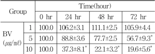
1 100.0 106.2±3.1 111.1±2.5 105.9±4.4 5 100.0 88.8±3.6 77.7±2.5 56.7±9.3* 10 100.0 37.3±8.1* 22.1±3.2* 19.6±5.6* Values are the mean ± SEM of three independent experiments performed in triplicate.
* : represents p<0.05 significant difference compared with control.
One-way analysis of variance followed by Tuckey test.
Table 1. Inhibition of DU-145 cell viability by BV
Fig. 1. Inhibition of DU-145 cell viability by BV Values are the mean ± SEM of three independent experiments performed in triplicate.
* : represents p<0.05 significant difference compared with control.
One-way analysis of variance followed by Tuckey test
dependent manner. To evaluate an effect of BV on the cell growth of DU-145 cells, I analyzed cell viability using WST-1 assay and direct cell counting. BV inhibited prostate cancer cell growth dose dependently during 72hr. The percentage of control significantly decreased by 5 ㎍/㎖ of BV at 72hr was 56.7±9.3%, which of control significantly decreased by 10 ㎍/㎖ of BV at 24, 48 and 72hr was 37.3±8.1, 22.1±3.2 and 19.6±5.6% (Table 1, Fig. 1).
B. Morphologic evaluation of apoptosis
To delineate whether the inhibition of cell growth by BV was due to increase of the induction of apoptosis, I evaluated change of the chromatin morphology of human prostate cancer cells using DAPI staining.
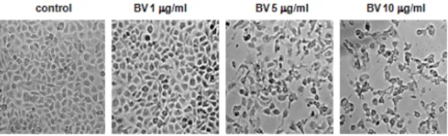
Consistent with the loss of viability, DU-145 cell demonstrated the characteristics of apoptotic morphologic change including cell shrinkage with irregular shape, cytoplasmic blebbing, chromatin condensation, DNA fragmentation and apoptotic bodies, etc (Fig. 2).
Fig. 2. Morphologic change of DU-145 cells treated by BV
Morphological changes were observed under microscope (magnification, 200×). The figures are representative of three experiments, with triplicate of each experiment.
C. Inhibition of NF-κB
It was demonstrated that BV negatively regulates NF-κB and suppress expression of inflammation related genes including iNOS and COX-2 in DU-145 cells. In addition, NF-κB is known to be inhibitory transcription factor of apoptosis. To investigate the hypothesis that BV can inactivate NF-κB and suppress the expression of inflammation related genes such as iNOS, COX-2 and cPLA2, and thereby prevent anti-apoptotic ability of NF-κB causing DU-145 cells go apoptosis, I assessed NF-κB
activity in the cells treated for different concen- tration of BV for 24 hr by EMSA and observed inflammation gene expression in the cells by western blot analysis. NF-κB was highly activated in this cell and inflammation related gene expression was also enhanced , however the activation of NF-κB and inflammatory gene expression were gradually decreased by the culture in the presence of BV in the cells(Fig. 3~5). In NF-kB activity, the density of control significantly decreased by 5 and 10㎍/㎖
of BV was 49.0±3.0 and 38.0±25.0% (Table 2, Fig.
3). In iNOS expression, the density of control significantly decreased by 5 and 10 ㎍/㎖ of BV was 53.0±13.0 and 10.0±3.0%. In COX-2 expression, the density of control significantly decreased by 1, 5 and 10 ㎍/㎖ of BV was 42.0±6.0, 37.0±4.0 and 8.0±
2.0%. In cPLA2 expression, the density of control sig- nificantly decreased by 1, 5 and 10 ㎍/㎖ of BV was 64.0±7.0, 21.0±4.0 and 24.0±12.0% (Table 3, Fig. 5).
Group Reiative density(%)
Control 100±12
BV (㎍/㎖)
1 88±15
5 *49±3*
10 *38±25*
Values are the mean ± SEM of three independent experiments performed in triplicate.
* : represents p<0.05 significant difference compared with control.
One-way analysis of variance followed by Tuckey test.
Table 2. Effect of BV on NF-κB activity in DU-145 Cells
Group
Apoptotic gene expression (relative density, %)
cPLA2 iNOS COX-2
BV (㎍/㎖)
0 100±15 100±10 100±10 1 *64±7* 65±5 *42±6* 5 *21±4* *53±13* *37±4* 10 * 24±12* *10±3* *8±2* Values are the mean ± SEM of three independent experiments performed in triplicate.
* : represents p<0.05 significant difference compared with control.
One-way analysis of variance followed by Tuckey test.
Table 3. Effects of BV on inflammatory gene expression in DU-145 cells
Fig. 5. Effects of BV on inflammatory gene expression in DU-145 cells Values are the mean ± SEM of three independent experiments performed in triplicate.
* represents p<0.05 significant difference compared with control.
One-way analysis of variance followed by Tuckey test Fig. 3. Effect of BV on NF-κB activity in
DU-145 cells
Activation of NF-κB was determined by electrophoretic mobility shift assay (EMSA), as described in materials and methods. Nuclear extracts from DU-145 cells with 1, 5 and 10 ㎍/㎖ of BV were incubated in binding reactions of 32P-labeled oligonucleotide containing the B sequence. NF- κB DNA binding activity was determined by EMSA.
* : represents p<0.05 significant difference compared with control.
One-way analysis of variance followed by Tuckey test.
Fig. 4. Effects of BV on inflammatory gene expression in DU-145 cells
DU-145 cells were treated with 1, 5 and 10 ㎍/㎖ of BV at 37°C for 24 hours. Equal amounts of total proteins (80g/lane) were subjected to 10% sodium dodecyl sulfate- polyacrylamide gel electrophoresis, and expression of cytosolic phospholipase A2(cPLA), cyclooxygenase 2 (COX-2), inducible nitric oxide synthase (iNOS), and β -actin (as an internal control) was detected by Western blotting using specific antibodies. Quantification of band intensities from 3 independent experimental results was determined by densitometry.
D. Inhibitory Effect of BV on NF-κB dependent Luciferase Activity in DU-145 Cells
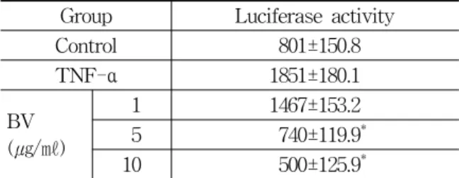
Transcriptional regulation involving the activation of NF-κB has been implicated in the expression of COX-2, cPLA2 and iNOS. To determine the role of BV in NF-κB dependent gene transcription, I conducted a transient transfection assay with a fusion gene containing SV40 promoter, 5 repeats of the consensus NF-κB binding sequence, and the luciferase reporter gene. DU-145 cells were transfected with this promoter-reporter gene construct, and transcriptional activities were measured after TNF- α stimulation with or without BV. As shown in Figure 6, cotreatment of the transfected cells with BV significantly inhibited the luciferase activity induced by TNF-αin DU-145 cells, which signifi- cantly decreased by 5 and 10㎍/㎖ of BV was 740.0±
119.9 and 500.0±125.9 (Table 4, Fig. 6).
Group Luciferase activity Control 801±150.8
TNF-α 1851±180.1
BV (㎍/㎖)
1 1467±153.2
5 *740±119.9* 10 *500±125.9* TNF-α : group treated by TNF-α in absence of BV.
Values are the mean ± SEM of three independent experiments performed in triplicate.
* : represents p<0.05 significant difference compared with control.
One-way analysis of variance followed by Tuckey test.
Table 4. Effect of BV on NF-κB dependent luciferase activity in DU-145 cells
Fig. 6. Effects of BV on TNF-α induced NF-κB dependent luciferase activity in DU-145 cells DU-145 cells were transfected with pNF-κB-Luc plasmid (5NF-κB) and then activated with TNF-α in absence of BV or presence of 1, 5 and 10 ㎍/㎖ of BV for 2 hours, and then the luciferase activity was determined. Values are the mean ± SEM of three independent experiments performed in triplicate. The level of induction was calculated relative to the luciferase activity in unstimulated transfected cells.
E. Mechanism of NF-κB inactivation
In the present study, to confirm whether BV also inhibits NF-κB through strong binding of BV to cysteine residues with sulfhydryl group in DU-145 cells, and to corroborate the above findings, transient transfection assay using a fusion gene containing pCMV promoter and p50 or IKKs mutant was conducted. DU-145 cells were transfected with these mutant genes constructs.
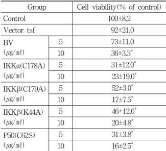
NF-κB activity and cell viability were then measured after stimulating the cells with BV (1, 5, 10 μg/㎖) for 24 hr. Contrary to the above son et al.’s and similar to Lee et al.s, present study revealed that BV inhibited cell viability and NF-κB activity dose dependently in the DU-145 cells transfected with p50 (C62S) or IKKs (IKKα (C178A), IKKβ (C179A), IKKβ (K44A)) mutant as well as in wild DU-145 cells or vector alone (Fig. 7). The percentage of contol significantly decreased by 10
㎍/㎖ of BV in the vector alone was 36±3.3, which of vector significantly decreased by 5 and 10 ㎍/㎖
of BV in the DU-145 cells transfected with IKKα (C178A) mutant was 31±12.0 and 23±19.0, which of
vector significantly decreased by 5 and 10 ㎍/㎖ of BV in the DU-145 cells transfected with IKKβ (C179A) or IKKβ (K44A) was 52±31.0 and 17±7.5, or 46±12.0 and 20±4.8%, which of vector significantly decreased by 5 and 10 ㎍/㎖ of BV in the DU-145 cells transfected with p50 (C62S) mutant was 31±
3.8 and 16±2.5% respectively (Table 5, Fig. 7).
Group Cell viability(% of control)
Control 100±8.2
Vector tsf 92±21.0 BV
(㎍/㎖)
5 73±11.0 10 *36±3.3* IKKα(C178A)
(㎍/㎖)
5 * 31±12.0* 10 23±19.0* IKKβ(C179A)
(㎍/㎖)
5 52±3.0* 10 17±7.5* IKKβ(K44A)
(㎍/㎖)
5 46±12.0* 10 20±4.8* P50(C62S)
(㎍/㎖)
5 31±3.8* 10 16±2.5*
Values are the mean ± SEM of three independent experiments performed in triplicate.
* : represents p<0.05 significant difference compared with control.
One-way analysis of variance followed by Tuckey test.
Table 5. Effect of BV on cell viability in the p50, IKKs mutant plasmid transfected DU-145 cells
Fig. 7. Effects of BV on NF-κB activity and cell viability in the p50, IKKs mutant plasmid transfected DU-145 cells
Values are the mean ± SEM of three independent experiments performed in triplicate.
* : represents p<0.05 significant difference compared with vector tsf.
One-way analysis of variance followed by Tuckey test.
Ⅳ. Discussion
The noteworthy findings in this study is the identification of anti-proliferative efficacy of BV against human testosterone refractory prostate carcinoma DU-145 cells. Most of the current available anti-cancer drugs exert their effect via induction of apoptosis in cancer cells15,16), which is considered as one of the major mechanisms for the targeted therapy of various cancers including prostate cancer15-18) .
Prostate cancer cells easily metastasize and don’t respond to cytotoxic chemotherapeutic agents due to resistance to apoptosis in the androgen refractory stage17). Therefore, the agents that induce apoptotic cell death of prostate cancer cells could be majorly used in controlling this hormone refractory malignant cancer18).
My data demonstrating an induction of apoptotic cell death in prostate cancer control suggest that comparatively low concentration (below 10 µg/㎖) of BV could be useful as a candidate preventing and treating this malignancy.
It has been well established that NF-κB is an important element in regulating cell growth or apoptosis of tumor cells, including prostate cancer cells19). Recently several studies have reported that NF-κB is constitutively activated in human prostate cancer tissue, androgen-insensitive human prostate carcinoma cells, and prostate cancer xenografts19,20). Moreover, compounds inhibiting NF-κB have shown to induce apoptotic cell death of DU-145 cells21,22). From the above, agents suppressing activities of NF-κB signal molecules may be potentially available in preventing and managing prostate cancer cell proliferation via induction of apoptotic prostate cancer cell death. In the present study, as DU-145 cells were exposed to BV(1~10
㎍/㎖), the cells were not grown, and died in a dose dependent manner. Moreover, BV inhibited prostate cancer cell growth significantly in 10 ㎍/㎖
of BV during 72 hr and characteristic apoptotic morphologic change sustained that inhibition of
DU-145 cell proliferation was due to apoptosis.
In NF-κB activity of the cells treated with different concentration of BV for 24 hr, NF-κB was highly activated in DU-145 cells, while activation of NF-κB was significantly decreased by the culture in the presence of 5-10 ㎍/㎖ of BV in the cells.
Meanwhile, Transcriptional regulation involving the activation of NF-κB has been implicated in cancer growth as well as the expression of COX-2 and iNOS. Consistent with the decreased activity of NF-κB down regulated by BV, Inflammation related gene expression and transcriptional activity was significantly decreased in the 5-10 ㎍/㎖ of BV treated cells. These data substantiated the hypothesis that BV can inactivate NF-κB and suppress the expression of inflammation related genes such as iNOS, COX-2 and cPLA2 , and thereby prevent anti-apoptotic ability of NF-κB causing DU-145 cells go apoptosis, suggesting that NF-κB signal may be significant contributor in BV-induced DU-145 cell death.
Although much efforts has been made to elucidate the mechanism on the molecular basis how BV affects on the NF-κB signal molecules in the previous reports12,13), it is not yet clear.
According to Son et al14), at the molecular level, SVT inhibited constitutively activated NF-κB signaling by impairing IκBα phosphorylation with inhibition of p50 translocation, and it bound with sulfhydryl group of cysteine residue in NF-κB, IKK α and IKKβ resulting in down regulation of NF-κB activity, moreover, abolished SVT-induced apoptotic cell death was found in the PC-3 cells transfected with mutant p50, IKKα and β in which the cysteine residue was replaced with other amino acids.
On the basis of the hypothesis that there is close structural analogy between SVT and BV, Lee et al12) and Oh et al13) tried to re-ascertain the above findings in their studies. They found that BV inhibited LNCaP or PC-3 cells proliferation and NF-κB activation. However, inconsistent with the previous Son’s report14), the reverse effect was not actually shown in the LNCaP or PC-3 cells transfected with p50 (C62S) or IKKs mutant.
In the present study, to confirm whether BV also inhibits activity of NF-κB, and to corroborate the above findings in another hormone refractory prostate cancer DU-145 cells, transient transfection assay using a fusion gene containing pCMV promoter and p50 or IKKs mutant was conducted.
NF-κB activity and cell viability were not reversely increased in this study either.
Consequently, although I could not elaborate on the reason why the abolition of son’s report13) was not reproduced in this study. My present findings actually revealed that anticancer effect against advanced DU-145 human hormone refractory prostate cancer cells of BV is associated with down-regulation of the constitutively over-expressed or NF-κB-dependent anti-apoptotic proteins via reduction of nuclear translocation of NF-κB proteins. It was suggested that BV could form a basis for making BV available as a novel agent for human prostate cancer prevention and/or inter- vention, andd that another mechanism study should be devised for the exact explanation.
Ⅴ. References
1. Hellerstedt BA, Pienta KJ. The current state of hormonal therapy for prostate cancer. CA Cancer J Clin. 2002 : 52, 154-79.
2. Luciana D. Apoptosis induction in DU-145 human prostate carcinoma cells. Tissue and Cell. 2005 : 37, 379-84.
3. Timme TL, Satoh T, Tahir SA, Wang H, The BS, Butler EB, Miles BJ, Amato RJ, Kadmon D, Thompson TC. Therapeutic targets for metastatic prostate cancer. Curr Drug Targets. 4. 2003 : 251-61.
4. Santos AF, Huang H, Tindall DJ. The androgen receptor: a potential target for therapy of prostate cancer. Steroids. 2004 : 69, 79-85.
5. Srilatha S, Aruna VK, Donna MP, David F.
Genistein potentiates the growth inhibitory effects of 1, 25-dihydroxyvitamin D3 in DU145
human prostate cancer cells: Role of the direct inhibition of CYP24 enzyme activity. Molecular and Cellular Endocrinology 241. 2005 : 49-61.
6. Brown RL, Haley TL, West KA, Crabb JW.
Pseudechetoxin: a peptide blocker of cyclic nucleotide-gated ion channels. Proc Natl Acad Sci USA. 1999 ; 96 : 7549.
7. Sher E, Giovannini F, Boot J, Lang B. Peptide neurotoxins, small-celllung carcinoma andneuro- logical paraneoplastic syndromes. Biochimie.
2000 ; 82 : 92736.
8. Van Lunteren E, Moyer M. Peptide toxin blockers of voltage-sensitive K+ channels:
Inotropic effects on diaphragm. J Appl Physiol.
1999 ; 3 : 100916.
9. Park HJ, Lee SH, Son DJ, Oh KW, Kim KH, Song HS, Kim GJ, Oh GT, Yoon DY and Hong JT. Antiarthritic effect of BV: inhibition of inflammation mediator generation by suppres- sion of NF-kappaB through interaction with the p50 subunit. Arthritis Rheum. 2004 ; 50 : 3504-15.
10. Holle L, Song W, Holle E, Wei Y, Wagner T, Yu X. A matrix metalloproteinase 2 cleavable melittin/avidin conjugate specifically targets tumor cells in vitro and in vivo. Int J Oncol.
2003 : 22, 93-8.
11. Park HJ, Lee YK, Song HS, Kim KJ, Son DJ, Lee JW and Hong JT. Melittin inhibits human prostate cancer cell growth through induction of apoptotic cell death. J Toxicol Pub Health. 2006
; 22(1) : 31-7.
12. HS Lee, HS Song. BV inhibits LNCaP cell proliferation through induction of apoptosis via inactivation of NF-κB. Journal of Acupuncture and Moxibustion. 2007 ; 25(2) : 59-74.
13. HJ Oh, HS Song. BV inhibits PC-3 cell prolif- eration through induction of apoptosis via in- activation of NF-kB. Journal of Acupuncture and Moxibustion. 2010 ; 27(3) : 1-14.
14. DJ Son, MH Park, SJ Chae et al. Inhibitory effect of snake venom toxin from Vipera lebetina turanica on hormone-refractory human prostate cancer cell growth: induction of
apoptosis through inactivation of nuclear factor KB. Mol Cancer Ther. 2007 ; 6(2) : 675-83.
15. Lowe SW, Lin AW. Apoptosis in cancer.
Carcinogenesis. 2000 ; 21 : 48595.
16. Guseva NV, Taghiyev AF, Rokhlin OW, Cohen MB. Death receptor-induced cell death in prostate cancer. J Cell Biochem. 2004 ; 91 : 7099.
17. Gurumurthy S, Vasudevan KM, Rangnekar VM.
Regulation of apoptosis in prostate cancer.
Cancer Metastasis Rev. 2001 ; 20 : 22543.
18. Kantoff PW. New agents in the therapy of hormone-refractory patients with prostate cancer. Semin Oncol. 1995 ; 22(1) : 324.
19. Suh J, Payvandi F, Edelstein LC et al.
Mechanisms of constitutive NF-kappaB activation in human prostate cancer cells. Prostate. 2002 ; 52 : 183200.
20. Raj GV, Sekula JA, Guo R, Madden JF, Daaka
Y. Lysophosphatidic acid promotes survival of androgen-insensitive prostate cancer PC3 cells via activation of NF-κB. Prostate. 2004 ; 61 : 10513.
21. Anton S, Andrew VS, Jozsef LV, Marta Z, Ren-Zhi C, Benjamin B, Brian D. Hammann, Patricia Armatis, Celia A. Kanashiro.Inhibition of human androgen-independent PC-3 and DU-145 prostate cancers by antagonists of bombesin and growth hormone releasing hormone is linked to PKC, MAPK and c-jun intracellular signalling. European Journal of Cancer 41. 2005 : 2735-44.
22. Kushlani G, Darrell KM, Richard ES, DDS AW.
Inhibition of nuclear factor B induces apoptosis following treatment with tumor necrosis factor and an antioxidant in human prostate cancer cells. Cancer Detection and Prevention 26. 2002 : 229-37.