Culture-Expanded Autologous Adipose-
Derived Mesenchymal Stem Cell Treatment for Osteonecrosis of the Femoral Head
Pil Whan Yoon, MD, Jong Yeal Kang, MD*, Chul-Ho Kim, MD
†, Soong Joon Lee, MD*, Jeong Joon Yoo, MD
‡, Hee Joong Kim, MD
‡, Sung Keun Kang, PhD
§, Ju Hyeon Min, BS
§, Kang Sup Yoon, MD*
,‡Department of Orthopedic Surgery, Asan Medical Center, University of Ulsan College of Medicine, Seoul,
*Department of Orthopedic Surgery, Seoul National University Boramae Hospital, Seoul,
†Department of Orthopedic Surgery, Gachon University Gil Medical Center, Incheon,
‡Department of Orthopedic Surgery, Seoul National University College of Medicine, Seoul,
§Biostar Stem Cell Research Institute, R Bio Co., Ltd., Seoul, Korea
Background: Outcomes of traditional treatment for osteonecrosis of the femoral head (ONFH) are not always satisfactory. Hence, cell-supplementation therapy has been attempted to facilitate necrotic-tissue regeneration. Adipose-derived mesenchymal stem cell (ADMSC) transplantation is potentially advantageous over bone marrow-derived MSC implantation, but its outcomes for ONFH remain unclear. The aim of this study was to determine 2-year radiological and clinical outcomes of culture-expanded autologous ADMSC implantation for ONFH.
Methods: Eighteen hips with necrotic lesions involving ≥ 30% of the femoral head were included. ADMSCs were harvested by liposuction and culture expanded for 3 passages over 3 weeks. With a 6-mm single drilling, ADMSCs were implanted into the necrotic zone. All patients underwent magnetic resonance imaging (MRI), single-photon emission computed tomography/com- puted tomography (SPECT/CT) at screening and 6 months, 12 months, and 24 months postoperatively. The primary outcome was the change in the size of necrotic area on MRI. Secondary outcomes were changes in clinical scores and radioisotope uptake on SPECT/CT. Conversion total hip arthroplasty (THA) was defined as the endpoint.
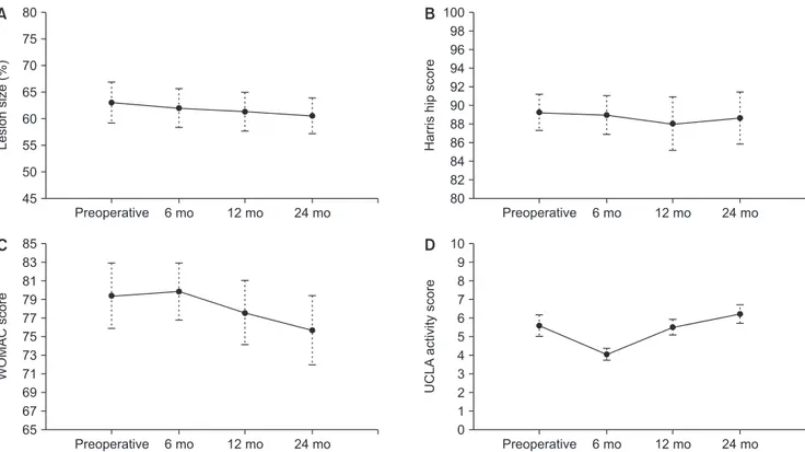
Results: Preoperatively, the necrotic lesion extent was 63.0% (38.4%–96.7%) of the femoral head. The mean Harris hip score was 89.2, the University of California at Los Angeles (UCLA) score was 5.6, and Western Ontario and McMaster Universities Arthritis index (WOMAC) was 79.4. Three patients underwent THA and 1 patient died in an accident. Finally, 11 patients (14 hips) were avail- able for ≥ 2-year follow-up. At the last follow-up, no surgery-related complications occurred, and 14 of 17 hips (82%) were able to perform daily activities without THA requirement. There was no significant decrease in lesion size between any 2 intervals on MRI.
However, widening of high signal intensity bands on T2-weighted images inside the necrotic lesion was observed in 9 of 14 hips (64%);
11 of 14 hips (79%) showed increased vascularity on SPECT/CT at 2 years postoperatively. No significant differences were observed between preoperative and 24-month mean Harris hip score (89.2 vs. 88.6), WOMAC (79.4 vs. 75.7), and UCLA score (5.6 vs. 6.2).
Conclusions: Our outcomes suggest that culture-expanded ADMSC implantation is a viable option for ONFH treatment without adverse events.
Keywords: Osteonecrosis of the femoral head, Stem cell, Core decompression, Lesion size, Magnetic resonance imaging
Copyright © 2021 by The Korean Orthopaedic Association
This is an Open Access article distributed under the terms of the Creative Commons Attribution Non-Commercial License (http://creativecommons.org/licenses/by-nc/4.0) which permits unrestricted non-commercial use, distribution, and reproduction in any medium, provided the original work is properly cited.
Clinics in Orthopedic Surgery • pISSN 2005-291X eISSN 2005-4408 Received May 22, 2020; Accepted June 22, 2020
Correspondence to: Kang Sup Yoon, MD
Department of Orthopedic Surgery, Seoul National University Boramae Hospital, 20 Boramae-ro 5-gil, Dongjak-gu, Seoul 07061, Korea Tel: +82-2-870-2311, Fax:+82-2-831-2826, E-mail: ksyoon@snu.ac.kr
Osteonecrosis of the femoral head (ONFH) is relatively common and leads to femoral head collapse and second- ary hip osteoarthritis. Although ONFH’s natural course varies with the location or size of the necrotic lesion, symptom development and disease progression are likely if > 30% of the femoral head is affected.1) If left untreated, it may require total hip arthroplasty (THA). Considering that ONFH occurs frequently in younger patients (age, 30–50 years),2,3) prevention or delay of femoral head col- lapse is the primary goal of surgical intervention.
Femoral head core decompression or multiple drill- ing had been widely used for ONFH treatment in the past.
These procedures aim to preserve the femoral head and facilitate necrotic tissue regeneration by revascularization and provision of channels for osteogenic cell recruitment from the proximal femur. However, the reported success rate of isolated core decompression is 30%–86%, and the results are unsatisfactory for larger lesions.4)
One of the reasons for the unsatisfactory outcome of isolated core decompression might be that osteoprogenitor cell numbers in the femoral head and proximal femur are insufficient for complete ONFH reconstruction or repair.5) Thus, many attempts have been made to facilitate necrotic lesion repair by stem cell implantation, as several authors have reported favorable outcomes of autologous bone marrow grafting for ONFH.5-9)
Adipose-derived mesenchymal stem cell (ADMSC) implantation has recently become a promising method for stem cell therapy because these cells have multi- mesenchymal lineage potential and can differentiate into osteoprogenitor cells under appropriate conditions.10) Moreover, ADMSC harvesting is relatively simple and cell lines can be easily expanded for sufficient cell numbers.
Thus, ADMSC implantation is promising for ONFH treat- ment. However, few studies have reported the efficacy of ADMSC implantation for ONFH treatment.
Therefore, the aim of the current study was to deter- mine the 2-year clinical and radiological outcomes of AD- MSC implantation for ONFH treatment. The authors hy- pothesized that ADMSC implantation would (1) decrease the necrotic lesion size on magnetic resonance imaging (MRI), (2) improve clinical scores, and (3) increase necrotic lesion radioisotope uptake on single-photon emission com- puted tomography/computed tomography (SPECT/CT).
METHODS
Study Design and Subjects
The phase I/IIa trial protocol was approved by the Korean Food and Drug Administration (KFDA). The study pro-
tocol was approved by the Institutional Review Board of Seoul National University Boramae Medical Center Insti- tutional Review Board (IRB No. 06-2010-128). Written in- formed consent was obtained from all participants before enrollment in the study. This study is registered with the International Clinical Trials Registry (NCT01643655).
The phase I cohort included 3 patients receiving injections of 1 × 10⁸ autologous ADMSCs in 3-mL saline.
Adverse event occurrence was evaluated for 28 days after the injection. Adverse events were defined according to the National Cancer Institute common terminology criteria for adverse events (NCI-CTCAE) grade 3 (dose-limiting toxicity) as follows: (1) allergic reaction/hypersensitivity, (2) injection-site reaction, (3) injection-site infection, (4) fibrosis-cosmesis, and (5) muscular/skeletal hypoplasia.
A safety review conducted on day 28 found no incidence of the abovementioned adverse events. Thus, the phase IIa study with 12 patients was commenced; each patient received phase I dosages. Thus, 15 patients (18 hips) were enrolled from July 2012 to January 2013.
Patients met the following eligibility criteria: (1) age 18–70 years; (2) nontraumatic ONFH; (3) hips without femoral head collapse or < 2 mm femoral head depression (Steinberg stage I, II, and IIIA); and (4) necrotic lesion size
≥ 30% of the femoral head area. Necrotic lesion size was measured on MRI as described previously.11) Exclusion cri- teria were (1) femoral head collapse ≥ 2 mm; (2) previous history of core decompression/multiple drilling; (3) histo- ry of bisphosphonate or parathyroid hormone therapy; (4) current consumption of immunosuppressants, cytotoxic agents, or corticosteroids; (5) inability to undergo MRI examination; (6) pregnancy or breastfeeding; (7) active infectious disease including human immunodeficiency vi- rus infection; (8) serious comorbidities that may affect the treatment process; (9) history of participation in another clinical trial ≤ 3 months; and (10) refusal to participate.
After providing informed consent, all patients underwent screening, including physical examination, blood and urine testing, chest X-ray, and electrocardi- ography. Anteroposterior and frog-leg hip X-rays, MRI, and SPECT/CT were also conducted at the initial visit.
Liposuction was performed 1 week afterward. When pa- tients were admitted for ADMSC implantation, baseline Harris hip score (HHS), Western Ontario and McMaster Universities Arthritis index (WOMAC), and University of California at Los Angeles (UCLA) scores were obtained.
Hip X-ray, MRI, and SPECT/CT were performed preop- eratively and at 6 months, 12 months, and 24 months post- operatively. HHS, WOMAC index, and UCLA score were also obtained at each postoperative visit.
Mesenchymal Stem Cell Preparation
Abdominal fat tissue samples were harvested by liposuc- tion under local anesthesia for ADMSC isolation 1 week after the patient screening. ADMSC preparation was con- ducted under standard conditions as previously described.
Harvested fat tissues were enzymatically digested and stro- mal vascular fraction cells were isolated and cultured in keratinocyte serum-free medium (Invitrogen Corp., Carls- bad, CA, USA) containing 0.2-mM ascorbic acid, 0.09-mM calcium, 5-ng/mL recombinant epidermal growth factor, and 5% fetal bovine serum. The cells were passaged until reaching 90% confluence and then culture-expanded for 3 passages over 3 weeks. Afterward, the cells were tested for cell number, viability, purity (CD31, CD34, CD45), iden- tity (CD 73, CD90), sterility, endotoxins, and mycoplasma.
Finally, 1 × 10⁸ ADMSCs in 3-mL saline were prepared in a syringe (Jointstem; R Bio, Seoul, Korea; http://www.rbio.
co.kr/).
Surgical Technique and Rehabilitation
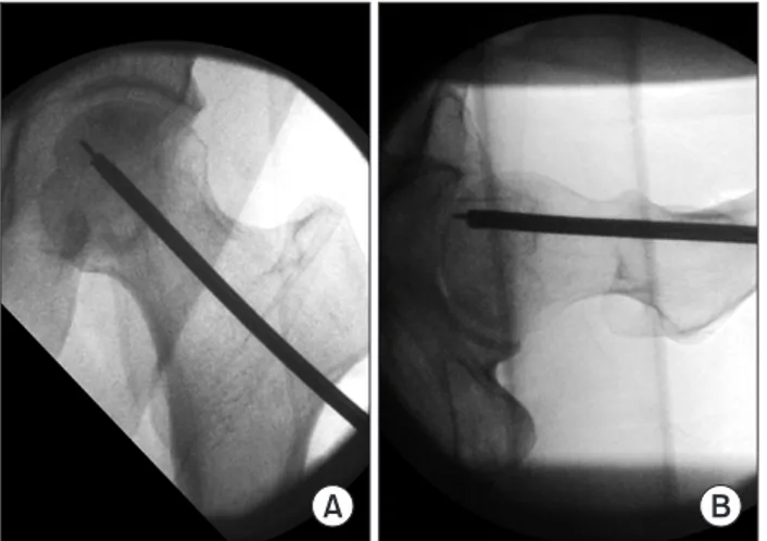
Under spinal anesthesia, the patient was placed supine on a fracture table. A longitudinal 5-cm skin incision was made just below the greater trochanter. Under fluoroscop- ic visualization, a guide pin was directed to the necrotic lesion center, which had been preoperatively identified on MRI. A 6-mm reamer was introduced along the guide pin, and reaming was performed just under the subchon- dral bone of the lesion (Fig. 1). Before ADMSC injection, the fracture table was tilted so that the ipsilateral greater trochanter faces upwards to prevent backing out of the injected cells. The syringe filled with ADMSCs was then connected to an 18-G spinal needle and 1×10⁸ ADMSCs in 3-mL saline were injected under fluoroscopic guidance.
After injection, the lateral cortex reamed channel opening was sealed with a collagen bone plug (TERUPLUG; Olym- pus Terumo Biomaterials Co., Tokyo, Japan). Ipsilateral, partial, crutch/cane-assisted weight-bearing was allowed for the first 6 weeks postoperatively. Afterward, total weight-bearing was gradually permitted as tolerated.
Outcome Measures
The primary outcome was the change in the necrotic lesion size on MRI as previously reported.11) Measure- ments were taken by 2 independent, blinded radiologists.
Secondary outcomes were changes in the clinical scores and SPECT/CT radioisotope uptake. Clinical assessments were performed using HHS. Patient activity level was assessed using WOMAC index and UCLA score at the last follow-up. In the original WOMAC scoring system, a higher score indicates a poorer status. For consistency with other scoring tools, the WOMAC index was reversed so that a higher score indicates better functional status.
Radioisotope uptake degree on SPECT/CT was evaluated qualitatively by an independent nuclear medicine expert and increased uptake was determined in a dichotomized fashion. All adverse events were recorded according to the NCI-CTCAE criteria version 4.0. Conversion to THA was defined as the endpoint and no further evaluation was made if the patient underwent THA.
Statistical Analysis
The change in the necrotic lesion size was determined by one-way analysis of variance with the Bonferroni post-hoc test. HHS, WOMAC index, and UCLA scores were com- pared preoperatively and 24 months postoperatively using the paired t-test. Statistical analyses were conducted using IBM SPSS ver. 20.0 (IBM Corp., Armonk, NY, USA). A p- value < 0.05 was considered statistically significant.
RESULTS
Demographic and Preoperative Characteristics of Patients From July 2012 to January 2013, 15 patients (13 men and 2 women; 18 hips) underwent ADMSC implantation and were followed up for 24 months. The mean age and body mass index were 43.8 years (range, 20–59 years) and 25.5
± 3.3 kg/m2, respectively. Seven patients had a history of heavy alcohol consumption, 2 received steroid therapy, 1 had caisson disease, and 6 were diagnosed with idiopathic ONFH. Fourteen hips were classified as Steinberg stage IIC and 4 as stage IIIA.12) Six hips were associated with pain, whereas 12 were asymptomatic and contralateral hip ONFH was incidentally found on diagnostic imaging.
A B
Fig. 1. Intraoperative anteroposterior (A) and lateral (B) fluoroscopic images showing guide pin and reamer placement at the predetermined target.
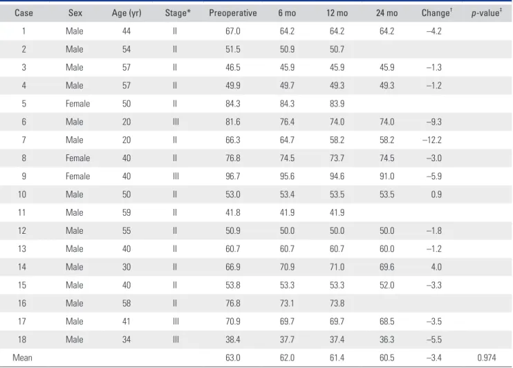
Six hips showed subchondral fractures with bone mar- row edema patterns outside the necrotic area on preop- erative MRI. The mean necrosis extent of the 18 lesions was 63.0% (38.4%–96.7%) of the total femoral head area.
The mean HHS was 89.2/100, the mean UCLA score was 5.6/10, and the mean WOMAC index was 79/96. During the follow-up period, 3 hips (18%) in 3 patients underwent THA at 13–16 months after the initial surgery because of femoral head collapse and pain aggravation, which dis- rupted daily activities. At 12 months after initial surgery, the mean HHS, UCLA score, and WOMAC index of 3 hips were 75/100, 5/10, and 61/96, respectively. One pa- tient with unilateral ONFH died in an unrelated accident during follow-up. Therefore, 24-month outcome data were available for 11 patients (14 hips).
Outcomes
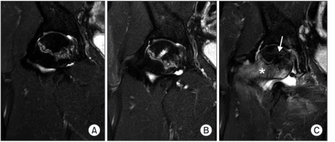
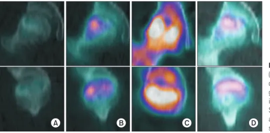
At the last follow-up, there were no surgery-related com- plications. MRI showed no significant lesion size changes between any 2 intervals (Table 1, Fig. 2). However, 1 pa- tient (case 6 and 7) showed definite lesion size decrease by 9.3% (case 6) and 12.2% (case 7) on MRI at 24 months after surgery. His plain radiographs also showed healed subchondral fracture and increased radiodensity of the femoral head (Fig. 3). Interestingly, widening of high signal intensity band on T2-weighted images inside the necrotic lesion was observed in 9/14 hips (64%), which might indicate increased vascularity and tissue regenera- tion (Fig. 4). This finding was not observed until the last follow-up in 3 hips after THA (Fig. 5). There were no sig- nificant differences between preoperative and 24-month postoperative HHS, WOMAC index, and UCLA score in 11 patients (14 hips) (Table 2, Fig. 2). SPECT/CT at 24
Table 1. Change in the Size of Necrotic Lesion over 2-Year Follow-up
Case Sex Age (yr) Stage* Preoperative 6 mo 12 mo 24 mo Change† p-value‡
1 Male 44 II 67.0 64.2 64.2 64.2 –4.2
2 Male 54 II 51.5 50.9 50.7
3 Male 57 II 46.5 45.9 45.9 45.9 –1.3
4 Male 57 II 49.9 49.7 49.3 49.3 –1.2
5 Female 50 II 84.3 84.3 83.9
6 Male 20 III 81.6 76.4 74.0 74.0 –9.3
7 Male 20 II 66.3 64.7 58.2 58.2 –12.2
8 Female 40 II 76.8 74.5 73.7 74.5 –3.0
9 Female 40 III 96.7 95.6 94.6 91.0 –5.9
10 Male 50 II 53.0 53.4 53.5 53.5 0.9
11 Male 59 II 41.8 41.9 41.9
12 Male 55 II 50.9 50.0 50.0 50.0 –1.8
13 Male 40 II 60.7 60.7 60.7 60.0 –1.2
14 Male 30 II 66.9 70.9 71.0 69.6 4.0
15 Male 40 II 53.8 53.3 53.3 52.0 –3.3
16 Male 58 II 76.8 73.1 73.8
17 Male 41 III 70.9 69.7 69.7 68.5 –3.5
18 Male 34 III 38.4 37.7 37.4 36.3 –5.5
Mean 63.0 62.0 61.4 60.5 –3.4 0.974
Values are presented as percentages.
*Preoperative Association Research Circulation Osseous (ARCO) stage. †Change in lesion size was calculated as: 100 × (size at 24 months – preoperative size)/preoperative size. ‡The p-value was calculated by repeated measures analysis of variance.
months showed increased radioisotope uptake around the necrotic margin in 79% hips (11/14) (Fig. 6). Radiographs and MRI evaluations of the 3 hips (18%) indicated symp-
tomatic subchondral fracture with femoral head collapse
< 2 mm at 24 months of follow-up.
Preoperative 80
75 70 65 60 55 50 45
6 mo 12 mo 24 mo
Lesionsize(%)
A B 100
98 96 94 92 90 88 86 84 82 80
6 mo 12 mo 24 mo
Harrishipscore
85 83 81 79 77 75 73 71 69 67 65
6 mo 12 mo 24 mo
WOMACscore
10 9 8 7 6 5 4 3 2 1 0
6 mo 12 mo 24 mo
UCLAactivityscore
C D
Preoperative
Preoperative Preoperative
Fig. 2. Line graphs showing the temporal pattern of change in the necrotic lesion size (A), Harris hip score (B), Western Ontario and McMaster Universities Arthritis index (WOMAC) score (C), and University of California at Los Angeles (UCLA) activity score (D). Whiskers indicate the 95%
confidence interval.
*
A B
C D
Fig. 3. The preoperative coronal T1 mag- netic resonance imaging (MRI) scan (A) and plain radiograph (B) showed a large necrotic lesion with a subchondral fracture (arrow) of the femoral head. The MRI scan (C) and plain radiograph (D) taken at 24 months after surgery showed a decreased lesion size (arrowhead) and increased radiodensity (asterisk) of the femoral head.
DISCUSSION
For ONFH treatment, several methods have been attempt- ed for femoral head preservation before collapse. Core de- compression and multiple drilling are attractive, relatively easy surgical options with acceptable outcomes. However, several cases result in failure, especially those with large necrotic lesions. There has been growing interest in cell supplementation along with core decompression. Since the first report by Hernigou and Beaujean,5) several studies have reported the outcomes of concentrated bone marrow aspirate or bone marrow-derived mesenchymal stem cell
(BMMSC) implantation for ONFH treatment.5-9,13) Howev- er, only 1 study reported the safety and efficacy of culture- expanded BMMSCs for ONFH treatment.9) To the best of our knowledge, the current study is the first to evaluate the safety and outcome of culture-expanded autologous AD- MSC implantation for ONFH treatment in humans.
Our results indicated that femoral head ADMSC implantation is safe without serious complications. As this was the first study to utilize culture-expanded AD- MSCs for ONFH treatment, no direct comparisons with other studies were possible. Most previous studies utilized BMMSCs for ONFH treatment.5-9,13,14) BMMSCs have both Table 2. Changes in the Clinical Scores over 2-Year Follow-up
Outcome score Preoperative 6 mo 12 mo 24 mo p-value*
Harris hip score 89.2 ± 8.2 88.9 ± 8.7 88.0 ± 12.5 88.6 ± 11.8 0.976
WOMAC score 79.4 ± 14.7 79.9 ± 13.3 77.6 ± 15.0 75.7 ± 15.5 0.810
UCLA score 5.6 ± 2.4 4.1 ± 1.3 5.5 ± 1.8 6.2 ± 14.2 0.123
Values are presented as mean ± standard deviation.
WOMAC: Western Ontario and McMaster Universities Arthritis index, UCLA: University of California at Los Angeles.
*The p-value was calculated by paired t-test for preoperative scores and scores at 24 months after surgery.
A B C D
Fig. 4. Coronal T2 magnetic resonance imaging scans showing widening of the high signal intensity band inside the necrotic lesion (arrows). (A) Preoperative. (B) Six months after surgery. (C) Twelve months after surgery. (D) Twenty-four months after surgery.
A B C
*
*
Fig. 5. Coronal T2 magnetic resonance imaging (MRI) scans (case 5) showing no definite widening of the high signal intensity band inside the necrotic lesion during follow-up. (A) Pre operative. (B) Six months after surgery. (C) Twelve months after surgery. The MRI shows a sub- chondral fracture (arrow) and bone marrow edema (asterisk).
angiogenic and osteogenic potential, and long-lasting re- construction repair can be expected with BMMSC implan- tation.14) However, several limitations associated with its use for ONFH treatment exist. First, an invasive procedure under general or spinal anesthesia is required to obtain bone-marrow cells and available quantities are limited.
Second, bone marrow mesenchymal stem cell isolation yield decreases with age and the potential of stem cells for differentiation and proliferation also decrease with patient age.15) On the other hand, large quantities of ADMSCs can be easily obtained using a less invasive procedure, and cell numbers obtainable from a given amount of adipose tis- sue do not decrease with age.16) Some studies reported that ADMSCs can induce paracrine and autocrine responses, which enhance osteogenic potential.17) The cells trans- duced by these mechanisms excrete bone morphogenetic protein, which may further enhance their osteogenic ac- tivity. A recent study reported that the proliferation capac- ity of ADMSCs is 4 times that of BMMSCs; ADMSCs also show better phenotypical characteristics, which indicates better bone differentiation potential.16) Thus, ADMSCs, in- stead of BMMSCs, are a more promising option for ONFH treatment. In this context, future studies are warranted to further reveal ADMSCs’ role for ONFH treatment despite our limited success.
Although lesion sizes remained almost the same 2 years postoperatively, 64% of the hips (11/17) survived without symptomatic femoral head collapse, which would require THA. Including 3 hips with subchondral fracture and femoral head collapse < 2 mm without significant associated symptoms, 82% hips did not require THA at the 2-year follow-up. This finding is especially important because only patients with > 30% femoral head lesion size were included. The mean preoperative lesion size was 63.0% in this population and the smallest lesion size was
38.4%. The decision to include only patients with large lesions was according to the findings of a previous study.1) At a 5-year follow-up of the natural course of ONFH, only 1 of 21 patients developed pain when the lesion size was < 30%, whereas 11 of 24 patients with a lesion size 30%–50% and 50 of 60 patients with a lesion size > 50%
developed pain.1) Necrotic lesion size is one of the most important factors in determining the prognosis of ONFH.1,18) Hungerford and Jones19) reported that small lesions (< 15%
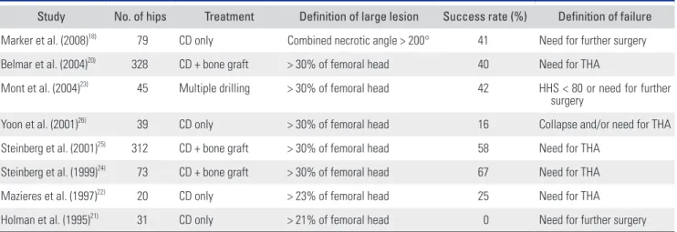
of the femoral head) were unlikely to progress and large lesions (> 30% of the femoral head) could not be suc- cessfully treated with core decompression. Indeed, many previous studies reported high failure rates of core de- compression for large lesions (Table 3).18,20-26) Although the present study did not include a control group, the 82% survival rate among hips with large necrotic lesions implies some contribution of implanted ADMSCs, in ad- dition to 6-mm drilling, to prevention of disease progres- sion.
One of the reasons this study failed to show lesion size decrease may be the large initial lesion size in the study population. Although no firm conclusions can be drawn about the reasons for no significant lesion size de- crease, we believe that the implanted cells failed to migrate throughout the entire necrotic region for effective repair with only a single drilling. We chose a 6-mm single drilling method for ADMSC implantation to minimize core de- compression or multiple drilling effect because KFDA had only approved it in order to prove the effect of ADMSC.
Increased radioisotope uptake might indicate increased vascular activity, but more effective methods to deliver the implanted cells to the entire necrotic tissue are needed.
Previous studies reported favorable outcomes with bone marrow or BMMSC implantation, but they included only patients with relatively small lesions7) or did not report
A B C D
Fig. 6. Coronal (first row) and sagittal (second row) single-photon emission computed tomography/computed tomo- graphy images showing increased uptake in the femoral head. (A) Preoperative. (B) Six months after surgery. (C) Twelve months after surgery. (D) Twenty-four months after surgery.
the lesion size.14,27) Therefore, direct comparisons between these studies and the present study are difficult. A previous study of bone marrow grafting reported that cases with lesions > 25% of the femoral head had significantly worse outcomes.5) In a study of a rabbit ONFH model, the group treated with adipose-derived stem cells showed higher bone trabecular volume on micro-CT, which suggests necrotic bone regeneration in the lesion.28) The outcomes of this study suggest that ADMSCs can also facilitate re- generation in human ONFH. However, further studies of ADMSC implantation in patients with smaller lesions are needed to clarify this issue.
In the current study, no significant changes in any of the clinical scores were observed before and 24 months postoperatively. This is probably because many hips were asymptomatic preoperatively. Pain was only noted in 6 of 18 hips, and 12 were incidentally diagnosed. The presence or absence of pain was not considered as inclusion or ex- clusion criteria in the current study, because it was unethi- cal to exclude patients with large necrotic lesions regard- less of pain as hips with large necrotic lesions are likely to collapse.19,29) Preoperative clinical scores of asymptomatic hips were already high; therefore, no room for further improvement existed because of a ceiling effect. There was some improvement in the scores of hips that were initially painful, but the difference was not statistically significance because of the small sample size. Further studies with larger sample sizes would enable subgroup analysis of hips with initial pain.
Our study has some limitations. First, there was no control group, which limits interpretation of outcomes.
Hence, future studies with control groups are required to further elucidate the efficacy of ADMSC implantation.
Second, the sample size was too small to conduct robust statistical analysis, which was inevitable considering that this was a phase I/IIa trial and only a small number of patients were enrolled. Because there were no procedure- related serious adverse events in this study, future studies with larger sample sizes are encouraged. Third, the fate of implanted ADMSCs was not examined. Although there was increased radioisotope uptake on SPECT/CT in many cases, it is unclear whether this change was induced by the implanted ADMSCs. In our cohort, the femoral head was drilled with a 6-mm reamer, which is smaller than that used for standard core decompression to minimize adverse effects. Nonetheless, it was not possible to verify whether the implanted cells functioned as expected.
At the last follow-up, there was progression of dis- ease in 6 out of 17 hips (36%), including 3 hips with symp- tomatic subchondral fractures and femoral head collapse
< 2 mm and 3 hips with THA. Our data may be insuf- ficient to guarantee the successful treatment of ONFH;
however, they can suggest that culture-expanded ADMSC implantation has a favorable safety profile with a low risk of systemic or surgical adverse events. Further compara- tive studies with larger sample sizes and longer follow-up are needed to establish the efficacy and benefit of culture- expanded ADMSC implantation for ONFH treatment over other joint preserving procedures.
CONFLICT OF INTEREST
One of the institutions (Seoul National University Borame Hospital, Seoul, Korea) received funding for this study from R Bio Co., Ltd., Seoul, Korea. No other potential con- flict of interest relevant to this article was reported.
Table 3. Summary of Previous Studies Reporting Success Rates of Core Decompression in the Hips with Large Necrotic Lesions
Study No. of hips Treatment Definition of large lesion Success rate (%) Definition of failure Marker et al. (2008)18) 79 CD only Combined necrotic angle > 200° 41 Need for further surgery Belmar et al. (2004)20) 328 CD + bone graft > 30% of femoral head 40 Need for THA
Mont et al. (2004)23) 45 Multiple drilling > 30% of femoral head 42 HHS < 80 or need for further surgery
Yoon et al. (2001)26) 39 CD only > 30% of femoral head 16 Collapse and/or need for THA Steinberg et al. (2001)25) 312 CD + bone graft > 30% of femoral head 58 Need for THA
Steinberg et al. (1999)24) 73 CD + bone graft > 30% of femoral head 67 Need for THA
Mazieres et al. (1997)22) 20 CD only > 23% of femoral head 25 Need for THA
Holman et al. (1995)21) 31 CD only > 21% of femoral head 0 Need for further surgery CD: core decompression, THA: total hip arthroplasty, HHS: Harris hip score.
REFERENCES
1. Nam KW, Kim YL, Yoo JJ, Koo KH, Yoon KS, Kim HJ. Fate of untreated asymptomatic osteonecrosis of the femoral head. J Bone Joint Surg Am. 2008;90(3):477-84.
2. Herndon JH, Aufranc OE. Avascular necrosis of the femoral head in the adult: a review of its incidence in a variety of conditions. Clin Orthop Relat Res. 1972;86:43-62.
3. Glimcher MJ, Kenzora JE. The biology of osteonecrosis of the human femoral head and its clinical implications. III.
Discussion of the etiology and genesis of the pathological sequelae; commments on treatment. Clin Orthop Relat Res.
1979;(140):273-312.
4. Lieberman JR, Conduah A, Urist MR. Treatment of osteo- necrosis of the femoral head with core decompression and human bone morphogenetic protein. Clin Orthop Relat Res.
2004;(429):139-45.
5. Hernigou P, Beaujean F. Treatment of osteonecrosis with autologous bone marrow grafting. Clin Orthop Relat Res.
2002;(405):14-23.
6. Gangji V, de Maertelaer V, Hauzeur JP. Autologous bone marrow cell implantation in the treatment of non-traumatic osteonecrosis of the femoral head: five year follow-up of a prospective controlled study. Bone. 2011;49(5):1005-9.
7. Gangji V, Hauzeur JP, Matos C, de Maertelaer V, Toungouz M, Lambermont M. Treatment of osteonecrosis of the femoral head with implantation of autologous bone-marrow cells: a pilot study. J Bone Joint Surg Am. 2004;86(6):1153-60.
8. Yoshioka T, Mishima H, Akaogi H, Sakai S, Li M, Ochiai N.
Concentrated autologous bone marrow aspirate transplanta- tion treatment for corticosteroid-induced osteonecrosis of the femoral head in systemic lupus erythematosus. Int Or- thop. 2011;35(6):823-9.
9. Zhao D, Cui D, Wang B, et al. Treatment of early stage os- teonecrosis of the femoral head with autologous implanta- tion of bone marrow-derived and cultured mesenchymal stem cells. Bone. 2012;50(1):325-30.
10. Mafi R, Hindocha S, Mafi P, Griffin M, Khan WS. Sources of adult mesenchymal stem cells applicable for musculoskel- etal applications: a systematic review of the literature. Open Orthop J. 2011;5 Suppl 2:242-8.
11. Kim YM, Ahn JH, Kang HS, Kim HJ. Estimation of the ex- tent of osteonecrosis of the femoral head using MRI. J Bone Joint Surg Br. 1998;80(6):954-8.
12. Steinberg ME, Steinberg DR. Classification systems for osteonecrosis: an overview. Orthop Clin North Am. 2004;
35(3):273-83.
13. Yan Z, Hang D, Guo C, Chen Z. Fate of mesenchymal stem
cells transplanted to osteonecrosis of femoral head. J Orthop Res. 2009;27(4):442-6.
14. Hernigou P, Poignard A, Zilber S, Rouard H. Cell therapy of hip osteonecrosis with autologous bone marrow grafting.
Indian J Orthop. 2009;43(1):40-5.
15. Giannoudis P, Tzioupis C, Almalki T, Buckley R. Fracture healing in osteoporotic fractures: is it really different?: a ba- sic science perspective. Injury. 2007;38 Suppl 1:S90-9.
16. Wyles CC, Houdek MT, Crespo-Diaz RJ, et al. Adipose- derived mesenchymal stem cells are phenotypically superior for regeneration in the setting of osteonecrosis of the femo- ral head. Clin Orthop Relat Res. 2015;473(10):3080-90.
17. Hemmingsen M, Vedel S, Skafte-Pedersen P, et al. The role of paracrine and autocrine signaling in the early phase of adipogenic differentiation of adipose-derived stem cells.
PLoS One. 2013;8(5):e63638.
18. Marker DR, Seyler TM, Ulrich SD, Srivastava S, Mont MA. Do modern techniques improve core decompression outcomes for hip osteonecrosis? Clin Orthop Relat Res.
2008;466(5):1093-103.
19. Hungerford DS, Jones LC. Asymptomatic osteonecrosis:
should it be treated? Clin Orthop Relat Res. 2004;(429):124- 30.
20. Belmar CJ, Steinberg ME, Hartman-Sloan KM. Does pain predict outcome in hips with osteonecrosis? Clin Orthop Relat Res. 2004;(425):158-62.
21. Holman AJ, Gardner GC, Richardson ML, Simkin PA.
Quantitative magnetic resonance imaging predicts clinical outcome of core decompression for osteonecrosis of the femoral head. J Rheumatol. 1995;22(10):1929-33.
22. Mazieres B, Marin F, Chiron P, et al. Influence of the volume of osteonecrosis on the outcome of core decompression of the femoral head. Ann Rheum Dis. 1997;56(12):747-50.
23. Mont MA, Ragland PS, Etienne G. Core decompression of the femoral head for osteonecrosis using percutaneous multiple small-diameter drilling. Clin Orthop Relat Res.
2004;(429):131-8.
24. Steinberg ME, Bands RE, Parry S, Hoffman E, Chan T, Hart- man KM. Does lesion size affect the outcome in avascular necrosis? Clin Orthop Relat Res. 1999;(367):262-71.
25. Steinberg ME, Larcom PG, Strafford B, et al. Core decom- pression with bone grafting for osteonecrosis of the femoral head. Clin Orthop Relat Res. 2001;(386):71-8.
26. Yoon TR, Song EK, Rowe SM, Park CH. Failure after core decompression in osteonecrosis of the femoral head. Int Or- thop. 2001;24(6):316-8.
27. Pak J. Regeneration of human bones in hip osteonecrosis and human cartilage in knee osteoarthritis with autologous adipose-tissue-derived stem cells: a case series. J Med Case Rep. 2011;5:296.
28. Abudusaimi A, Aihemaitijiang Y, Wang YH, Cui L, Mai- maitiming S, Abulikemu M. Adipose-derived stem cells en-
hance bone regeneration in vascular necrosis of the femoral head in the rabbit. J Int Med Res. 2011;39(5):1852-60.
29. Mont MA, Zywiel MG, Marker DR, McGrath MS, Delanois RE. The natural history of untreated asymptomatic osteo- necrosis of the femoral head: a systematic literature review. J Bone Joint Surg Am. 2010;92(12):2165-70.