대한족부족관절학회지: 제10권 제1호 2006
J Korean Foot Ankle Soc. Vol. 10. No. 1. pp.7-10, 2006
- 7 -
∙Address for correspondence Il Soo Eun, M.D.
Department of Orthopaedic Surgery, Pusan Medical Center 1330, Geoje-2dong, Yeonje-gu, Busan, 611-072, Korea Tel: +82-51-607-2866 Fax: +82-51-507-2551 E-mail: [email protected]
저명한 불안정성을 가진 만성 족관절 염좌 환자의 족관절 골성 병변에 대한 분석
부산의료원 정형외과, 부산대학교 의과대학 정형외과교실*
정철용․은일수․김병철․최성종․류총일*․김종균․최현수
Analysis of Ankle Bony Abnormality in the Patients with Chronic Ankle Sprain and Marked Ankle Instability
Chul Yong Jung, M.D., Il Soo Eun, M.D., Byung Cheol Kim, M.D., Sung Jong Choi, M.D., Chong Il Yoo, M.D.*, Jong Kyun Kim, M.D., Hyun Soo Choi, M.D.
Department of Orthopaedic Surgery, Pusan Medical Center, Busan;
Department of Orthopaedic Surgery, Pusan National University College of Medicine*, Busan, Korea
=Abstract=
Purpose: We analyzed the ankle bony abnormality of patients with marked ankle instability who had chronic ankle sprain more than 3 years.
Materials and Methods: We evaluated the chronic ankle sprain (more than 3 years) patients with marked ankle instability tested by varus stress test and anterior draw test from March 2000 to December 2005. Eighty-nine patients (104 ankle) were evaluated and there were 38 males and 51 females. The mean age of patient at the time of diagnosis was 34.5 (range, 18 to 56 years). The average duration of morbidity was 7 years and 3 months (range, 3 years and 3 months to 21 years). The patients who had history of dislocation, fracture, malalignment, operated patients, and rheumatoid ones were excluded. Plain radiographs of AP, lateral, oblique and mortise view were checked.
Results: Radilologic abnormalities were found at 74 ankles (71%) among 104 ankles. Frequent sequences of location were anterior talotibial osteophyte, medial malleolar osteophyte, Os subfibulare, lateral malleolar osteophyte. Posteior osteophyte, ankle arthritis, talar articular defect were rarely found.
Conclusion: Seventy-one percent among patients with chronic ankle sprain and marked ankle instability showed more than one radiologic abnormalities. Thus, more exclusive and accurate ankle examination should be performed in these patients.
Key Words: Chronic ankle sprain, Ankle instability, Ankle bony abnormality
서 론
관절 염좌는 가장 흔히 발생하는 스포츠 손상 중 하나로 서 자연 치유 질환으절의 내반 당김 검사 및 전방 당김 검사
정철용․은일수․김병철․최성종․류총일․김종균․최현수
- 8 -
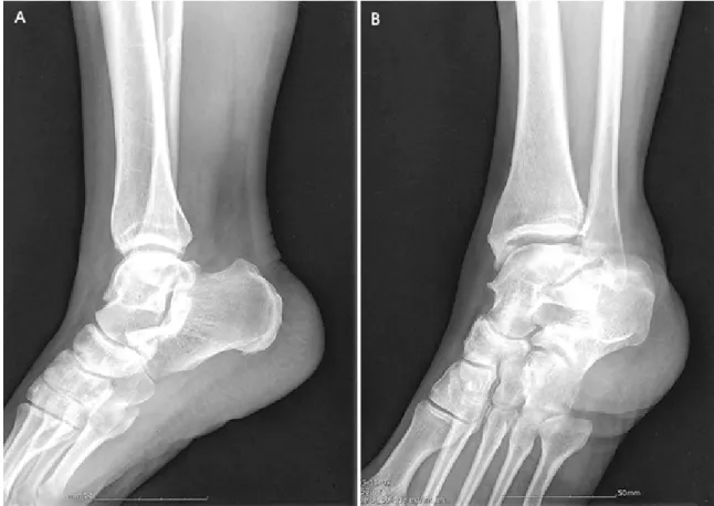
Figure 2. Standard lateral radiograph (A) and Oblique radiograph (B) of an ankle. The standard lateral view do not show any abnormality.
But, on the Anteromedial Oblique view bony spurs are visible on the anteromedial tibial rim and on the talar neck.
를 통하여 판단하였높은 비율로 발생하고 있으며 그 빈도가 점차 증가하고 있는 추세이다11). 이는 비단 직업적인 운동 선수에서뿐만 아니라 일반인들의 스포츠 참여와 다양한 스 포츠, 그리고 스포츠 활동 인구의 증가로 인해 그 손상의 빈 도가 증가되고 있다.
족관절의 염좌는 자연 치유 질환으로 보존적 치료에 잘 반응하여 기능의 대부분이 회복된다고 여겨진다10,11,18). 족 관절 염좌시 외측부인대, 삼각인대 그리고 원위 경비 인대 결합에 인대 손상이 발생할 수 있으며 이중 가장 많게는 외 측 인대 복합체, 특히 전방 거비인대와 종비인대에 손상을 입게 된다6-8). 3도 이상의 외측부 인대 손상시 족관절 불안 정이 발생할 수도 있으나 Brostrom5) 술식 등을 이용하여 재건술을 시행함으로서 좋은 결과를 보이고 있다.
그러나 최근 족관절 염좌 후 수개월 내지 수년이 지나 족 관절의 불안정, 만성적 동통, 반복되는 부종과 구축 및 연골 손상, 관절염, 재발성 염좌 등의 잔류 증상이 발생한다는 결 과가 다수의 연구에서 보고 되고 있으며, 그 비율은 30~
70%까지이다1,14,19,21). DiGlovannie 등9)은 족관절의 만성 염좌 및 불안정을 가진 환자에서 외측 측부 인대의 손상 이
외에 15개의 동반 병변을 확인한 논문을 발표하였다. 그 중 비골 건 활액막염(pe서 골성 병변이 발견되어 매우 높은 발 견 빈도를 보였다.
족관절의 전방 골극은 반복적인 과 족저 굴곡 및 내반 자 극이 전방 관절 낭을 반복적으로 견인함으로써 관절낭의 부 착 부위에서 골극이 형성된다는 이론과 족관절의 불안정으 로 인한 과 배굴 굴곡이 발생하고 이로 인하여 반복적이고 직접적인 미세 충돌이 골극이 형성한다는 이론이 있다3,17). 보통 족관절의 전방 골극은 족관절 측면상에서 검사를 한 다. 그러나 전내측 골극은 표준 측면상에서 발견되지 않는 경우가 많다. 이는 전내측 경골 환에 의하여 겹쳐지기 때문 이다. 따라서 전내측 골극은 발을 30° 외회전시킨 상태에서 방사선 방향을 45°로 촬영한 사면상에서 가장 잘 보인다.
반면 전외측 골극은 사면상으로는 잘 보이지 않고 표준 측 면상에서 잘 보인다20). 본 연구의 경우 전방 골극을 가진 65 예 중 19예는 측면상에서는 발견되지 않았으나 사면상에서 발견되었다(Fig. 2). 따라서 족관절 전방 골극은 족관절의 측면상과 사면상을 같이 촬영하여 검사하여야 할 필요성이 있다20). 불안정을 동반한 만성 족관절 염좌에 있어서 전방
저명한 불안정성을 가진 만성 족관절 염좌 환자의 족관절 골성 병변에 대한 분석
- 9 - 골극은 60~70%의 높은 빈도로 발견된다는 보고가 있으며 본 연구에서도 104족관절 중 65족관절(62%)에서 발견되어 비슷한 발견 빈도를 보였다9).
족관절 염좌로 인한 족관절 내측 병변은 삼각인대 손상, 내측 관절낭 손상, 경골 내측과 골절 및 외상성 활액막염 등 이 있다16). 특히 만성적으로 외전 손상이 발생할 경우에는 경골 내과와 거골사이의 충돌로 인한 골극 형성과 연골 손 상이 발생하게 되며4), 족관절의 불안정이 있을 경우 이러한 현상은 더욱 저명하게 나타난다13). 본 연구에서는 모든 환 자가 족관절 불안정을 동반하고 있는 경우로 내과 골극이 50예(48%), 거골 연골 결손이 5예(5%)에서 발견되었다. 거 골 연골 결손은 전내측 부위와 후내측 부위에서 주로 발생 하고, 빈도는 족관절의 염좌시 0~7%까지 보고되고 있다
2,4). 본 연구에서 발견된5예는 모두 거골의 후내측 부위에 서 발견되었다.
비골 하 부 골은 비골의 외하방에서 떨어져 있는 골조직 으로 전거비인대가 부착되어 있다. 비골 하 부 골의 발생은 정상 발육 과정 중에서 부골화 중심이 2차 골화 중심으로부 터 떨어져 나와 유합되지 않은 상태로 성장되어 발생하는 정상 소견이라는 견해와 족관절의 전거비인대 견열 골절 손 상의 불유합으로 발생한다는 견해가 있다12,16). 전자의 경우 대부분 무증상이고 우연히 발견되는 양상을 보이며 양측성 으로 나타나는 반면, 후자의 경우 족관절의 불안정성을 초 래하고 동통을 야기하며 제거하였을 시 동통이 사라지는 양 상을 보인다. Brostrom은 급성 족관절 염좌의 8%에서 전거 비 인대의 견열 골절손상이 발생한다고 하였다5,6). 본 연구 에서 발견된 29예의 비골하 부골 중 4예에서 양측성으로 관 찰되었고 그 외에는 모두 편측성으로 발견되었는데 모두 골 성 이상 소견으로 분류하였다.
외과 골극은 18예(17%)에서 발견되어 전방 골극의 64예 (62%), 내측 골극의 50예(48%) 및 비골하 부골의 29예 (28%)보다 드물게 관찰 되었다. 그러나 외과 골극이 관찰된 경우 동반 골극의 발견 빈도가 18예 중 13예(72%)로 전방 골극의 28%, 내측 골극의 26% 및 비골하 부골의 38%보다 높게 발견되었다.
초기 족관절 염좌 수상 후 병‧의원을 내원하여 석고 고정 시행이나 압박붕대 착용 등의 적절한 치료의 시행 여부에 따라 두 군으로 나누었을 때 초기 적절한 치료를 시행한 군 이 34명, 적절한 치료를 시행하지 않은 군이 55명이었다.
각 군에서 골성 병변이 나타난 예가 각각 14명(41%)과 47명 (85%)으로 초기에 적절한 치료를 시행하지 않은 군에서 비 교적 높은 빈도의 골성 병변이 발견되었다. 각 군의 증상 유 병 기간은 4.3년과 3.9년이고 나이는 35세와 38세로 유의
한 차이는 없었으나 모 집단이 작고 골성 병변 발현에 관련 되는 그 외의 요인들이 많아 이의 상관관계는 지속적인 관 찰 및 추가 연구가 요구된다. 골성 병변이 나타난 군과 나타 나지 않은 군으로 나누었을 때 두 군의 증상 유병기간은 각 각 7.4년과 4.3년으로 골성 병변이 나타난 군에서 유병 기 간이 길게 나타났다. 이러한 결과는 초기의 적절한 치료를 하지 않았을 경우 및 유병 기간이 길 경우 예후에 좋지 않다 는 다른 보고들과 비슷한 결과를 나타내었다1,13,15).
결 론
족관절 염좌는 대부분 기능적 제한 없이 치유되는 질병 이나 만성적 염좌 및 불안정성이 동반되었을 때는 다양한 골성 병변이 발생할 수 있다. 본 연구에서는 3년 이상 반복 되는 족관절 염좌의 병력을 가지고 있으면서 불안정성이 나 타나는 환자들의 족관절에 발생한 골성 병변을 분석한 결과 104족관절 중 74족관절(71%)에서 하나 이상의 골성 병변을 관찰할 수 있었다. 이중 전방 골극이 65족관절(62%)로 가장 흔히 관찰되었다. 따라서 이러한 환자들에 대해서는 족관절 의 방사선 사진상 이상 소견에 대한 면밀한 관찰이 필요할 것으로 사료된다.
REFERENCES
1. Anandacoomarasamy A and Barnsley L: Long term out- comes of inversion ankle injury. Br J Sports Med, 39: e14, 2005.
2. Berndt AL and Harty M: Transchondral fractures (osteochondritis dissecans) of the talus. J Bone Joint Surg, Am, 41-A: 988-1020, 1959.
3. Biedert R: Anterior ankle pain in sports medicine: etiology and indications for arthroscopy. Arch Orthop Trauma Surg, 110: 293-297, 1991.
4. Biedert R: Osteochondral lesions of the talus. Unfall- chirurg, 92: 199-205, 1989.
5. Brostrm L: Sprained ankle. Acta Chir Scand, 132: 551- 565, 1966.
6. Brostrom L: Sprained ankle: I. Anatomic lesions in recent sprains. Acta Chir Scand, 128: 483-495, 1964.
7. Cass JR, Morrey BF and Chao EYS: Three-dimensional kinematics of ankle instability following serial sectioning of lateral collateral ligaments. Foot Ankle, 5: 142-149, 1984.
8. Cawley PW and France EP: Biomechanics of the lateral ligaments of the ankle: an evaluation of the effects of axial load and single plane motions on ligament strain patterns.
Foot Ankle, 12: 92-99, 1991.
9. DIGiovanni BF, Fraga CJ, Cohen BE and Shereff MJ:
정철용․은일수․김병철․최성종․류총일․김종균․최현수
- 10 - Associated injuries found in chronic lateral ankle instability. Foot Ankle Int, 21: 809-815, 2000.
10. Garrick JG and Requa RK: The epidemiology of foot and ankle injuries in sports. Clin Podiatr Med Surg, 6:
629-637, 1989.
11. Gerber JP, Williams GN, Scoville CR, Arciero RA and Taylor DC: Persistent disability associated with ankle sprains: a prospective examination of an athletic popu- lation. Foot Ankle Int, 19: 653-660, 1998.
12. Griffiths JD and Menelaus MB: Symptomatic ossicles of the lateral malleolus in children. J Bone Joint Surg Br, 69:
317-319, 1987.
13. Harrington KD: Degenerative arthritis of the ankle secondary to long-standing lateral ligament instability. J Bone Joint Surg Am, 61: 354-361, 1979.
14. Konradsen L, Bech L, Ehrenbjerg M and Nickelsen T:
Seven years follow-up after ankle inversion trauma. Scand J Med Sci Sports, 12: 129-135, 2002.
15. Maffulli N, Lepore L and Francobandiera C: Traumatic lesions of some accessory bones of the foot in sports activity. J Am Podiatr Med Assoc, 80: 86-90, 1990.
16. Martin DF, Curl WW and Baker CL: Arthroscopic treatment of chronic synovitis of the ankle. Arthroscopy, 5:
110-114, 1989.
17. Ogilvie-Harris DJ, Mahomed N and Demaziere A:
Anterior impingement of the ankle treated by arthroscopic removal of bony spurs. J Bone Joint Surg, 75: 437-440, 1993.
18. Trevino SG, Davis P and Hecht PJ: Management of acute and chronic lateral ligament injuries of the ankle.
Orthop Clin North Am, 25: 1-16, 1994.
19. van Dijk CN, Bossuyt PM and Marti RK: Medial ankle pain after lateral ligament rupture. J Bone Joint Surg, 78:
562-567, 1996.
20. van Dijk CN, Wessel RN, Tol JL and Maas M: Oblique radiograph for the detection of bone spurs in anterior ankle impingement. Skeletal Radiol, 31: 214-221, 2002.
21. Williams GN, Molloy JM, DeBerardino TM, Arciero RA and Taylor DC: Evaluation of the Sports Ankle Rating System in young, athletic individuals with acute lateral ankle sprains. Foot Ankle Int, 24: 274-282, 2003.