146
KISEP Case Report 臨床耳鼻:第 ••••••••••••••••••••••••••••••••••••••••••••••••••••••••••••••••••••••• 17 卷 第 1 號 2006
J Clinical Otolaryngol 2006;17:146-149
Micro-Titanium Mesh를 이용한 상악동 전벽 골결손 재건술 1예
가천의과대학교 이비인후-두경부외과학교실
강일규·김선태·김선영·정주현·차흥억
Reconstruction of Anterior Wall Bony Defect of Maxillary Sinus with Micro-Titanium Mesh:A Case Report
Il-Gyu Kang, MD, Seon Tae Kim, MD, Sun Young Kim, MD, Joo Hyun Jung, MD and Heung Eog Cha, MD
Department of Otolaryngology-Head & Neck Surgery, Gil Medical Center, Gachon Medical School, Incheon, Korea-
ABSTRACT
-A maxillary fracture may be caused by high velocity or other high power injuries to the face. In cases of a simple wall fracture, plate, screw and free bone graft are preferred methods of repair. However, in cases with complex wall fractures or wall defects, these methods are not adequate because of problems such as permanent bony defects and facial depression. Therefore, we used the micro-titanium mesh as reconstructive material for a maxillary wall defect. The micro-titanium mesh was found to have morphological stability and biocompatibility. The mesh struc- ture prevented soft tissue ingrowths to the bone defect area. Therefore, micro-titanium mesh can be used success- fully for cases where indicated for repair of maxillary sinus defects. So we successfully reconstructed the maxillary sinus anterior wall defect with surgical mesh.
(J Clinical Otolaryngol 2006;17:146-149)KEY WORDS
: Surgical mesh·Maxillary fracture.
서 론
상악골은 두개의 두개저와 하악골 사이에 위치해 비골 을 지지하고, 안면근이 부착되며, 상악동의 외벽 및 안와 저의 골판으로서의 기능과 안모의 외형을 구성하는 역할 을 한다. 따라서, 상악골의 결손은 기능적, 심미적인 문제 를 야기할 수 있고, 광범위한 결손은 섬유성 조직에 의한 반흔성 연조직으로 치유되게 된다.
1)또한 낭종의 형성이
나 만성상악동염등의 합병증이 초래될 수도 있다. 따라 서 상악동의 광범위한 손상 및 결손 시 내부구조의 해부 학적인 재건이 필요하다.
상악골의 재건 방법으로 다양한 재료들이 사용되고 있 다. 스테인레스강 철사를 이용한 철사 결박, 악간 고정 그리고 두개골에 의한 안면골의 현수법 및 플레이트와 나 사를 이용한 고정이 흔하게 이용되며, 이러한 재료들은 전형적인 Le Fort 분류에 따른 변형의 수복에는 효과적 이다. 하지만 분쇄성, 다발성 골절에는 한계가 있다. 따 라서 이러한 경우에는 안면부 수복,
2)안면부 보철,
3)하 악골 골절,
4)신경 외과술
5)등에 주로 쓰였던 Titanium mesh를 사용할 수 있다. Titanium mesh는 뼈와 연부 조직 모두와 잘 결합되고 단단한 지지 구조물로 이용될 수 있어 매우 유용하다.
1)저자는 외상 후 발생한 상악동 전벽의 광범위한 골결손 부위를 Titanium mesh를 이용
논문접수일:2006년 4월 20일심사완료일:2006년 6월 26일
교신저자:차흥억, 405-760 인천광역시 남동구 구월동 1198 가천의과대학교 이비인후-두경부외과학교실 전화:(032) 460-3770·전송:(032) 467-9044 E-mail:[email protected]
강일규 외:Micro-Titanium Mesh를 이용한 상악동 전벽 골결손 재건술 1예
147
하여 성공적으로 재건하였기에 문헌 고찰과 함께 보고하 고자 한다.
증 례
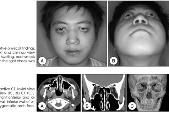
19세 남자 환자로 내원 2일 전 외상 후 발생한 양측 안와부의 부종 및 반상 출혈, 우측 협부의 통증을 주소 로 내원하였다. 내원 당시 시행한 이학적 검사상 양측 안 와부의 부종 및 반상 출혈, 우측 결막하 출혈이 관찰되 었고, 복시 및 안구 운동 장애는 관찰되지 않았으며, 우 측 협부의 부종이 관찰되었으나 함몰 소견은 관찰되지 않 았다(Fig. 1). 방사선학적 검사 상 안구 전산화 단층 촬 영 및 안면부 삼차원 전산화 단층 촬영 상에서 우측 안 와 하부의 골절 및 우측 상악골의 전벽의 함몰 골절 및 외측 벽의 선상 골절 소견 관찰되었고, 우측 혐골궁의 골 절도 의심되었으며, 좌측상악골의 외측벽에도 선상 골절 소견이 관찰되었다(Fig. 2).
이에 대한 수술적 처치의 계획으로 우선 우측 안와 하 부 골절은 Medpor barrier sheet의 삽입을 이용한 안 와 하부 재건술을 계획하였고, 우측 상악골 전벽의 함몰 골절에 대해서는 골성 지지 구조의 부재와 추후 있을 수
있는 안구 함몰의 가능성 등을 고려해 micro-titanium mesh를 이용한 재건을 고려하였으며, 우측 상악골 외측 선상 골절의 경우 transantral approach를 통해 정복을 시도하고자 계획하였다. 우측 협골궁 골절의 교정은 Gillie 접근법을 통하여 확인한 후 교정하고자 하였고, 좌측 상 악골의 외측벽의 선상골절이 의심되었으나 환자 외관상의 변화가 없고 부정교합 등의 기능적 이상도 없어 보존적 인 치료를 시행하기로 하였다.
수상후 10일째 수술을 시행하였고, 치은 협부 절개를 시행하여 우측 상악동 전벽 골절 부위를 확인하였다. 상 악동 전벽이 광범위하게 함몰된 소견이 관찰되었고, 골 절의 양상이 분쇄골절의 소견을 보여 미세 플레이트를 이 용한 골간 수복이 불가능한 상태였으며, 일부에서 골결 손 소견이 관찰되었다. 또한 수복 과정에서 점막 골막과 분쇄 골간 분리가 되어 정상적인 수복이 어려운 상황이 었고, 수복 후에 안면부 함몰이 예상되었다.
따라서 광범위한 골결손 부위를 덮어주고 안면부의 연 조직 함몰을 예방할 수 있는 micro-titanium mesh를 이 용하였다.
1)Micro-titanium mesh를 이용하여 상악골 전벽의 형태로 만든 뒤 골결손 부위를 재건하였고, 미니 플레이트를 이용하여 약해진 상악골을 고정하였다. Tran -
A B C
Fig. 2. Preoperactive CT (axial view (A), coronal view (B), 3D CT (C))
:These show right anterior and la- teral maxillary wall, inferior wall of or- bit and right zygomatic arch frac- ture.
B A
Fig. 1. Preoperative physical findings, frontal view (A) and chin up view (B):periorbital swelling, ecchymosis and swelling on the right cheek was seen.
J Clinical Otolaryngol 2006;17:146-149
148
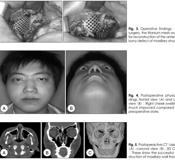
santral approach를 통해 우측 상악골 외측 골절 교정 을 시도하였으나 외측 골절의 수복은 용이치 않았다. 따 라서, 우측 상악골 외측 골절의 수복은 골절 정도가 심 하지 않고, 수복이 용이치 않아 지켜보기로 하고, 우측 안 와 하벽 골절은 medpor를 이용하여 정복하였으며, 우 측 협골궁은 수상 후 이미 골절 부위가 유합된 상태로 외관상의 함몰 소견 없고, 술전 개구 장애 등의 기능적 이상이 없어 수술적 정복은 시행하지 않고 수술을 마쳤 다(Fig. 3). 환자는 발열 등의 합병증 없이 술후 10일째 퇴원하였다(Fig. 4). 외래 통원 치료 하던 중 술후 2개 월째 시행한 안면부 전산화 단층 촬영상 micro-titanium mesh는 수복 직후의 모습 그대로 잘 유지되고 있었고, 안와 골절부의 수복 역시 잘 유지되고 있었다(Fig. 5).
고 찰
안면 외상 시 상악동 결손이 있는 경우에 재건을 해야 하는 경우는 치료 없이는 적절한 기능과 외양이 유지되 지 않을 때라고 할 수 있다. 상악골 골절의 치료에 있어 서 중요한 것은 적절한 기능과 외양을 복원하는 것이며 기능적인 측면에서 가장 중요한 것은 치열궁을 원래대로 환원시켜 수상 전의 교합을 얻는 것과 안구함몰이나 외 안근의 기능부전이 생기지 않도록 수상 전의 안와내 용 적을 유지시키는 것이다.
6)성공적인 상악동 수술을 위한 기본적인 요구조건은 호 흡 상피의 보존과 충분한 환기, 부비동으로의 배출로 확
Fig. 3. Operative findings:During surgery, the titanium mesh was used for reconstruction of the anterior wall bony defect of maxillary sinus, Rt.A B
Fig. 4. Postoperative physical fin- dings, frontal view (A) and chin up view (B):Right cheek swelling was much improved compared to the preoperative state.
A B C
Fig. 5. Postoperactive CT (axial view (A), coronal view (B), 3D CT (C))
:These show the successful recon- struction of maxillary wall fracture.
강일규 외:Micro-Titanium Mesh를 이용한 상악동 전벽 골결손 재건술 1예
149
보에 있다. 이러한 상악골 수술시 기존의 골부가 모자라 는 경우, 상악동의 결손부를 재건해 주는 방법에 여러 가 지가 있다.
Hackermann 등
7)은 상악동 결손부의 재건에 동결건 조 뇌막 이식을 처음 이용했고, 현재에도 안와저 골절의 치료에 이용되고 있다. 그러나 Creutsfeldt-Jakob di - sease의 전염우려로 완벽한 멸균처리가 요구되고 있다.
Remagen과 Prezmecky는 hydroxyapatite로 골결손 부위를 메운 후 이식편의 소실을 방지하기 위하여 lyo - philized dura로 덮어주었다고 보고한 바 있다.
8)9)골결 손부가 작은 공동형 골결손에는 장골능으로부터 채취한 망상골(particulated marrow and cancellous bone:
PMCB)이 시도되기도 했다.
10)Micro-titanium mesh의 경우 재료가 유연하므로 복 잡한 형태의 결손의 치료에 유리하고, 두께가 얇고 견고 하므로 정확한 체적의 형성과 장기간의 형태 유지에 유 리하며 나사를 이용하여 고정하므로 술후 위치 이동이 없 다는 장점이 있다.
1)Sugar 등
11)12)은 Titanium mesh를 이용하여 안와 내벽의 재건술을 시행하여 좋은 결과를 얻 었다고 보고한 바 있다.
저자들은 micro-titanium mesh를 이용하여 상악골 결손부위의 재건을 시도하였는데, Titanium의 표면은 공 기중에서 산화막을 형성하고 부식에 대한 저항성 및 조 직에 대한 생체적합성이 뛰어나며 micro-titanium mesh 는 시술 중 변형이 가능하여 수술이 용이하였다. 또한 mesh가 가지고 있는 구멍으로 유체의 이동이 가능하나 연조직의 개재는 성공적으로 막을 수 있어 이는 상악동 이 호흡상피로 치유되게 하며 환기에도 도움을 주며,
1)금속성 강도로 안모의 외형유지 역시 가능하다.
9)그리고 추후 제거할 필요가 없기에 다양한 상악골 결 손부에 훌륭히 적용 될 수 있다.
따라서 저자들은 상악골의 결손증례에서 연조직의 복 합 결손이 아닌 단순 골결손 증례의 경우 micro-tita -
nium mesh를 이용한 재건술이 상악골의 해부학적인 구 조의 재건에 효과적이라고 사료되며, 이상과 같은 골결 손 증례에서 훌륭한 임상 결과를 얻을 수 있었다.
중심 단어
:티타늄 메쉬·상악동 골결손.
REFERENCES
1) Kim SG, Choi YS, Choung PH, Lee CH. The reconstruction of the maxillary defect using micro-titanium mesh. J Korean Assoc Oral Maxillofac Surg 2000;26:197-203.
2) Blake GB, Macfarlane MR, Hilton JW. Titanium in recons- truction of the skull and face. Br J Plast Surg 1990;43:528-35.
3) Albrektsson T, Branemark PI, Jacobsson M, Tjellstrom A.
Present clinical application of osseointegrated percutaneous implants. Plast Reconstr Surg 1987;79:721-30.
4) Cobetto GA, McClary SA, Zallen RD. Treatment of man- dibular fractures with malleable titanium mesh plates: A re- view of 120 cases. J Oral Maxillofac Surg 1983;41:597-600.
5) Blair GAS, Fannin TF, Gordon DS. Titanium-strip cranio- plasty. Br Med J 1976;2:907.
6) Kim JS. Maxillofacial trauma. In: Jang SO, Yim HH, Lee JK, Lee CH, Yang SK, Cho JS, editors. Otolaryngology Head and Neck Surgery. Iljokac;2002. p.1158.
7) Hackermann G, Machtens E, B ning K. Die Deckung des fa- cialen Kieferh hlenfensterers mit lyophilisierter Dura. Dtsch Zahnaarzit Z 1976;31:265.
8) Chen M, Zingg M, Laedrach K, Raveh J. Early surgical intervention for orbital floor fractures: A clinical evaluation of lyophilized dura and cartilage reconstruction, J Oral Ma- xillofac Surg 1992;50:935-41.
9) Sailer HF. Transplantation of Lyophilized Cartilage in Maxi- llofacial Surgery: Experimental Foundations and Clinical Success. Basel, NewYork: Karger;1963.
10) Shirota T, Ohino K, Motohashi M, Michi K. Histologic and microradiologic comparison of block and particulate can- cellous bone and marrow grafts in reconstructed mandibles being considered for dental implant placement. J Oral Maxi- llofac Surg 1996;54:15-20.
11) Sugar AW, Kuriakose M, Walshaw ND. Titanium mesh in orbital wall reconstruction. Int J Oral Maxillofac Surg 1992;
21:140-4.
12) Wolfe SA. Application of craniofacial surgical precepts in orbital reconstruction following trauma and tumor removal.
J Maxillofac Surg 1982;10:212-23.