Introduction
Periodontal disease (PD) is the most common osteolytic
infection of alveolar bone seen in humans worldwide. PD is a common, chronic immunoinflammatory disease initiat- ed by subgingival bacteria and results in the inflammatory destruction of periodontal tissues, including the alveolar bone periodontal ligament, and gingivae. It is characteriz- ed by gradual destruction of periodontal tissue, in the end leading to tooth loss. One major mechanism by which PD exerts systemic effects is through the generation of oxida- tive stress [1-4]. In recent years, evidence has emerged to
Antioxidant Effect of Eugenol in Human Periodontal Ligament Fibroblasts
Yong-Ho Kim, Bong-Soo Park
Department of Oral Anatomy, School of Dentistry, Pusan National University, Korea
(Received 2 March 2015, revised 21 March 2015, accepted 24 March 2015, Published Online 30 March 2015)
Abstract: Periodontal disease (PD) is the most common osteolytic disease of alveolar bone, oral infection seen in humans worldwide. PD is a common, chronic immunoinflammatory disease initiated by a complex subgingival bacterial and results in the inflammatory destruction of periodontal tissues, including the alveolar bone periodontal ligament, and gingivae. The effects of eugenol on periodontal ligament fibroblasts (PDLF) cell under oxidative injury have not been fully studied. Despite many studies in regard to the antioxidant effect of eugenol, the protective effect of eugenol against oxidative damage to PDLF cell, as well as the relationship between eugenol and apoptosis, has not been investigated so far. The aim of this study was to assess the protective effect of eugenol against H2O2-induced oxidative stress in PDLF cell.
Cell lines were separately grown as monolayers at 5% CO2and 37�C humidified atmosphere using appropriate media supplemented with 10% fetal bovine serum, 2 mM glutamine and 100 μg/mL penicillin-streptomycin.
DMEM/F12 was used as the culture medium for periodontal ligament fibroblast cells.
The viability of the PDLF cells which induced by the different concentrations of H2O2(control, 50, 100, 200, 400 μM) for 24 h was detected by MTT assay. Cell viability was significantly reduced in a H2O2-concentration dose-dependent manner. The mitochondria-dependent pathway of apoptosis is regulated by Bcl-xl family, such as the anti-apoptotic protein Bcl-xl, pro-apoptotic protein Bak. With H2O2injury, the protein level of Bak was up- regulated while the protein level of Bcl-xl was down-regulated. In group treated H2O2and eugenol, the ratio was reduced and the expression of Bak decreased at the same time, indicating that eugenol can attenuate apoptosis through mitochondrial related pathway in PDLF cells.
Therefore, although the findings of this study are limited to an in vitro interpretation, we suggest that eugenol preconditioning may have a beneficial effect in the recovery of periodontal ligament from oxidative stress.
Keywords : Antioxidant, Eugenol, Oxidative stress, Periodontal ligament fibroblasts
The author (s) agree to abide by the good publication practice guideline for medical journals.
The author (s) declare that there are no conflicts of interest.
Correspondence to : Bong-Soo Park (Department of Oral Anatomy, School of Dentistry, Pusan National University)
E-mail : [email protected]
http://dx.doi.org/10.11637/kjpa.2015.28.1.45 Original Article
ⓒ 2015 Korean Association of Physical Anthropologists
This is an Open Access article distributed under the terms of the Creative Commons Attribution Non-Commercial License (http://creativecommons.org/ licenses/by-nc/3.0) which permits unrestricted non-commercial use, distribution, and reproduction in any medium, provided the original work is properly cited.
ISSN 2287-626X (Online)∙ISSN 1225-150X (Print)
implicate reactive oxygen species (ROS) oxidative stress and the pathogenesis of periodontal disease in humans. The presence of excess reactive oxidants is thought to provide a basis for the progression of various diseases [5]. Euge- nol, 2-methyoxypheol, which is contained in cloves as well as in cinnamon and other aromatic spices is used as a sup- plement or a therapeutic ingredient in various medications and foods. Eugenol is used to treat digestive disorders and skin infections and is found in insect attractants and in UV absorbers [6]. It is a beneficial antioxidant when ingested in moderate amounts reducing the level of free radicals.
However, there are some reports that excessive doses of undiluted eugenol oil can cause symptoms. According to some studies, eugenol in excessive doses can be consider- ed poison [7,8]. Previous studies reported that oxidative stress can directly induce cell death or apoptosis in various cell types, including osteoblasts, intestinal endothelial cells, and hepatocytes [9-11]. Studies demonstrated that eugenol has an antiapoptotic effect on in vivo and in vitro [12-14].
In ancient times, natural products were the main source of health care products. In modern medicine, they are still major sources of new drug development [15,16]. The eff- ects of eugenol on periodontal ligament fibroblasts (PDLFs) exposed to oxidative injury has been widely investigated its protective effect against oxidative damage of PDLFs, as well as its relationship with apoptosis, has not been studied.
The aim of this study was to assess the protective effect of eugenol against H2O2-induced oxidative stress in PDLFs cell.
Materials and Methods
1. Reagents
The Human periodontal Ligament Fibroblasts (PDLF) was purchased from Lonza (Basel, Switzerland). The fol- lowing chemicals and reagents were obtained from the in- dicated companies: eugenol, Hoechst 33342 was purchas- ed from Sigma. The following reagents were obtained com- mercially: 3-[4,5-dimethylthiazol-2-yl]2,5-diphenyl tetra- zolium bromide (MTT), apoptosis detection kit was obtain- ed from Biovision (Milpitas, CA, USA). Antibodies used in the study were as follows: cleaved Caspase 3 (1 : 1,000), : Bcl-xl (1 : 1,000), : Bax (1 : 1,000), Santa Cruz. Secondary antibodies against rabbit (1 : 3,000), and mouse (1 : 3,000),
immunoglobulins were purchased from Bio-Rad.
2. Cell culture
Cell lines were separately grown as monolayers at 5%
CO2and 37�C humidified atmosphere using appropriate media supplemented with 10% fetal bovine serum, 2 mM glutamine and 100 μg/mL penicillin-streptomycin. DMEM /F12 was used as the culture medium for periodontal liga- ment fibroblast cells. Cells were passaged 3 times a week by treating with trypsin-EDTA and used for experiments after 5 passages.
3. Treatment of eugenol
Eugenol which were made by dissolving them in DMSO were kept frozen at -20�C until use. The stock was dilut- ed to their concentration with DMEM when needed. Prior to eugenol treatment cells were grown to about 80% confl- uence and then exposed to eugenol at different concentra- tions (0, 50, 100, 200, 400 μM) for 24 h. Cells grown in medium containing an equivalent amount of DMSO with- out eugenol served as control. The groups were randomly divided into the following groups: Control, eugenol, H2O2, H2O2++eugenol.
4. MTT assay
Cell viability assay was measured using a quantitative colorimetric assay MTT solution, showing the mitochon- drial activity of living cells. PDLF cells (3×10⁴) were seed in 96-well plates. After drug treatment as indicated, cells were incubated with 300 μL MTT (final concentration 0.5 mg/mL) for 1.5 h at 37�C. The reaction was terminated by addition of 200 μL DMSO. Cell viability was measured by an ELISA reader (Tecan, Männedorf, Switzerland) at 570 nm excitatory emission wavelength.
5. Flow cytometer analysis
Cells were seeded into a 6-well plate at 1×10⁶ cells/mL and incubated overnight. Cells treated with eugenol were incubated for various time points. In each time point, the harvested cells were washed with PBS containing 1% bo- vine serum albumin and centrifuged at 2,000 rpm for 10 min. The cells were resuspended ice-cold 95% ethanol with 0.5% Tween 20 to a final concentration of 70% ethanol.
Fixed cells were pelleted, and washed in 1% BSA-PBS solution. Cells were resuspended in 1 mL PBS containing 20 μg/mL RNase A, incubated at 4�C for 30 min, washed once with BSA-PBS, and resuspended in PI solution (10 μg/mL). After cells were incubated at 4�C for 5 min in the dark, DNA content were measured on a CYTOMICS FC500 flow cytometry system (Beckman Coulter, FL,CA,USA) and data was analyzed using the Multicycle software which allowed a simultaneous estimation of cell-cycle parameters and apoptosis.
6. Immunofluorescent staining to detect cytochrome c, AIF translocation
The cells were plated on coverslips and treated with eu- genol. After 24 h, the cells were stained with 50 nM Mito- Tracker Red at 37�C for 30 min. After washing two times with PBS, the cells were fixed with 4% paraformaldehyde (PFA) in PBS for 15 min and washed three times with PBS.
After permeabilization with Triton X-100 and blocking 1% BSA in PBS, the cells were incubated with primary an- tibodies in 1% BSA overnight at 4�C. After washing with PBS, cells were incubated with FITC-conjugated secon- dary antibodies in 1% BSA-PBS for 60 min and rinsed in PBS. Fluorescent images were observed and analyzed using a Zeiss LSM 750 laser-scanning confocal microscope (Göettingen, Germany).
7. Western blot analysis
Cells (2×10⁶) were washed twice in ice-cold PBS, resu- spended in 200 μL ice-cold solubilizing buffer [300 mM NaCl, 50 mM Tris-Cl (pH 7.6), 0.5% Triton X-100, 2 mM PMSF, 2 μL/mL aprotinin and 2 μL/mL leupeptin] and in- cubated at 4�C for 30 min. The lysates were centrifuged at 14,000 rpm for 15 min at 4�C. Protein concentrations of cell lysates were determined with Bradford protein assay (Bio-Rad, Richmond, CA, USA) and 20 μg of proteins were resolved by 10% SDS/PAGE. The gels were trans- ferred to Polyvinylidene fluoride (PVDF) membranes (Millipore, Billerica, MA, USA) and reacted with appro- priate primary antibodies. Immunostaining with secondary antibodies was detected using SuperSignal West Femto (Pierce, Rockford, IL, USA) enhanced chemilumines- cence substrate and detected with Alpha Imager HP (Alpha Innotech, Santa Clara, USA).
Result
1. Eugenol improved the cell viability of H2O2-induced apoptosis in PDLFs
The effect of eugenol on PDLFs was investigated over a wide concentration range. Eugenol suppressed H2O2-de- Fig. 1.Chemical structure of eugenol.
% of cell viability% of cell viability
120 100 80 60 40 20 0
(A)
(B)120
100 80 60 40 20 0
*
*
*
*
* *
0 μM 25 μM 50 μM 75 μM 100 μM
Concentration of eugenol (μM)
0 μM 50 μM 100 μM 200 μM 400 μM Concentration of H2O2(μM)
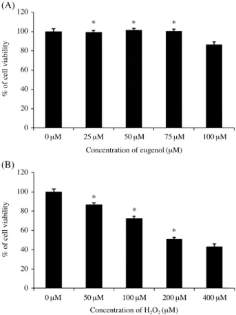
Fig. 2.Effect of eugenol preconditioning on cell viability. (A) The normal PDLF cells were treated with different concentrations (0, 25, 50, 75, 100 μM) of eugenol for 24 h. (B) PDLF cells were treated H2O2with various doses (0, 50, 100, 200, 400 μM). Cell viability was determined by 3-(4,5-dimethylthiazol-2-yl)-2, 5-diphenyltera- zolium bromide (MTT) assay. The values are denoted as mean SE of three independent experiments. *P⁄0.05 versus untreated sam- ples.
CH3O
OH
pendent programed cell death, pointing to its potential as a potent antioxidant. The chemical structure of eugenol is shown Fig. 1. The PDLF cells with various doses of euge- nol (below 100 μM) and exposed the cells to H2O2injury and then we measured cell viability by the MTT assay (Fig.
2A). Alteration of cell viability was not observed in euge-
nol treatment group (p⁄0.05). The viability of the PDLF cells which induced by the different concentrations of eu- genol (0, 25, 50, 75, 100 μM) for 24 h was detected by MTT assay. However, the cell viability was significantly reduc- ed in a H2O2-treated in PDLFs dose-dependent manner (Fig. 2B).
2. Eugenol protected against H2O2-induced apoptosis in PDLFs
To investigate the protective effect of eugenol, the PDLFs cells were treated 50 μM of eugenol for 24 h and exposed to H2O2(100 μM). After 24 h, the cells were assayed for cell viability. Treatment of the cells with 50 μM of eugenol significantly increased the cell viability compared to that observed in the cells exposed exclusively to H2O2(Fig. 3).
3. Eugenol treatment led to a decrease in H2O2- induced apoptosis in the PDLFs
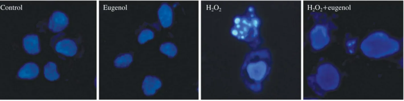
The changes in nuclear morphology were assessed by Hoechst 33342 staining after the H2O2treatment. The con-
120 100 80 60 40 20 0
*
*
*
% of cell viability
Eugenol 0 μM 50 μM 0 μM 50 μM
H2O2 0 μM 0 μM 100 μM 100 μM
Fig. 3.Protective Effect of eugenol on PDLF cell viability. Cells were pretreated with eugenol (50 μM) after 24 h treated with differ- ent concentrations of H2O2(100 μM) for 24 h, other group treated single H2O2. Cell viability was analyzed using the MTT assay.
Fig. 4.Nucleus condensation signally was observed on PDLF cells stimulated with H2O2. Apoptotic bodies in the H2O2++eugenol group were markedly reduced.
Fig. 5.H2O2-induced apoptosis in PDLF cells. Cells were treated with eugenol (50 μM) for 24 h, and ratio of apoptotic cells was determin- ed by flow cytometry analysis.
Control Eugenol H2O2 H2O2++eugenol
Control Eugenol H2O2 H2O2++eugenol
4.3% 25.8% 13.3% 17.6%
FL3 Lin FL3 Lin FL3 Lin FL3 Lin
0 1023 0 1023 0 1023 0 1023
291 330 223 205E
F
G H
trol PDLF nuclei had a normal round shape. However, when the cells were exposed to H2O2for 24 h, nuclear con- densation and fragmentation appeared. The eugenol treat- ment rescued the H2O2-induced nuclear morphological change (Fig. 4). Treatment of the cells with eugenol allevi- ated the cell damage. Based on these results eugenol signi- ficantly reduced apoptosis in the H2O2exposed PDLFs.
4. H2O2induced mitochondrial dysfunction and caspase-mediated apoptosis
Mitochondria play a key role in the intrinsic pathway of apoptosis and dissipation of the mitochondrial membrane potential (Δψm) is associated with mitochondrial dysfunc- tion [17-19]. Thus, we verified the change of Δψm in H2O2- Fig. 6.H2O2induced translocation of cytochrome c from mitochondria into cytosolic fraction in PDLF cells. Cells were incubated with 50 μM eugenol for 24 h and then stained with MitoTracker (red), cytochrome c (green), and DAPI (blue) to visualize mitochondria, cyto- chrome c, nuclei respectively.
Control
Eugenol
H2O2
H2O2++eugenol
DAPI Cytochome c Mito tracker Merge
induced apoptosis using DiOC6. H2O2triggered the loss of Δψm in the PDLFs, as compared to the controls. The loss of Δψm resulted in the release of cytochrome c into the cytosolic fraction. We next investigated the transloca- tion of cytochrome c using immunofluorescent staining.
Eugenol treated group reduced compared H2O2-induced cause release cytochrome c and AIF (Figs. 6, 7).
5. Effect of eugenol treatment on apoptosis activation
The activation of cleaved caspase 3 is a key upstream event in the initiation and execution of apoptosis.
Cleaved caspase 3 was up-regulated in the H2O2group, and decreased in the eugenol and H2O2++eugenol groups.
The mitochondria-dependent pathway of apoptosis is regu- lated by the Bcl-xl family, such as the anti-apoptotic pro- Fig. 7.Cells were incubated with 50 μM eugenol for 24 h and then stained with MitoTracker (red), AIF (green), and DAPI (blue) to visualize mitochondria, cytochrome c, nuclei respectively. Images were observed by Confocal microscopy.
DAPI AIF Mito tracker Merge
Control
Eugenol
H2O2
H2O2++eugenol
tein Bcl-xl and the pro-apoptotic protein Bak. With H2O2 injury, the protein level of Bak was up-regulated and the protein level of Bcl-xl was down-regulated. In the H2O2++ eugenol group, the level was reduced and the expression of Bak decreased at the same time, indicating that eugenol attenuated apoptosis through a mitochondrial related path- way in PDLFs (Fig. 8).
Discussion
The objective of the current study was to test the hypo- thesis that eugenol, a natural plant constituent widely used in food products and dental materials, protects against oxi- dative stress and apoptosis caused by H2O2[20]. This study has three principal findings. First, the eugenol treatment conferred increased protection of PDLFs in oxidative in- jury (Fig. 2A). Second, the eugenol treatment protected human PDLFs against oxidative stress induced apoptosis.
Using western blot analysis, we showed that the eugenol treatment decreased cleaved caspase 3 levels via a caspase- dependent pathway, and that it reduced the ratio of Bak and Bcl-xl which are associated with a mitochondrial re- lated pathway. The activation of upstream regulators of
cleaved caspase 3 is key to the initiation and induction of apoptosis [21,22]. It is known that the mitochondria-depen- dent apoptotic pathway is regulated by Bcl-xl protein fam- ily, such as the anti-apoptotic protein Bcl-xl and pro-apo- ptotic protein Bak, which are critical downstream regula- tors in caspase activation [14,23]. Third, we found that eu- genol reduced apoptotic cell death in PDLF cells that ROS played a crucial role in this process [24]. Western blot an- alysis showed a cleaved caspase 3 and Bak, known pro- apoptotic proteins, and Bcl-xl a known anti-apoptotic pro- tein [25-27]. The results showed that the anti-apoptotic effect of eugenol was mediated by a mitochondria related pathway. In summary, the present study, eugenol treatment less than 100 μM have not shown the cytotoxic effect in PDLFs and among of eugenol concentrations, 50 μM treat- ment groups showed that to stimulate the expression of anti-apoptotic proteins under oxidative stress. This study was performed to investigate the effects of eugenol on the periodontal healing process. Although the findings of this study are limited to an in vitro interpretation, we suggest that eugenol preconditioning may have a beneficial effect on the recovery of periodontal ligament from oxidative stress.
Fig. 8.Eugenol changed expressions of apoptosis-related proteins in PDLF cells. Cells were treated with eugenol for the indicated levels of Bcl-xl, cleaved caspase 3, Bak were analyzed by western blotting.
250 200 150 100 50 0 Cleaved caspase 3 protein expressionBcl-xl protein expression
Bak protein expression
120 100 80 60 40 20 0 400
300 200 100 0
Bcl-xl
Cleaved caspase 3
Bak
GAPDH
20 kDa
17 kDa
30 kDa
37 kDa
Control Eugenol H2O2 H2O2++eugenol Control Eugenol H2O2 H2O2++eugenol
Cleaved caspase 3
Bak Bcl-xl
Control Eugenol H2O2 H2O2++eugenol Control Eugenol H2O2 H2O2++eugenol
References
1. D’Aiuto F, Nibali L, Parkar M, Patel K, Suvan J, Donos N.
Oxidative stress, systemic inflammation, and severe perio- dontitis. J Dent Res 2010; 89:1241-6.
2. Waddington RJ, Moseley R, Embery G. Reactive oxygen species: a potential role in the pathogenesis of periodontal diseases. Oral Dis 2000; 6:138-51.
3. Baltacioglu E, Yuva P, Aydin G, Alver A, Kahraman C, Karabulut E, et al. Lipid peroxidation levels and total oxidant /antioxidant status in serum and saliva from patients with chronic and aggressive periodontitis. Oxidative stress index:
a new biomarker for periodontal disease? J Periodontol 2014; 85:1432-41.
4. Patel SP, Pradeep AR, Chowdhry S. Crevicular fluid levels of plasma glutathione peroxidase (eGPx) in periodontal health and disease. Arch Oral Biol 2009; 54:543-8.
5. Oktay S, Chukkapalli SS, Rivera-Kweh MF, Velsko IM, Holliday LS, Kesavalu L. Periodontitis in rats induces sys- temic oxidative stress that is controlled by bone-targeted antiresorptives. J Periodontol 2015; 86:137-45.
6. Yoo CB, Han KT, Cho KS, Ha J, Park HJ, Nam JH, et al.
Eugenol isolated from the essential oil of Eugenia caryoph- yllata induces a reactive oxygen species-mediated apopto- sis in HL-60 human promyelocytic leukemia cells. Cancer Lett 2005; 225:41-52.
7. Toda M, Kawabata J, Kasai T. Alpha-glucosidase inhibitors from clove (Syzgium aromaticum). Biosci Biotechnol Bio- chem 2000; 64:294-8.
8. Lee DY, Kim SS, Kim KY, Lee WB, Kim DK, Kim KH, et al. Studies on sigmal transduction mechanism of alcohol- induced neuronal cell death and protective effect. Korean J Phys Anthropl 2004; 17:31-43
9. Basaran-Kucukgergin C, Bingul I, Tekkesin MS, Olgac V, Dogru-Abbasoglu S, Uysal M. Effects of carnosine, taurine, and betaine pretreatments on diethylnitrosamine-induced oxidative stress and tissue injury in rat liver. Toxicol Ind Health 2014.
10. Fatokun AA, Tome M, Smith RA, Darlington LG, Stone TW. Protection by the flavonoids quercetin and luteolin against peroxide- or menadione-induced oxidative stress in MC3T3-E1 osteoblast cells. Nat Prod Res 2014; 1-6.
11. Nakata K, Sato N, Hirakawa K, Asakura T, Suzuki T, Zhu R, et al. Pattern recognition analysis of proton nuclear ma- gnetic resonance spectra of extracts of intestinal epithelial cells under oxidative stress. J Nippon Med Sch 2014; 81:
236-47.
12. Chami N, Bennis S, Chami F, Aboussekhra A, Remmal A.
Study of anticandidal activity of carvacrol and eugenol in vitro and in vivo. Oral Microbiol Immunol 2005; 20:106-11.
13. Ou HC, Chou FP, Lin TM, Yang CH, Sheu WH. Protective
effects of eugenol against oxidized LDL-induced cytotoxi- city and adhesion molecule expression in endothelial cells.
Food Chem Toxicol 2006; 44:1485-95.
14. Lee HS, Nam KK, Yook KH, Ahn JS, Noh MS, Kim DJ, et al. Alpha-tocopherol prevents H2O2-induced tight junction occlidin disruption in blood-brain barrier. Korean J Phys Anthrop 2006; 19:223-33.
15. Balunas MJ, Kinghorn AD. Drug discovery from medicinal plants. Life Sci 2005; 78:431-441.
16. Newman DJ, Cragg GM, Snader KM. Natural products as sources of new drugs over the period 1981-2002. J Nat Prod 2003; 66:1022-37.
17. Hsu PC, Huang YT, Tsai ML, Wang YJ, Lin JK, Pan MH.
Induction of apoptosis by shikonin through coordinative modulation of the Bcl-2 family, p27, and p53, release of cytochrome c, and sequential activation of caspases in hu- man colorectal carcinoma cells. J Agric Food Chem 2004;
52:6330-7.
18. Lu CC, Yang JS, Chiang JH, Hour MJ, Lin KL, Lee TH, et al. Cell death caused by quinazolinone HMJ-38 challenge in oral carcinoma CAL 27 cells: dissections of endoplasmic reticulum stress, mitochondrial dysfunction and tumor xen- ografts. Biochim Biophys Acta 2014; 1840:2310-20.
19. Wang Q, Frolova AI, Purcell S, Adastra K, Schoeller E, Chi MM, et al. Mitochondrial dysfunction and apoptosis in cu- mulus cells of type I diabetic mice. PLoS One 2010; 5:
e15901.
20. Lakhani SA, Masud A, Kuida K, Porter GA, Jr., Booth CJ, Mehal WZ, et al. Caspases 3 and 7: key mediators of mito- chondrial events of apoptosis. Science 2006; 311:847-51.
21. Cepero E, King AM, Coffey LM, Perez RG, Boise LH. Ca- spase-9 and effector caspases have sequential and distinct effects on mitochondria. Oncogene 2005; 24:6354-66.
22. Oltvai ZN, Milliman CL, Korsmeyer SJ. Bcl-2 heterodime- rizes in vivo with a conserved homolog, Bax, that accelera- tes programmed cell death. Cell 1993; 74:609-19.
23. Sedlak TW, Oltvai ZN, Yang E, Wang K, Boise LH, Thom- pson CB, et al. Multiple Bcl-2 family members demonstrate selective dimerizations with Bax. Proc Natl Acad Sci U S A 1995; 92:7834-8.
24. Liu YN, Wang YX, Liu XF, Jiang LP, Yang G, Sun XC, et al. Citreoviridin induces ROS-dependent autophagic cell death in human liver HepG2 cells. Toxicon 2015; 95:30-7.
25. Renault TT, Manon S. Bax: Addressed to kill. Biochimie.
2011; 93:1379-91.
26. Lindsay J, Esposti MD, Gilmore AP. Bcl-2 proteins and mitochondria--specificity in membrane targeting for death.
Biochim Biophys Acta. 2011; 18134:532-9.
27. Shroff EH, Snyder C, Chandel NS. Role of Bcl-2 family members in anoxia induced cell death. Cell Cycle. 2007; 6:
807-9.
치주인대 섬유모세포에서 유지놀의 산화스트레스 억제 효과
김용호, 박봉수
부산대학교 치의학전문대학원 구강해부학교실
간추림 : 치아주위조직 질환은 전세계적으로 구강감염 원인이 되는 질환 중 가장 흔히 관찰되며 골용해를 동반 한다. 치주염의 발생은 치은, 치주인대, 치조골을 서서히 파괴하는 것을 특징으로 하며 결국에는 치아를 잃게 된 다. 기존의 연구결과에서 치주질환의 기전은 염증성 세균에 의해 형성된 산화스트레스가 주된 원인으로 알려져 있다. 유지놀은 항상화 작용으로 산화스트레스를 억제하는 물질로 여러 차례 보고된바 있으며 본 연구의 목적 은 치주인대 섬유모세포에서 H2O2에 의해 유도된 산화스트레스에 대한 유지놀의 보호 효과를 평가하고자 수행 되었다.
유지놀의 항산화 효과를 평가하기 위해 세포생존율 분석, 세포주기분석, 면역형광염색법, western blot분석을 이용하여 실험하였다.
세포생존율은 H2O2단독으로 처리한 그룹보다 유지놀 전처리한 그룹에서 세포생존율이 더욱 높게 나타났으 며 세포자멸사와 관련된 단백질 cleaved caspase 3, Bak, Bcl-xl은 H2O2단독 처리한 그룹과 H2O2처리 후 유지놀 을 처리한 그룹을 비교하였을 때 H2O2단독처리 한 그룹에서는 cleaved caspase 3와 Bak의 단백질 발현이 높게 나타났으며 Bcl-xl의 발현은 낮게 나타났다.
본 연구결과는 산화스트레스를 받은 치주인대세포의 세포자멸사를 유지놀이 억제하며 또한 산화스트레스에 의한 치주조직 손상에 대한 치료제 후보군에 유지놀이 높은 가능성이 있음을 제시한다.
찾아보기 낱말 : 산화스트레스, 유지놀, 치주인대세포, 항산화
교신저자 : 박봉수 (부산대학교 치의학전문대학원 구강해부학교실) 전자우편 : [email protected]