© 2016 The Korean Academy of Medical Sciences.
This is an Open Access article distributed under the terms of the Creative Commons Attribution Non-Commercial License (http://creativecommons.org/licenses/by-nc/4.0) which permits unrestricted non-commercial use, distribution, and reproduction in any medium, provided the original work is properly cited.
pISSN 1011-8934 eISSN 1598-6357
Peeled and Piled Volume Models of the Kidney that Show Actual Morphology
Beom Sun Chung,
1Min Suk Chung,
1Byeong-Seok Shin,
2and Koojoo Kwon
21Department of Anatomy, Ajou University School of Medicine, Suwon, Korea; 2Department of Computer Science and Information Engineering, Inha University, Incheon, Korea
http://dx.doi.org/10.3346/jkms.2016.31.10.1514 • J Korean Med Sci 2016; 31: 1514-1515
IMAGES IN THIS ISSUE
Basic Medical Sciences
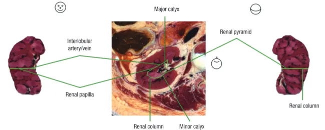
Fig. 1. The peeled (-12 mm) model of right kidney reconstructed by computer software (left and right). The level of peeling is indicated with green line on the reference sectioned image (center). The renal cortex (renal column) is red, while the renal medulla (renal pyramid, renal papilla) is pink. The renal column includes the interlobular artery and vein.
Head shapes are appended to represent the viewpoints of the volume model and the sectioned image: anterior view (left), horizontal view (center), and posterior view (right).
Interlobular artery/vein
Renal papilla
Renal column Minor calyx Major calyx
Renal pyramid
Renal column
Fig. 2. The piled (+12 mm) model of right kidney. The followings are found on the anterior surface of the kidney: liver (superolateral), inferior vena cava and renal vein (supero- medial), ascending colon (inferolateral), and the duodenum (inferomedial); On the posterior surface, we identify the followings: 12th rib (superior), latissimus dorsi (lateral), erec- tor spinae (medial), quadratus lumborum (inferolateral), psoas major (inferomedial), and external oblique muscle (posterior).
Liver
Inferior vena cava
Right renal vein
Ascending
colon Duodenum
Ascending
colon Duodenum Right
renal vein
12th rib Latissimus
dorsi
Quadratus lumborum Erector
spinae Psoas major
External oblique muscle
External oblique muscle Latissimus
dorsi
Chung BS, et al. • Volume Model of the Kidney
http://jkms.org
1515
http://dx.doi.org/10.3346/jkms.2016.31.10.1514
Current learning tools such as photographs, cadavers, and com- puter models are not sufficient for learning the comprehensive anatomy of the kidney and its surroundings. The objective of this research was to aid medical students by presenting tutorial software that contains peeled and piled volume models of the kidney. The sectioned images of donated female cadaver (age at death, 26 years; cause of death, stomach cancer; kidney pa- thology, none) were stacked to reconstruct three-dimensional volume model (voxel size, 0.2 mm) (1). Peeling and piling of the model were continuously carried out (intervals, 0.4 mm) to ex- plore the inside and outside of the kidney (2).
For convenient browsing, 61 peeled and piled models (from -12 mm to +12 mm) in anterior, posterior, medial, and lateral views were put into software (Fig. 1 and 2). “Browsing software (Male - Peeled and piled kidney) (118 MB)” can be download- ed from the website (anatomy.co.kr) or directly from the ad- dress (http://vkh.ajou.ac.kr/Browsing_software_(kidney)_set- up.exe) without charge or registration.
ACKNOWLEDGMENT
Raw data of the Visible Korean Human were acquired by the assistance from the Korea Institute of Science and Technology Information. Raw data of the Visible Korean Human were ac- quired by the assistance from the Korea Institute of Science and
Technology Information.
ORCID
Beom Sun Chung http://orcid.org/0000-0002-3644-9120 Min Suk Chung http://orcid.org/0000-0002-0527-9763 Byeong-Seok Shin http://orcid.org/0000-0001-7742-4846 Koojoo Kwon http://orcid.org/0000-0002-2467-5809 REFERENCES
1. Park HS, Choi DH, Park JS. Improved sectioned images and surface mod- els of the whole female body. Int J Morphol 2015; 33: 1323-32.
2. Kwon K, Shin DS, Shin BS, Park HS, Lee S, Jang HG, Park JS, Chung MS.
Virtual endoscopic and laparoscopic exploration of stomach wall based on a cadaver’s sectioned images. J Korean Med Sci 2015; 30: 658-61.
Address for Correspondence:
Koojoo Kwon, PhD
Department of Computer Science and Information Engineering, Inha University, 27 Inhang-ro, Jung-gu, Incheon 22332, Korea E-mail: [email protected] Funding: This work was funded by the National Research Foundation of Korea (NRF) grant funded by the Korea government (MSIP) (No. 2015R1A2A2A01008248, 2015R1A5A7037630). This research was supported by Basic Science Research Program through the National Research Foundation of Korea (NRF) funded by the Ministry of Education (No. 2015R1D1A4A01020277) .