pISSN 2288-0585⋅eISSN 2288-6850
Clinical Utility of Fecal Immunochemical Transferrin Test in Gastrointestinal Bleeding Detection
Jong-Mi Lee1, Mi Jung Park1, Woong Heo1, Kang Gyun Park1, Yong Gyu Park2, Seung Beom Han3, Young-Seok Cho4, Yeon-Joon Park1
Departments of 1Laboratory Medicine, 2Medical Life Science, 3Pediatrics, 4Gastroenterology, Seoul St. Mary’s Hospital, College of Medicine, The Catholic University of Korea, Seoul, Korea Background: Gastrointestinal (GI) bleeding can result
from various conditions, including ulcers, neoplasms and infectious enterocolitis. The aim of this study was to evaluate the utility of the fecal immunochem- ical transferrin test compared with the fecal Hb test in various clinical settings.
Methods: A total of 1,116 clinical stool specimens submitted for fecal occult blood testing were pro- spectively examined using both FIT Hb and FIT Tf kits (AlfresaPharma, Japan). To verify the specificity of the two tests, stool specimens from 265 health check-up examinees were also included.
Results: A review of medical records revealed that 396 patients had clinical conditions associated with GI bleeding. FIT Hb and FIT Tf results were positive in 156 (39.4%) and 137 (34.6%) cases, respectively, and an additional 194 (49.0%) cases tested positive with either FIT Hb or FIT Tf. The two tests showed a
moderate strength of agreement (kappa value; 0.56).
Colitis (n=71) was associated with the most GI bleed- ings, followed by acute gastroenteritis (n=29), GI ul- cers (n=27) and GI cancers (n=15). While the first two groups had higher positive rates on FIT Tf, pa- tients in the latter two groups had higher positive rates on FIT Hb. Notably, four of nine specimens from premature babies tested positive only on FIT Tf.
The specificity of FIT Hb and FIT Tf was 100% and 99.6%, respectively.
Conclusion: Concurrent use of FIT Hb and FIT Tf im- proved the detection rate of occult GI bleeding, espe- cially in patients with infectious GI disease (such as colitis or gastroenteritis) and in premature babies.
(Ann Clin Microbiol 2018;21:51-57)
Key Words: Fecal occult blood test, Hemoglobin, Prematurity, Transferrin
51
Received 31 January, 2018, Revised 18 May, 2018, Accepted 24 June, 2018
Correspondence: Yeon-Joon Park, Department of Laboratory Medicine, Seoul St. Mary’s Hospital, College of Medicine, The Catholic University of Korea, 222 Banpo-daero, Seocho-gu, Seoul 06591, Korea. (Tel) 82-2-2258-1640, (Fax) 82-2-2258-1719, (E-mail) [email protected]
ⓒ The Korean Society of Clinical Microbiology.
This is an Open Access article distributed under the terms of the Creative Commons Attribution Non-Commercial License (http://creativecommons.org/licenses/by-nc/4.0) which permits unrestricted non-commercial use, distribution, and reproduction in any medium, provided the original work is properly cited.
INTRODUCTION
Gastrointestinal (GI) bleeding can occur due to various con- ditions including ulcer disease, esophagitis, varices, vascular le- sions, neoplasm, diverticula, hemorrhoids, fissures, inflam- matory bowel disease and infectious colitis [1]. Fecal occult blood tests (FOBTs) represent simple and non-invasive methods for the detection of GI bleeding. The three classes of FOBTs in- clude: guaiac-based tests, heme-porphyrin tests, and im- munochemical tests, all of which have limited sensitivities and specificities [2]. Recently, the fecal immunochemical test for he- moglobin (FIT Hb) has been widely used due to its higher sen- sitivity compared with older forms of FOBTs and the test re-
sults are not influenced by food or medications [3,4]. FIT Hb is based on antibodies against the protein component of intact human globin in feces [5]. However, the globin is easily de- graded by enterobacteria and digestive enzymes, and therefore, less sensitive for detection of upper GI bleeding. Transferrin (Tf), which is a serum β-globulin that binds and transports iron [6], can leaks into the GI tract during GI bleeding and it can be detected in feces. It is more stable than hemoglobin to diges- tive enzymes [7], and it can be a potential marker for GI bleed- ing [3,8]. Several studies have shown that the combined use of both fecal Tf and Hb as biomarkers results in higher rates of positivity in colorectal cancer screening [3,9]. However, its utili- ty in various clinical conditions of GI bleeding has not been
Table 1. Demographic characteristics of 265 health check-up examinees and 1,116 patients
Demographic characteristics
Health check-up
examinees Patients
Male/female 146/119 (55%/44.9%) 598/518 (54%/46%)
Age 44 (21-79) 56 (0-99)
FIT Hb 3.0 (0-22) 5.0 (0-120,000)
FIT Tf 1.0 (0-131) 2.0 (0-18,300)
Values are presented as number or median (range), ages (years), FIT Hb (ng/mL), FIT Tf (ng/mL).
Fig. 1. Correlation between FIT Hb and FIT Tf results. The division bar indicates the Hb and Tf cut-off concentrations. The trend line is shown in blue. The data are plotted on logarithmic axes. The test results of FIT Hb and FIT Tf range between 0.1-120,000 ng/mL and 0.1-18,300 ng/mL, respectively.
investigated. Therefore, in this study, we evaluated the benefit of FIT Tf in clinical specimens for FOBT. In addition, we eval- uated the specificities of FIT Hb and FIT Tf by examining the stool specimens obtained from patients undergoing health check-up.
MATERIALS AND METHODS
1. Study subjects
From August to October 2016, a total of 1,381 stool speci- mens (1,116 specimens from patients and 265 specimens from health check-up) submitted to clinical microbiology laboratory for examination of occult blood were included. The demo- graphic characteristics of the participants are listed in Table 1.
We reviewed the medical records of all patients to identify the clinical conditions. This study was approved by the institutional review board of Seoul St. Mary’s Hospital.
2. Fecal immunochemical test
Fecal Hb and Tf were measured using CE-cleared FIT Hemoglobin NS-Prime test and FIT Transferrin NS-Prime test (AlfresaPharma, Osaka, Japan), respectively, on the NS-Prime analyzer according to the manufacturers’ instructions. Briefly, the surface of the fecal specimen was scraped using the collec- tor stick with the gutters, and the collector stick was inserted in- to the collector body and the container was shaken to dissolve the feces from the gutters of the stick. After leaving the contain- er for 30 min, it was placed into the equipment. Specimen val- ues were calculated using the Discrete Clinical Chemistry ana- lyzer NS-Prime. In each run, high and low positive controls and blank control were included. The positive thresholds of FIT Hb and FIT Tf were 100 ng/mL (20 μg/g feces) and 50 ng/mL (10 μg/g feces), respectively.
3. Statistical analysis
Statistical analyses were performed using MedCalc (v.16.4.3).
To compare the positive rates of the two tests, the McNemar test was used and statistical significance was established at P<
0.05. Spearman correlation analysis was used to evaluate the correlation between assays, and inter-rater agreement (kappa value) was calculated.
RESULTS
1. Results from the general health check-up
The median values of FIT Hb and FIT Tf were 3.0 (25%-75%: 2.0-5.0) and 1.0 (25%-75%: 0.0-2.0) ng/mL, re- spectively (Table 1). All the specimens tested were negative for FIT Hb and only a single specimen was positive for FIT Tf (131 ng/mL). However, colonoscopy and esophagogastroduo- denoscopy examination of the examinee showed no specific lesions. Therefore, the specificities of FIT Hb and FIT Tf were 100% (95% CI: 98.6-100.0) and 99.6% (95% CI: 97.9-100.0), respectively. Of the 265 health check-up examinees, 132 sub- jects underwent colonoscopy and colon polyps were found in 72 cases. Pathological examinations were performed in 54 cases, all of which showed non-advanced adenoma.
2. Results from the clinical specimens
The demographic characteristics of the 1,116 patients and the median values of FIT Hb and FIT Tf are listed in Table 1. Fig. 1
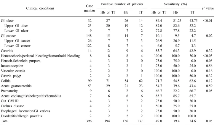
Table 3. Comparison of FIT Hb and Tf results based on clinical conditions
Clinical conditions Case
number
Positive number of patients Sensitivity (%)
P value
Hb or Tf Hb Tf Hb or Tf Hb Tf
GI ulcer 32 27 26 14 84.4 81.25 43.75 <0.01
Upper GI ulcer 23 20 19 12 87.0 82.6 52.2
Lower GI ulcer 9 7 7 2 77.8 77.8 22.2
GI cancer 148 15 14 7 10.1 9.5 4.7 0.02
Upper GI cancer 26 7 7 3 26.9 26.9 11.5
Lower GI cancer 122 8 7 4 6.6 5.7 3.3
Gastritis 14 12 9 6 85.7 64.3 42.9 0.32
Hematochezia/perianal bleeding/hemorrhoid bleeding 8 8 8 4 100.0 100.0 50.0 <0.05
Henoch-Schonlein purpura 4 3 3 0 75.0 75.0 0.0 0.08
Intussusception 4 3 2 1 75.0 50.0 25.0 0.56
Vascular ectasia 2 2 2 0 100.0 100.0 0.0 0.16
Gastric polyp 2 2 2 1 100.0 100.0 50.0 0.32
Colitis 99 71 54 62 71.7 54.5 62.6 0.12
Acute gastroenteritis 53 29 21 23 54.7 39.6 43.4 0.59
Prematurity 9 6 2 6 66.7 22.2 66.7 0.05
Acute cholangitis/cholecystitis/hemobilia 7 6 6 6 85.7 85.7 85.7
Gut GVHD 4 3 2 2 75.0 50.0 50.0
Crohn's disease 4 2 1 1 50.0 25.0 25.0
Esophageal laceration/GI varices 4 3 2 2 75.0 50.0 50.0
Duodenitis/allergic proctitis 2 2 2 2 100.0 100.0 100.0
Total 396 194 156 137 49.0 39.4 34.6 0.05
Abbreviations: GI, gastrointestinal; GVHD, graft-versus-host disease; Hb, hemoglobin; Tf, transferrin.
Table 2. Correlation between FIT Hb and FIT Tf results involving 1,116 clinical specimens
Method
FIT Hb
Positive Negative Total
FIT Tf Positive 101 50 151
Negative 69 896 965
Total 170 946 1,116
shows the distribution of the two test results, which revealed moderate correlation (ρ=0.601, P<0.0001). Positive results for FIT Hb and FIT Tf were found in 170 (15.2%) and 151 (13.5%) patients, respectively (Table 2), and 220 (19.7%) samples were positive for either test, and 101 (9.1%) samples tested positive for both. Therefore, the positive percent agreement (PPA) was 9.1% (101/1,116) and negative percent agreement (NPA) was 80.3% (896/1,116). The two test results showed moderate strength of agreement (kappa value; 0.56).
A review of the patients’ medical records indicated that 396 patients had clinical conditions that caused GI bleeding (Table 3). The two largest groups were GI cancer (n=148) and colitis (n=99), followed by acute gastroenteritis (AGE, n=53), GI ulcer
(n=32), gastritis (n=14) and so on. However, while 10.1%
(15/148) of specimens from GI cancer patients tested positive for either FIT Hb or FIT Tf, the majority (71.7%, 71/99) of specimens from colitis patients showed positive results.
Therefore, colitis (n=71) was the largest group showing positive test results, followed by AGE (n=29), GI ulcer (n=27) and GI cancer (n=15) (Table 3).
Based on clinical conditions, FIT Hb showed higher positive rates in 8 groups (GI cancer, GI ulcer, gastritis, hematochezia/
perianal bleeding/hemorrhoid bleeding, Henoch-Schönlein pur- pura, intussusception, vascular ectasia, and gastric polyp) en- compassing 71 cases; the positivity of FIT Hb was significantly higher than that of FIT Tf (91.6% vs. 45.1%, P<0.01). The FIT Tf showed higher positive rate in 3 groups (colitis, AGE and prematurity) encompassing 106 cases (Table 3); the positivity of FIT Tf was significantly higher than that of FIT Hb (72.6% vs.
85.9%, P=0.05).
Notably, although the number of specimens derived from pre- mature births was small (n=9), the four specimens showed pos- itive results only with FIT Tf . They were from four babies who had clinical conditions suggesting GI bleeding; one baby (gestational age (GA) of 32 weeks and 5 days) who showed FIT
Table 4. Evaluation of FIT Hb and FIT Tf results involving 1,116 clinical specimens
Method Sensitivity (%) Specificity (%) Positive predictive value (%)
Negative predictive
value (%) Acurracy (%)
FIT Hb 39.4 (34.6-44.4) 98.1 (96.8-98.9) 91.8 (86.7-95.0) 74.6 (73.1-76.1) 77.2 (74.7-79.7) FIT Tf 34.6 (29.9-39.5) 98.1 (96.8-98.9) 90.7 (85.1-94.4) 73.2 (71.7-74.6) 75.5 (72.9-78.0) FIT Hb or Tf 49.0 (44.0-54.0) 96.4 (94.8-97.6) 88.2 (83.5-91.7) 77.5 (75.7-79.1) 79.6 (75.7-79.1) Tf of 3,077 ng/mL had necrotizing enterocolitis (NEC), and
eventually underwent surgery for an ileus. The other baby (GA of 26 weeks 1 day) who showed a FIT Tf value of 545 ng/mL had a gastrostomy tube with bloody drainage. The remaining two babies (GA of 34 weeks 6 days for both) were diagnosed with enterocolitis-related symptoms of bloody loose stool (224 ng/mL, 151 ng/mL of FIT Tf, respectively). Two other cases of NEC (GA of 23 weeks 2 days for both) showed positive results in both tests. The level of FIT Tf was 5,104 ng/mL and 5,757 ng/mL and that of FIT Hb was 4,487 ng/mL and 1,345 ng/mL, respectively.
In the remaining 5 groups encompassing 21 patients, the two tests showed equal positive rates. Taken together, among the 396 cases who had clinical conditions associated with possible GI bleeding, the combination of two tests increased the sensi- tivity up to 49.0% (194/396) compared with that of FIT Hb only (39.4%, 156/396) with a small decrease in specificity (from 98.1% to 96.4%) (Table 4).
The distribution of the clinical conditions in the remaining 720 patients was as follows: hematologic disease (n=259); vari- ous infections (n=110) including pneumonia, sepsis, urinary tract infections, and acute pyelonephritis, brain stroke (n=69);
non-GI tract cancer (n=60); renal disease (n=31); heart disease (n=26); and various other conditions (n=165). Among the 720 patients, the patient with hemorrhagic cystitis and another with post kidney transplantation state with iron deficiency anemia showed positive results with both tests. We regarded these two cases as true positives. Discordant positive results were ob- served in 22 cases (12 cases with FIT Hb and 10 cases with FIT Tf, respectively).
DISCUSSION
First, FIT Hemoglobin NS-Prime and FIT Transferrin NS-Prime (AlfresaPharma) showed high specificity in health check-ups (100% and 99.6%, respectively), which was higher than that of the other kits (Inverness Clearview ULTRA iFOB,
Alere Clearview iFOB Complete, Polymedco OC-Light iFOB, Quidel QuickVue, iFOB, PolyMedco OC FIT-CHEK) evaluated in previous studies (88-95%) [10,11]. Younger age in our study (mean age of 43.9 years) compared with previous studies (mean age of 56.9 years and 64.2 years) might also have resulted in a higher specificity.
Among the 1,116 clinical specimens, the overall positive rate of FIT Hb was slightly higher than that of FIT Tf (15.2% vs.
13.5%) (P=0.08). Combining the two tests, 220 (19.7%) cases tested positive with either FIT Hb or FIT Tf. The two tests showed moderate strength of agreement (kappa value; 0.56) and the combination of the two tests increased the positive rate up to 19.7%. Using the same method as this study, Takashima et al. [9] also reported that the positive rate increased from 6.7%
to 10.1% following the addition of FIT Tf to FIT Hb in color- ectal screening of 12,255 healthy subjects.
According to the patients’ medical records, GI cancer (n=148) was the largest group undergoing FOBT, however, the positive rate was low (10.1%, 15/148), because most of the patients (98.0%, 145/148) were on the follow-up after therapeutic resection. Among the 15 specimens, which showed positive re- sults with either test, the positive rate of FIT Hb was sig- nificantly higher than that of FIT Tf (93.3%, 14/15 vs. 46.7%, 7/15, respectively). This result was in line with a previous study of Chen et al. [12], in which IFOBT (WHPM, Inc., Irwindale, CA, USA) showed higher sensitivity than fecal Tf dipstick for the detection of colorectal cancer and premalignant lesions (96%
vs. 92% for colorectal cancer, 58% vs. 50% for precancerous le- sion). However, Sheng et al. [13] reported that the sensitivity of fecal Tf dipstick was higher than that of IFOBT in colorectal cancer and premalignant lesions (76% vs. 61%). The differences in these studies might be derived from the difference in target population because while the tests were done as a cancer screening in previous studies, in our study, the tests were done as a follow up study after therapeutic resection. The other rea- son might be the difference in the test kits used.
The second and the third largest groups including colitis and
AGE accounted for 7.2% (154/1,116) of all the clinical speci- mens submitted for FOBT, suggesting that GI symptoms such as abdominal pain or diarrhea were also a major indication of FOBT. The positive rate was the highest in these two groups (72.3% and 54.7%) and FIT Tf showed higher positive rates than FIT Hb (87/154 vs. 77/154). The higher positive rate of FIT Tf might be attributed to the higher stability of Tf and re- sistance to digestive enzymes and bacterial degradation [7]. A bloody stool can be seen in bacterial diarrhea but not in viral diarrhea [1], and therefore, FOBT may aid clinicians in the identification of patients requiring hospitalization [14], pathogen testing, and initiation of optimal antibiotic therapy. Further eval- uation using FIT Tf involving enterocolitis of various causes is needed.
To our knowledge, this is the first study that evaluated fecal transferrin in specimens from premature babies. In premature babies, NEC, infectious colitis, congenital gut anomalies, bleed- ing disorders, vascular malformations, and specific medications are the common causes associated with GI bleeding [15,16].
Four out of nine cases showed positive results only with FIT Tf and two cases tested positive in both tests (positive rates of FIT Tf and FIT Hb were 100% and 33.3%, respectively, P=0.05). In addition, the fecal Tf levels of the latter two cases (5,104 ng/mL and 5,757 ng/mL) were much higher than those of the other cas- es (3,077 ng/mL, 545 ng/mL, 224 ng/mL and 151 ng/mL).
Although Tf concentration is known to be lower in younger children and lower GA [17,18], FIT Tf was still more sensitive than FIT Hb in detecting fecal occult blood in premature babies.
Our finding is in line with a previous study where routine FOBT targeting of fecal Hb adopted by many NICUs, did not predict NEC in very low-birth weight neonates [19]. We pre- sume that it might be related to differences in globin compo- nents of fetal and adult Hb, because fetal Hb consists of 60-90%
of total Hb until 1 month of age [20]. Considering the relatively high prevalence (5-6%) of NEC in babies with very low birth weight (<1,500 grams), further studies investigating the utility of fecal TF in this population are imperative.
In summary, based on our study, both FIT Hb and FIT Tf were highly specific, and showed moderate correlation (rs= 0.601, P<0.05) and low PPA (9.1%). According to the clinical conditions, FIT Hb was more sensitive to detecting non-in- fectious GI bleeding such as GI ulcer, cancer and anal bleeding, while FIT Tf was more sensitive for infectious GI bleeding such as colitis and acute gastroenteritis. In addition, the concen- trations of both Hb and Tf can vary from person to person, so
the use of both markers will be helpful to diminish the effect of individual variation [7]. Therefore, we hypothesize that test- ing FIT Hb alone is not sufficient to detect GI bleeding, and the concurrent use of FIT Tf is essential to improve the detection of GI bleeding, especially in patients with colitis, gastroenteritis and premature babies.
ACKNOWLEDGMENTS
Asan Pharm. Co. Ltd. (Korea) supported this study but was not involved in either data collection or manuscript preparation.
We are grateful to Dr. Seongah Kim and Dr. Ilou Park for re- viewing medical records. This work was supported in part by a grant from the Korea Health Industry Development Institute (grant number: HI 16C0443).
REFERENCES
1. Camilleri M and Murray JA. Diarrhea and Constipation. In:
Kasper D, Fauci A, Hauser S, Longo D, Jameson JL, Loscalzo J, eds. Harrison's Principles of Internal Medicine. 19th ed, New York; McGraw-Hill Education, 2015:264-74.
2. Young GP, Symonds EL, Allison JE, Cole SR, Fraser CG, Halloran SP, et al. Advances in fecal occult blood tests: the FIT revolution. Dig Dis Sci 2015;60:609-22.
3. Demian WLL, Collins S, Fowler C, McGrath J, Antle S, Moores Z, et al. Evaluation of the analytical performance of the novel NS-Prime system and examination of temperature stability of fecal transferrin compared with fecal hemoglobin as biomarkers in a colon cancer screening program. Pract Lab Med 2015;2:29-36.
4. Jin P, Sheng JQ, Wu ZT, Meng MM, Wang X, Wang XW, et al.
Combined fecal transferrin test and immuno fecal occult blood test for detecting colorectal cancer and advanced adenoma in asymptomatic and symptomatic populations. J Cancer Sci Ther 2012;4:243-48.
5. Fletcher RH, Ransohoff DF, Imperiale TF. Comparison of a brush-sampling fecal immunochemical test for hemoglobin with a sensitive guaiac-based fecal occult blood test in detection of colorectal neoplasia. Cancer 2007;109:1925-6; author reply 1926.
6. Aisen P and Listowsky I. Iron transport and storage proteins.
Annu Rev Biochem 1980;49:357-93.
7. Uchida K, Matsuse R, Miyachi N, Okuda S, Tomita S, Miyoshi H, et al. Immunochemical detection of human blood in feces. Clin Chim Acta 1990;189:267-74.
8. Miyoshi H, Oka M, Sugi K, Saitoh O, Katsu K, Uchida K.
Accuracy of detection of colorectal neoplasia using an immuno- chemical occult blood test in symptomatic referred patients:
comparison of retrospective and prospective studies. Intern Med 2000;39:701-6.
9. Takashima Y, Shimada T, Yokozawa T. Clinical benefit of measuring both haemoglobin and transferrin concentrations in faeces: demonstration during a large-scale colorectal cancer screening trial in Japan. Diagnosis 2015;2:53-9.
10. Levy BT, Bay C, Xu Y, Daly JM, Bergus G, Dunkelberg J, et al.
Test characteristics of faecal immunochemical tests (FIT) compared with optical colonoscopy. J Med Screen 2014;21:
133-43.
11. Imperiale TF, Ransohoff DF, Itzkowitz SH, Levin TR, Lavin P, Lidgard GP, et al. Multitarget stool DNA testing for colorectal-cancer screening. N Engl J Med 2014;370:1287-97.
12. Chen JG, Cai J, Wu HL, Xu H, Zhang YX, Chen C, et al.
Colorectal cancer screening: comparison of transferrin and immuno fecal occult blood test. World J Gastroenterol 2012;18:2682-8.
13. Sheng JQ, Li SR, Wu ZT, Xia CH, Wu X, Chen J, et al.
Transferrin dipstick as a potential novel test for colon cancer screening: a comparative study with immuno fecal occult blood test. Cancer Epidemiol Biomarkers Prev 2009;18:2182-5.
14. Lai CC, Ji DD, Wu FT, Mu JJ, Yang JR, Jiang DD, et al. Etiology and risk factors of acute gastroenteritis in a Taipei emergency department: clinical features for bacterial gastroenteritis. J Epidemiol 2016;26:216-23.
15. Maayan-Metzger A, Ghanem N, Mazkereth R, Kuint J. Charac- teristics of neonates with isolated rectal bleeding. Arch Dis Child
Fetal Neonatal Ed 2004;89:F68-70.
16. Rayhorn N, Thrall C, Silber G. A review of the causes of lower gastrointestinal tract bleeding in children. Gastroenterol Nurs 2001;24:77-82; quiz 82-3.
17. Lackmann GM, Schnieder C, Bohner J. Gestational age-dependent reference values for iron and selected proteins of iron metabolism in serum of premature human neonates. Biol Neonate 1998;74:
208-13.
18. Soldin OP, Bierbower LH, Choi JJ, Choi JJ, Thompson-Hoffman S, Soldin SJ. Serum iron, ferritin, transferrin, total iron binding capacity, hs-CRP, LDL cholesterol and magnesium in children;
new reference intervals using the Dade Dimension Clinical Chemistry System. Clin Chim Acta 2004;342:211-7.
19. Pickering A, White R, Davis NL. Routine fecal occult blood testing does not predict necrotizing enterocolitis in very low birth weight neonates. J Neonatal Perinatal Med 2016;9:171-8.
20. Orkin SH, Nathan DG, Ginsburg D, Look AT, Fisher DE, Lux S.
Nathan and Oski’s Hematology of Infancy and Childhood. 7th ed.
Philadelphia: Saunders/Elsevier; 2009:1778.
=국문초록=
위장관 출혈 검출을 위한 대변 트렌스페린 면역화학검사의 임상적 유용성 평가
가톨릭대학교 의과대학 서울성모병원 1진단검사의학교실, 2의과학교실, 3소아과학교실, 4소화기과학교실 이종미1, 박미정1, 허 웅1, 박강균1, 박용규2, 한승범3, 조영석4, 박연준1
배경: 위장관 출혈의 원인은 궤양, 종양, 장염 등 다양하다. 본 연구에서는 다양한 임상 환자의 검체에서 대변 트렌스페 린검사의 유용성을 대변 헤모글로빈검사와 비교하였다.
방법: 대변잠혈반응검사가 의뢰된 1,116개의 환자 검체와 265개의 건강인 검체를 이용해 헤모글로빈과 트렌스페린에 대한 면역화학검사(NS-Prime test, AlfresaPharma, Japan)를 각각 시행하였다.
결과: 건강인을 대상으로 한 헤모글로빈과 트렌스페린의 면역화학검사의 특이도는 각각 100%, 99.6%였다. 환자들의 의
무기록 확인 결과 396명의 환자들이 장출혈과 관련된 임상소견을 보였고 이중 194건(49.0%)이 둘 중 하나 이상의 검사에
서 양성이었다. 헤모글로빈과 트렌스페린 대변면역화학검사의 양성건수는 각각 156건(39.4%), 137건(34.6%)이었다. 임상
양상에 따른 분류에서 대장염과 급성장염이 각각 71명, 29명이었고, 위장관계 궤양과 암이 각각 27명, 15명이었다. 이 중 대장염과 급성장염은 트렌스페린 면역화학검사의 양성률이 더 높았고, 위장관계 궤양과 암은 헤모글로빈 면역화학검
사에서 더 높은 양성률을 보였다. 특히 9건의 미숙아 환자 검체 중 4건에서는 대변 트렌스페린 면역화학검사에서만 양성
을 보였다.
결론: 대변잠혈반응검사 시, 특히 대장염, 급성 장염, 미숙아의 경우, 대변 헤모글로빈 단독 검사보다 대변 트렌스페린을 함께 검사해야 검출률을 높일 수 있을 것으로 생각된다. [Ann Clin Microbiol 2018;21:51-57]
교신저자 : 박연준, 06591, 서울시 서초구 반포대로 222
가톨릭대학교 의과대학 서울성모병원 진단검사의학교실 Tel: 02-2258-1640, Fax: 02-2258-1719
E-mail: [email protected]