• 검색 결과가 없습니다.

Multifilament Cable Wire versus Conventional Wire for Sternal Closure in Patients Undergoing Major Cardiac Surgery

N/A
N/A
Protected

Academic year: 2021

Share "Multifilament Cable Wire versus Conventional Wire for Sternal Closure in Patients Undergoing Major Cardiac Surgery"

Copied!
7
0
0

로드 중.... (전체 텍스트 보기)

전체 글

(1)

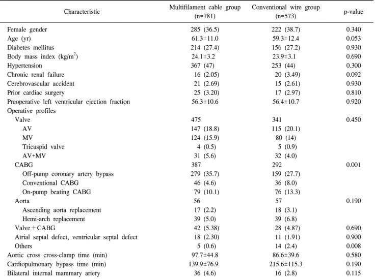
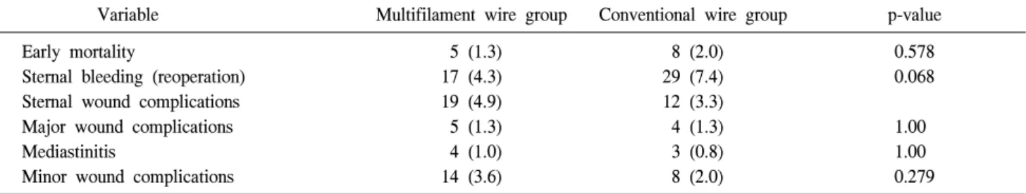
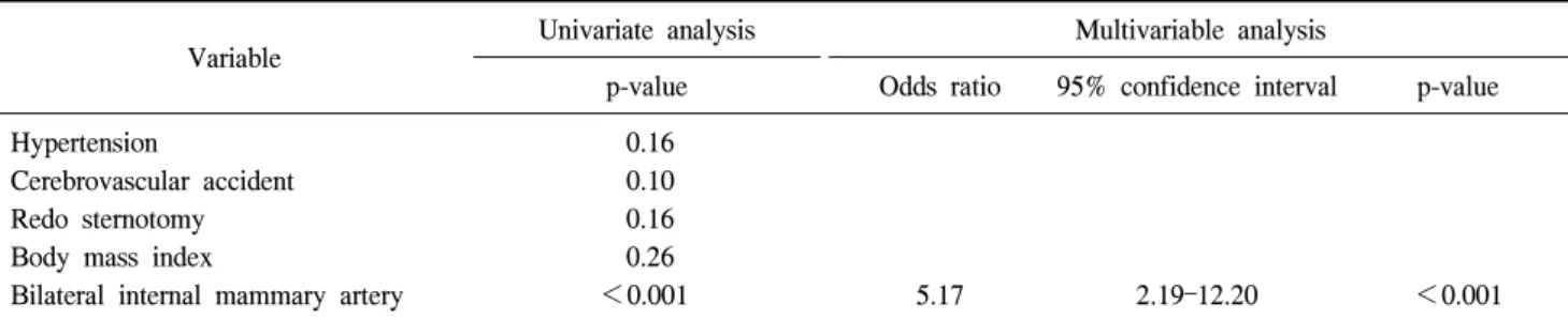
ISSN: 2233-601X (Print) ISSN: 2093-6516 (Online)

Department of Thoracic and Cardiovascular Surgery, Asan Medical Center, University of Ulsan College of Medicine Received: September 11, 2014, Revised: November 28, 2014, Accepted: December 1, 2014, Published online: August 5, 2015

Corresponding author: Joon Bum Kim, Department of Thoracic and Cardiovascular Surgery, Asan Medical Center, University of Ulsan College of Medicine, 88 Olympic-ro 43-gil, Songpa-gu, Seoul 138-736, Korea

(Tel) 82-2-3010-3580 (Fax) 82-2-3010-6966 (E-mail) [email protected]

C

The Korean Society for Thoracic and Cardiovascular Surgery. 2015. All right reserved.

CC

This is an open access article distributed under the terms of the Creative Commons Attribution Non-Commercial License (http://creative- commons.org/licenses/by-nc/4.0) which permits unrestricted non-commercial use, distribution, and reproduction in any medium, provided the original work is properly cited.

Multifilament Cable Wire versus Conventional Wire for Sternal Closure in Patients Undergoing Major Cardiac Surgery

You Na Oh, M.D., Keong Jun Ha, M.D., Joon Bum Kim, M.D., Sung-Ho Jung, M.D., Suk Jung Choo, M.D., Cheol Hyun Chung, M.D., Jae Won Lee, M.D.

Background: Stainless steel wiring remains the most popular technique for primary sternal closure. Recently, a mul- tifilament cable wiring system (Pioneer Surgical Technology Inc., Marquette, MI, USA) was introduced for sternal clo- sure and has gained wide acceptance due to its superior resistance to tension. We aimed to compare conventional steel wiring to multifilament cable fixation for sternal closure in patients undergoing major cardiac surgery. Methods:

Data were collected retrospectively on 1,354 patients who underwent sternal closure after major cardiac surgery, us- ing either the multifilament cable wiring system or conventional steel wires between January 2009 and October 2010. The surgical outcomes of these two groups of patients were compared using propensity score matching based on 18 baseline patient characteristics. Results: Propensity score matching yielded 392 pairs of patients in the two groups whose baseline profiles showed no significant differences. No significant differences between the two groups were observed in the rates of early mortality (2.0% vs. 1.3%, p=0.578), major wound complications requiring re- construction (1.3% vs. 1.3%, p>0.99), minor wound complications (3.6% vs. 2.0%, p=0.279), or mediastinitis (0.8%

vs. 1.0%, p=1.00). Patients in the multifilament cable group had fewer sternal bleeding events than those in the con- ventional wire group, but this tendency was not statistically significant (4.3% vs. 7.4%, p=0.068). Conclusion: The surgical outcomes of sternal closure using multifilament cable wires were comparable to those observed when con- ventional steel wires were used. Therefore, the multifilament cable wiring system may be considered a viable op- tion for sternal closure in patients undergoing major cardiac surgery.

Key words: 1. Multifilament cable wire 2. Conventional wire 3. Sternal closure 4. Cardiovascular surgery 5. Complication

INTRODUCTION

Median sternotomy, which was re-introduced by Julian in 1957, remains the standard method of accessing the heart and great vessels [1]. It provides excellent exposure of the heart, which allows a range of heart operations to be performed,

and is well tolerated by most patients [2]. However, wound complications, such as sternal instability or infection, may oc- cur after median sternotomy. These complications have been reported to occur in 0.3%–5.0% of cases, and are associated with a significant mortality rate of 14%–47%, with an espe- cially high risk of mortality in cases of mediastinitis [3].

http://dx.doi.org/10.5090/kjtcs.2015.48.4.265

(2)

Furthermore, the rate of re-exploration for bleeding following cardiac surgery has been reported to be approximately 2.0%–

7.0%, and the causes of postoperative bleeding can be mul- ti-factorial [4]. A stable sternal approximation is believed to be the most important factor for preventing these complications.

Various bone fixation materials and sternal closure techniques have been developed in order to maximize sternal stability;

however, the advantages and disadvantages of each technique have been poorly described to date. The current standard technique for sternal closure is cerclage with stainless steel wires. Recently, a multifilament cable wiring system (Pioneer Surgical Technology Inc., Marquette, MI, USA) was intro- duced for sternal closure, and has become increasingly popu- lar due to its superior resistance to tension. However, no comparative studies have been conducted to analyze the out- comes of these two different sternal closure techniques. There- fore, we aimed to compare conventional steel wiring to fix- ation using multifilament cables for sternal closure in patients undergoing major cardiac surgery.

METHODS 1) Patients

From January 2009 to October 2010, 1,354 patients under- went major cardiac surgery via median sternotomy at the Asan Medical Center, Seoul, Korea. Among these patients, 781 (57.7%) underwent sternal closure using the multifilament cable wiring system, while the remaining 573 patients (42.7%) underwent sternal closure using conventional stainless steel wires. Data were collected through a retrospective chart review. The deci- sion to use multifilament cable wires in a given patient was influenced by that patient’s demographic profile (age, gender, diabetes status, and osteoporosis status) and surgical factors (the type of surgery, the presence of an intraoperative sternal fracture, and the use of the bilateral internal thoracic artery).

However, the choice was ultimately made at the discretion of the operating surgeon, reflecting his or her individual attitude regarding the use of multifilament cable wires.

2) Surgical technique

The multifilament cable group consisted of 781 patients, in whom 1.0-mm multifilament stainless cables (Pioneer Surgical

Technology Inc.) were deployed in a figure-of-eight fashion around the sternum. In 755 of these patients (96.7%), conven- tional stainless steel wires were added around the manubrium and the lower part of the sternal body. A median of two multifilament cable wires were used for sternal closure (range, 1–4), while a median of three conventional stainless steel wires were used in the procedure (range, 1–8).

The conventional wire group consisted of 573 patients in whom a median of seven standard stainless steel wires (range, 4–12) were used for sternal closure in a simple interrupted fashion: two to three pieces were used around the manubrium and four to five pieces were used around the sternal body.

The steel wires were tightened by twisting with a large nee- dle driver.

3) Definitions and follow-up

The primary outcomes of interest were early death and sternotomy-related complications, including sternal bleeding, sternal wound complications, and mediastinitis. Sternal bleed- ing was defined as postoperative bleeding requiring re-explor- ation that was ultimately determined to have originated in the sternum. Sternum wound complications were divided into ma- jor and minor wound complications. Major wound complica- tions were defined as infections or dehiscence that required a major procedure, such as flap interposition, to be performed in the operating room. Among patients with major wound complications, those who showed sternal instability with bony involvement or had live organisms isolated from the media- stinum were diagnosed with mediastinitis. Minor wound com- plications were defined as less severe problems that were treated with medication or bedside wound revision and showed no sign of bacterial involvement.

Clinical follow-up was conducted every three to six months on an outpatient basis. All patients underwent follow-up, and the median follow-up duration was 27.5 months (interquartile range, 22.4–32.9 months).

4) Statistical analysis

Categorical variables are shown as frequencies and percen- tages, and continuous variables are expressed as means±

standard deviation or medians with ranges. In order to de-

crease the effect of treatment selection bias and potential con-

(3)

Table 1. Baseline patient demographics

Characteristic Multifilament cable group (n=781)

Conventional wire group

(n=573) p-value

Female gender 285 (36.5) 222 (38.7) 0.340

Age (yr) 61.3±11.0 59.3±12.4 0.053

Diabetes mellitus 214 (27.4) 156 (27.2) 0.930

Body mass index (kg/m

2

) 24.1±3.2 23.9±3.1 0.690

Hypertension 367 (47) 253 (44) 0.300

Chronic renal failure 16 (2.05) 20 (3.49) 0.092

Cerebrovascular accident 21 (2.69) 15 (2.61) 0.930

Prior cardiac surgery 25 (3.20) 17 (2.97) 0.810

Preoperative left ventricular ejection fraction 56.3±10.6 56.4±10.7 0.920

Operative profiles

Valve 475 341 0.450

AV 147 (18.8) 115 (20.1)

MV 124 (15.9) 80 (14)

Tricuspid valve 4 (0.5) 5 (0.9)

AV+MV 31 (5.6) 32 (4.0)

CABG 387 292 0.001

Off-pump coronary artery bypass 279 (35.7) 159 (27.7)

Conventional CABG 46 (4.6) 36 (8.0)

On-pump beating CABG 79 (10.1) 76 (13.3)

Aorta 56 57 0.190

Ascending aorta replacement 17 (2.2) 18 (3.1)

Hemi-arch replacement 39 (5.0) 39 (6.8)

Valve+CABG 42 (5.38) 28 (4.87) 0.690

Atrial septal defect, ventricular septal defect 18 (2.30) 11 (1.91) 0.900

Others 5 (0.6) 14 (2.4) 0.008

Aortic cross cross-clamp time (min) 97.7±44.8 86.6±39.6 0.580

Cardiopulmonary bypass time (min) 139.9±76.9 215.6±115.3 0.190

Bilateral internal mammary artery 36 (4.6) 16 (2.8) 0.115

Values are presented as number (%) for categorical variables and as mean±standard deviation for continuous variables.

AV, aortic valve; MV, mitral valve; CABG, coronary artery bypass grafting.

founding, we carried out adjustments for the differences in the baseline characteristics between the two groups by apply- ing propensity score matching. The propensity scores were calculated using multiple logistic regression analysis. The ad- equacy of the propensity score model was evaluated using the C-statistic and the Hosmer-Lemeshow test. The model yielded a C-statistic of 0.592 and a Hosmer-Lemeshow goodness-of- fit p-value of 0.466, which indicated that the model was well-calibrated with reasonable discrimination. In order to identify propensity score-matched pairs (one to one matches), the greedy 5-to-1 digit matching algorithm was used. After the propensity score matching, the data of the two groups were compared using the paired t-test or the Wilcoxon signed

rank test for continuous variables, and using the McNemar test or the marginal homogeneity test for categorical variables.

All reported p-values were two-sided, and p-values <0.05 were considered to indicate statistical significance. SAS ver.

9.1 (SAS Institute Inc., Cary, NC, USA), was used for stat- istical analyses.

RESULTS

Table 1 summarizes the baseline characteristics, comorbid-

ities, and procedure-related parameters of all patients. In com-

parison with the patients in the conventional wire group, the

patients in the multifilament cable group tended to be older

(4)

Table 2. Baseline patient demographics (propensity-matched)

Characteristic Multifilament cable group

(n=392)

Conventional wire group

(n=392) p-value

Female gender 159 (40.6) 162 (41.3) 0.828

Age (yr) 60.6±12.1 60.1±12.0 0.588

Diabetes mellitus 112 (28.6) 107 (27.3) 0.691

Body mass index (kg/m

2

) 23.9±3.3 23.9±3.1 0.974

Hypertension 182 (46.4) 168 (42.9) 0.315

Chronic renal failure 12 (3.1) 13 (3.3) 0.839

Cerebrovascular accident 9 (2.3) 12 (3.1) 0.518

Prior cardiac surgery 9 (2.3) 14 (3.6) 0.398

Preoperative left ventricular ejection fraction 56.0±11.2 56.9±9.8 0.839

Operative profiles

Valve 0.996

AV 84 (21.4) 84 (21.4)

MV 57 (14.5) 58 (14.8)

Tricuspid valve 4 (1.0) 3 (0.8)

AV+MV 21 (5.4) 22 (5.6)

CABG 0.992

Off-pump coronary artery bypass 110 (28.1) 108 (27.6)

Conventional CABG 33 (8.4) 33 (8.4)

On-pump beating CABG 54 (13.8) 52 (13.3)

Aorta 0.835

Ascending aorta replacement 11 (2.8) 13 (3.3)

Hemi-arch replacement 27 (6.9) 30 (7.7)

Valve+CABG 20 (5.1) 21 (5.4) 0.873

Atrial septal defect, ventricular septal defect 5 (1.3) 7 (1.8) 0.923

Others 5 (1.3) 1 (0.3) 0.217

Aortic cross-clamp time (min) 47.8±48.6 48.4±52.2 0.877

Cardiopulmonary bypass time (min) 154.5±82.0 98.4±148.0 0.183

Bilateral internal mammary artery 17 (4.3) 14 (3.6) 0.629

Values are presented as number (%) for categorical variables and as mean±standard deviation for continuous variables.

AV, aortic valve; MV, mitral valve; CABG, coronary artery bypass grafting.

Table 3. Early operative outcomes

Variable Multifilament wire group Conventional wire group p-value

Early mortality 5 (1.3) 8 (2.0) 0.578

Sternal bleeding (reoperation) 17 (4.3) 29 (7.4) 0.068

Sternal wound complications 19 (4.9) 12 (3.3)

Major wound complications 5 (1.3) 4 (1.3) 1.00

Mediastinitis 4 (1.0) 3 (0.8) 1.00

Minor wound complications 14 (3.6) 8 (2.0) 0.279

Values are presented as number (%).

(61.3±11.0 years vs. 59.3±12.4 years; p=0.053), had a lower prevalence of chronic renal failure (2.1% vs. 3.5%; p=0.092), and were more likely to undergo off-pump coronary artery bypass graft surgery (Table 1).

Propensity score matching yielded 392 pairs of patients for

whom no significant difference existed in the baseline profile,

including age, body mass index, history of previous cardiac

surgery, and the rate of bilateral internal mammary artery use

(5)

Table 4. Multivariable risk factor analysis for sternal wound complications (logistic regression)

Variable Univariate analysis Multivariable analysis

p-value Odds ratio 95% confidence interval p-value

Hypertension 0.16

Cerebrovascular accident 0.10

Redo sternotomy 0.16

Body mass index 0.26

Bilateral internal mammary artery <0.001 5.17 2.19–12.20 <0.001

Only variables with p<0.30 on univariate logistic regression analysis were incorporated into the multivariable analysis.

Table 5. Management of sternal wound complications and mediastinitis

Variable Multifilament cable group Conventional wire group p-value

Vacuum-assisted closure application or wet dressing 4 (1.0) 6 (1.5) 0.752

Sternal reconstruction 7 (1.8) 3 (0.8)

Titanium plate+tissue flap 2 (0.5) 0 (0.0) 0.499

Muscle flap 4 (1.0) 3 (0.8) 1.00

Omental flap 1 (0.3) 0 1.00

Values are presented as number (%).

between the two matched groups (Table 2).

The occurrence of postoperative complications is summar- ized in Table 3. In the propensity score-matched cohort, no significant differences were observed between the two groups regarding the rates of early mortality (1.3% in the multifila- ment cable group vs. 2.0% in the conventional wire group;

p=0.578), major wound complications requiring reconstruction (1.3% in the multifilament cable group vs. 1.3% in the con- ventional wire group; p=1.00), minor wound complications (3.6% in the multifilament cable group vs. 2.0% in the con- ventional wire group; p=0.279), or mediastinitis (1.0% in the multifilament cable group vs. 0.8% in the conventional wire group; p=1.00). The patients in the multifilament cable group tended to have fewer sternal bleeding events than those in the conventional wire group, but this discrepancy only reached a marginal level of statistical significance (4.3% vs. 7.4%;

p=0.068). On multivariable analysis, the use of the bilateral internal mammary artery was the only significant risk factor for sternal wound complications (odds ratio, 5.17; 95% con- fidence interval, 2.19–12.20; p<0.001) (Table 4).

The management of sternal wound complications or media- stinitis is summarized in detail in Table 5. Sternal wound complications requiring sternal reconstruction occurred in 10

patients; two patients underwent sternal fixation with a tita- nium plate and autologous tissue flap, while the other eight patients received an autologous tissue flap, using either omen- tum (n=1) or muscle (n=7). No significant differences were observed between the two groups in the rate of sternal wound complications requiring vacuum-assisted closure, the use of wet dressing, or sternal reconstruction.

DISCUSSION

Complications from median sternotomy have been reported to occur in approximately 0.3%–5.0% of cases, and are asso- ciated with significant morbidity and mortality [5]. The loos- ening and failure of sternal fixation is associated with com- promised wound healing, and may lead to sternal wound de- hiscence. Infection is also associated with wire loosening [6].

Although a variety of sternum closure techniques have been

developed, the primary consideration in choosing a technique

should be its effectiveness in ensuring the rigid closure of the

sternum, which restores the stability of the sternum and pro-

motes its primary healing. Several sternum closure techniques

have been reported to prevent sternal dehiscence, but no con-

sensus yet exists about an ideal method. Wangsgard et al. [7]

(6)

reported that the figure-of-eight stainless-steel cable system and the dynamic sternal fixation plate system are expected to show a lower failure rate both in situations involving lateral distraction and in situations with longitudinal shear cyclic loading, in comparison to the figure-of-eight stainless-steel wire system. Furthermore, the figure-of-eight stainless-steel cable system has the best ability to resist failure, especially in high cycle numbers. Khasati et al. [8] compared two different sternal closure methods (simple wire versus figure-of-eight), and found that the figure-of-eight method had no significant bene- fit over the simple wire method. Cohen and Griffin [9] per- formed a biomechanical study of three sternal closure techni- ques (figure-of-eight stainless steel wires, stainless steel plates, and figure-of-eight stainless steel cables). Both the plate sys- tem and the stainless steel cable system showed significant advantages over the figure-of-eight stainless steel wire system [9]. Although many previous studies have reported sternum closure techniques that have been claimed to be ideal for sternal fixation, few comparative clinical studies have been conducted to analyze the outcomes of different sternal closure techniques in practice.

The Pioneer sternal cable has been reported to have a fa- tigue strength approximately ten times stronger than standard steel wires [10]. The cable wire hugs the bone uniformly in a figure-of-eight fashion, which may decrease the possibility of loosening, micro-motion, potential postoperative malunion, and resultant compromised wound healing. In this context, it is worth noting that the multifilament cable group in this study experienced less sternal bleeding, even though this as- sociation did not attain statistical significance. We speculate that this finding may be due to the greater fatigue strength and the higher resistance to stress of the multifilament cable wires. The resultant higher tension in the bone union and more stable fixation of the sternum may lead to a lower like- lihood of bleeding from the sternal bone marrow and adjacent structures [7]. The cable wires are also easy to handle and may decrease the time required for sternum closure. However, despite all these putative advantages, surgeons must avoid over-tensioning cables, as they may break, and the multifila- ment cable wire may cut through soft bone tissue that is not protected and immobilized.

In the present study, 1,354 elective open heart surgery pa-

tients were separated into two groups depending on which sternal closure technique was employed. The size of the co- hort was sufficiently large to power the statistical analysis.

Propensity scoring was used to match important differences in the baseline risk profiles that may otherwise have confounded the outcomes. Since multiple surgeons performed the relevant operations in our institution, rigorous statistical adjustments were made to minimize the effects of individual surgeons on the outcome. In this study, multifilament cable wires were not found to have a clear clinical advantage over conven- tional steel wires. Instead, the results of our study showed no significant differences in the clinical outcomes between the two groups, except for a slight tendency for less sternal bleeding in the multifilament cable group, as described above.

The multifilament cable wires may intuitively be thought to hold the sternum more tightly, leading to less bleeding than was observed among patients in whom conventional wires were used. However, further studies are necessary to improve our understanding of the mechanism by which multifilament cable wires result in less bleeding, as well as to identify the subsets of patients in which the benefits of this technique can be maximized.

This study has several limitations. The accuracy of this ret- rospective review depended on the availability of information in the patients’ medical records. A particularly important qualifi- cation is that we were not able to determine the precise ori- gin of sternal bleeding. The non-randomized design may have affected our results due to unmeasured confounders, proce- dural bias, or detection bias, even with the use of rigorous statistical adjustment. Moreover, conventional stainless steel wires were also used to close the sternum in the cable wire group, which rendered this cohort heterogeneous to some ex- tent, thus potentially making our results somewhat ambiguous.

In conclusion, the clinical outcomes of sternal closure us- ing multifilament cable wires were comparable to those ob- served when conventional stainless steel wires were used.

Therefore, the new multifilament cable fixation system may

be considered a viable option for sternal closure in patients

undergoing major cardiac surgery.

(7)

CONFLICT OF INTEREST

No potential conflict of interest relevant to this article was reported.

ACKNOWLEDGMENTS

This study was supported by a Grant of the Samsung Vein Clinic Network (Daejeon, Anyang, Cheongju, Cheonan; Fund No. KTCS04-029).

REFERENCES

1. Dalton ML, Connally SR, Sealy WC. Julian’s reintroduction of Milton’s operation. Ann Thorac Surg 1992;53:532-3.

2. McGregor WE, Payne M, Trumble DR, Farkas KM, Magovern JA. Improvement of sternal closure stability with reinforced steel wires. Ann Thorac Surg 2003;76:1631-4.

3. Losanoff JE, Collier AD, Wagner-Mann CC, et al. Biomecha- nical comparison of median sternotomy closures. Ann Thorac Surg 2004;77:203-9.

4. Yavuz S, Eris C, Turk T. Re-exploration for excessive bleed- ing after coronary artery bypass surgery: how early is bet- ter? Eur J Cardiothorac Surg 2007;32:819-20.

5. El Oakley RM, Wright JE. Postoperative mediastinitis: clas- sification and management. Ann Thorac Surg 1996;61:1030-6.

6. Schimmer C, Reents W, Berneder S, et al. Prevention of sternal dehiscence and infection in high-risk patients: a pro- spective randomized multicenter trial. Ann Thorac Surg 2008;86:1897-904.

7. Wangsgard C, Cohen DJ, Griffin LV. Fatigue testing of three peristernal median sternotomy closure techniques. J Cardiothorac Surg 2008;3:52.

8. Khasati N, Sivaprakasam R, Dunning J. Is the figure-of-eight superior to the simple wire technique for closure of the ster- num? Interact Cardiovasc Thorac Surg 2004;3:191-4.

9. Cohen DJ, Griffin LV. A biomechanical comparison of three sternotomy closure techniques. Ann Thorac Surg 2002;73:

563-8.

10. Pioneer Surgical Technology Inc. Pioneer sternal cable sys- tem: cardiothoracic: cable system [Internet]. Marquette (MI):

Pioneer Surgical Technology Inc.; c2015 [cited 2014 Dec 27].

Available from: http://www.pioneersurgical.com/cardiothoracic/

Cable-System/Pioneer-Sternal-Cable-System/.

수치

Table 1. Baseline patient demographics
Table 3. Early operative outcomes
Table 4. Multivariable risk factor analysis for sternal wound complications (logistic regression)

참조

관련 문서

Endoscopic spine surgery has become a practical, minimally invasive technique for decompression in patients with spinal disc herniation or stenosis.. However,

This showed that the surgical outcome was successfulin 269 patients (98.89%),butthe revision surgery due to surgicalrelapse was performed in three patients.Of 152 patients

The purpose of this study was to identify the frequency and related factors of advanced airway management for patients with cardiac arrest by the

Result: During the study period, 482 patients had elevated cardiac troponin-T levels. The positive predictive value for the.. The diagnostic importance of

3.1 Comparison of ISO Quality levels and weldability of laser-arc hybrid welded joints by wire for heat resistant steel varying travel speed in 10kW · ·

7.3 Switching systems (Routers in Backbone) 7.4 Signaling systems... Dual-tone multi-frequency

Previous studies suggested using the poste- rior condylar axis (PCA) [12], Whiteside’s line [18] or the transepicondylar axis (TEA) [9, 11, 14, 20] as a reference axis

Thus, in the present study, we aimed to compare protein pro- files between achalasia patients and healthy controls using the serum proteomic analysis for the detection of