INTRODUCTION
Fulminant hepatic failure carries a very high mortality of 80-90% (1). Orthotopic liver transplantation has now become the treatment of choice of fulminant hepatic failure, with a survival rate of 50-75% after 1 yr (2). But, spontaneous sur- vival rate of patients with fulminant hepatic failure ranges from 15% to 25% (3). Therefore, a liver assist device to help the liver recover spontaneously can be an alternative method to replace orthotopic liver transplantation or a bridge to patients waiting for a suitable donor in preventing the occurrence of irreversible neurologic damage. Several devices have been developed and employed clinically.
To prove the functional support of liver assist devices in ful- minant hepatic failure, it is necessary to establish a standard- ized hepatic failure model which closely correlates with clini- cal situations and can be reproduced in large animals. There are several animal models of hepatic failure. The surgical mod- els include total hepatectomy, partial or complete devascu- larization, and portacaval shunt with 70% partial hepatec- tomy (4-7). The pharmacological models include D-galac- tosamine, acetaminophen, and carbon tetrachloride (4, 8-12).
The aim of these models is to simulate acute fulminant hepat- ic failure that results in the rapid onset of hepatic coma and death. Even though the application of liver assist devices in human has been performed, the additional development of technology and demonstration of safety, efficacy, and mech-
anism of action should be greatly accelerated by testing a clini- cally relevant large animal model.
Ischemic injury is one of the reasons for hepatic failure (6).
For pigs, the tolerable period of hepatic ischemia varies from 35 to 180 min (6, 14-16). If the time of hepatic ischemia exceeds the tolerable period, hepatocytes can be injured irre- versibly and necrosis can occur, too. In the present study, we have evaluated a model of fulminant hepatic failure induced by clamping the portal vein and the hepatic artery intermit- tently to cause reperfusion injury to the liver and then by lig- ating hepatic artery at the end of the last clamping.
MATERIALS AND METHODS Animals
Female white pigs, weighing from 17 kg to 25 kg, were used for this experiment. One day before surgery, 20 g mag- nesium sulfate was administered orally for bowel cleansing and then pigs were fasted. All the experiments were conducted under the supervision of a veterinarian according to local insti- tutional guidelines for the care and use of laboratory animals.
Operative procedure
All operative procedures were performed at the “Clinical Kuhn Uk Lee, Long-xian Zheng*, Yong Beom Cho, Ki-Ho Kim, Jongwon Ha, Kyung-Suk Suh, Sung Eun Jung
Department of Surgery, Seoul National University College of Medicine, Seoul, Korea; Department of Surgery*, The 2nd Affiliated Hospital of Harbin Medical University, Harbin, PR China
Address for correspondence Kuhn Uk Lee, M.D.
Department of Surgery, Seoul National University College of Medicine, 28 Yongon-dong, Jongno-gu, Seoul 110-744, Korea
Tel : +82.2-2072-2312, Fax : +82.2-766-3975 E-mail : [email protected]
*This study was supported by a grant of the Korea Health 21 R&D project, Ministry of Health & Welfare, Republic of Korea (02-PJ3-PG6-EV09-0001).
427
An Experimental Animal Model of Fulminant Hepatic Failure in Pigs
The objective of this study was to develop an experimental animal model of fulmi- nant hepatic failure to test the efficacy of the bioartificial liver system. The portal vein and the hepatic artery were clamped intermittently and then the hepatic artery was ligated (ligation group, n=5). Pigs whose hepatic arteries were not ligated after clamping were assigned to the non-ligation group (n=5). The biochemical changes in blood, histologic alterations of the liver and neurologic examination for pigs were checked up. All animals died within 17 hr in the ligation group. On the other hand, all animals survived more than 7 days in the non-ligation group. In the ligation group, the levels of ammonia, lactic acid and creatinine showed a progressively increas- ing pattern. Prothrombin time was also prolonged gradually. Cytoplasmic conden- sation and nuclear pyknosis of hepatocytes were detected histologically at autop- sy. Neurologic findings such as decreased pain sensation, tachypnea and no light reflex of pupils were observed. The findings shown in the ligation group are similar to the clinical features of fulminant hepatic failure in human and this animal model is reproducible. Therefore, this can be a suitable animal model to evaluate the effi- cacy of the bioartificial liver system for treating fulminant hepatic failure.
Key Words : Models, Animal; Liver Failure, Acute; Hepatic Artery; Hepatic Encephalopathy
Received : 26 July 2004 Accepted : 22 December 2004
Research Institute, Seoul National University Hospital. Pre- medication was performed with an intramuscular injection of ketamine chloride (25 mg/kg). After injecting Pentothal sodium (2 mg/kg), the animals were intubated and ventilat- ed with a mixture of nitrous oxide, oxygen, and enflurane.
One catheter was placed in the external jugular vein for infu- sion and another catheter was introduced into the femoral artery for both blood pressure monitoring and blood sam- pling. The abdomen was opened by a midline incision. The common bile duct, the hepatic artery, and the portal vein were exposed. A sling was laid around the hepatic artery and the portal vein for clamping. It took 25 min for the 1st and 2nd clamping and 30 min for the 3rd and 4th. The hepatic artery was ligated after the 4th clamping (ligation group, n=5).
Pigs whose hepatic artery was not ligated after clamping were assigned to the non-ligation group (n=5). The inter- vals between each clamping were 30 min and total clamp- ing time was 110 min.
During the operation, lactated Ringer’s solution was given
and 8.4% NaHCO3and calcium gluconate were administered for the maintenance of physiologic status. Five furosemide injections (10 mg) were totally given every 90 min to make them excrete potassium. The anesthesia was discontinued after the operation was over. The animals were kept in isolated cages and carefully controlled and their behaviors were registered.
Clinical assessment
The behaviors of the animals after awaking from anesthe- sia were controlled continuously. The following criteria were registered: abilities to get up, to walk, and to respond to pain- ful stimuli, and also light reflex of pupils. The purpose of these observations was to detect the signs of hepatic encepha- lopathy.
Biochemical measurements
Blood sampling was done four times: before clamping, after
Ammonia (g/dL)
1,600 1,400 1,200 1,000 800 600 400 200 0
Baseline 4 hr 7 hr Before death Time
ligation non-ligation
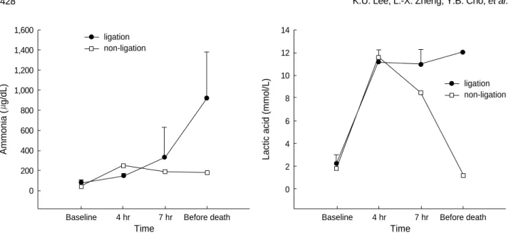
Fig. 1.Evolution of ammonia and lactic acid (mean±SD) before clamping, after 4 hr and 7 hr since the first clamping, and before death (in non- ligation group, 20 hr after clamping).
Lactic acid (mmol/L)
14 12 10 8 6 4 2 0
Baseline 4 hr 7 hr Before death Time
ligation non-ligation
Fig. 2.Evolution of total bilirubin, AST, and ALT (mean±SD) before clamping, after 4 hr and 7 hr since the first clamping, and before death (in non-ligation group, 20 hr after clamping).
Total bilirubin (mg/100 mL)
3.5 3.0 2.5 2.0 1.5 1.0 0.5 0.0
Baseline 4 hr 7 hr Before death Time
ligation non-ligation
AST (U/L)
4,000
3,000
2,000
1,000
0
Baseline 4 hr 7 hr Before death Time
ligation non-ligation
ALT (U/L)
160 140 120 100 80 60 40 20 0
Baseline 4 hr 7 hr Before death Time
ligation non-ligation
4 hr and 7 hr since the first clamping respectively, and lastly before death (in non-ligation group, 20 hr after clamping).
And the following parameters were measured from the experi- ment: glucose, aspartate transaminase (AST), alanine transami- nase (ALT), total bilirubin, blood urea nitrogen (BUN), cre- atinine, ammonia, potassium, lactic acid, prothrombin time (PT) and pH. The PT was not checked in the time when it has passed 7 hr since the first clamping.
Histologic evaluations
Specimens of the liver were taken three times: before clamp- ing, after 4 hr since the first clamping, and immediately after death (in non-ligation group, 20 hr after clamping). By means of liver biopsy, liver tissues were fixed by 1 cm thickness in 10% formalin. After fixation, 27 m sections of each biopsy were stained with hematoxylin eosin and examined by light microscopy.
Statistical analysis
Data were expressed as mean values±standard deviation (SD). Biochemical data were compared to the baseline level by use of Wilcoxon signed rank test. The level of statistical significance was set at p<0.05.
RESULTS Survival
In the ligation group, the first animal died from hyperkale- mia at 6.5 hr after ligation of the hepatic artery. The reason was that 8.4% NaHCO3and calcium gluconate were not adminis- tered. The remaining 4 animals were administered and died after the duration of 14.4±2.1 hr (range: 12.5-17.0). In the non-ligation group, all five animals have survived for more
Glucose (mg/100 mL)
400
300
200
100
0
Baseline 4 hr 7 hr Before death Time
ligation non-ligation
Fig. 4.Evolution of glucose and PT (mean±SD) before clamping, after 4 hr and 7 hr since the first clamping, and before death (in non- ligation group, 20 hr after clamping). The PT was not checked in the third measure after the clamping, that is, in the time when it has passed 7 hr since the first clamping.
PT (sec)
16
14
12
10
8
6
4
Baseline 4 hr Before death Time
ligation non-ligation
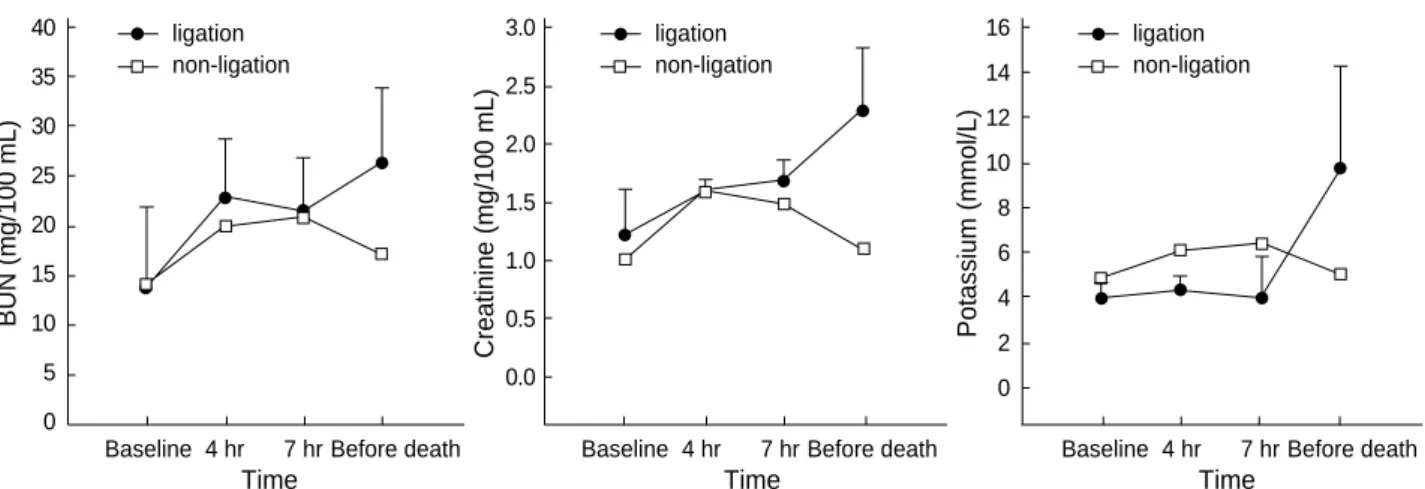
Fig. 3.Evolution of BUN, creatinine, and potassium (mean±SD) before clamping, after 4 hr and 7 hr since the first clamping, and before death (in non-ligation group, 20 hr after clamping).
BUN (mg/100 mL)
40 35 30 25 20 15 10 5 0
Baseline 4 hr 7 hr Before death Time
ligation non-ligation
Creatinine (mg/100 mL)
3.0 2.5 2.0 1.5 1.0 0.5 0.0
Baseline 4 hr 7 hr Before death Time
ligation non-ligation
Potassium (mmol/L)
16 14 12 10 8 6 4 2 0
Baseline 4 hr 7 hr Before death Time
ligation non-ligation
than 7 days.
Clinical assessment
The animals in the non-ligation group did not show marked behavioral abnormalities. The first abnormality observed in
the ligation group was usually ataxic gait and impaired bal- ance. And then drowsiness, a decrease in pain sensations, and the posture like walking in the air spontaneously at supine position were followed in 10 hr after ligating the hepatic artery.
The loss of sensation, absence of light reflex, and nonreactive coma developed after 10 hr since ligating the hepatic artery.
Biochemical assessment
Metabolic acidosis was observed after intermittent clamp- ing of the portal vein and the hepatic artery, but it was cor- rected by administration of sodium bicarbonate. In the non- ligation group, the levels of ammonia, lactic acid, AST, ALT, and creatinine gradually declined after showing increased val- ues initially, whereas BUN and potassium remained within the normal range. In the ligation group, the levels of ammo- nia, lactic acid, and creatinine showed a progressively increas- ing pattern. The levels of ammonia, lactic acid, total biliru- bin, and BUN increased after 4 hr of hepatic ligation. How- ever, the difference were not significant statistically. The in- crease of AST, ALT, creatinine, and potassium was not sig- nificant statistically, either. PT recovered normal range after slight prolongation in the non-ligation group, but it was gra- dually prolonged in the ligation group (Fig. 1-4).
Fig. 5. (A) Hematoxylin-eosin-stained specimen before clamping the portal triad in pigs (×100). Image shows normal portal tract in the pig liver. (B) Specimen after 4 hr since the first clamping in the non-ligation group (×100). Widened portal spaces with infil- tration of a few inflammatory cells and ischemic changes of hepa- tocytes in porto-to portal space are shown but there are no necrot- ic hepatocytes. The sinusoidal spaces are dilated. (C) Specimen after 20 hr since the first clamping in the non-ligation group (×100).
There are no histologic differences from those of (B).
A B
C
Fig. 6.Hematoxylin-eosin-stained specimen after 4 hr since ligat- ing the hepatic artery in pigs (×100). The findings are similar to those of the non-ligation group but, cytoplasmic condensation and unclear pyknosis in focal areas are found.
Histopathology
The histologic features in the non-ligation group which were checked before clamping, after 4 hr and 20 hr since the first clamping, respectively were shown in Fig. 5. There were no histologic differences: widened portal spaces with infiltra- tion of a few inflammatory cells, ischemic changes of the hepa- tocytes in porto-to-portal space, and dilated sinusoidal spaces.
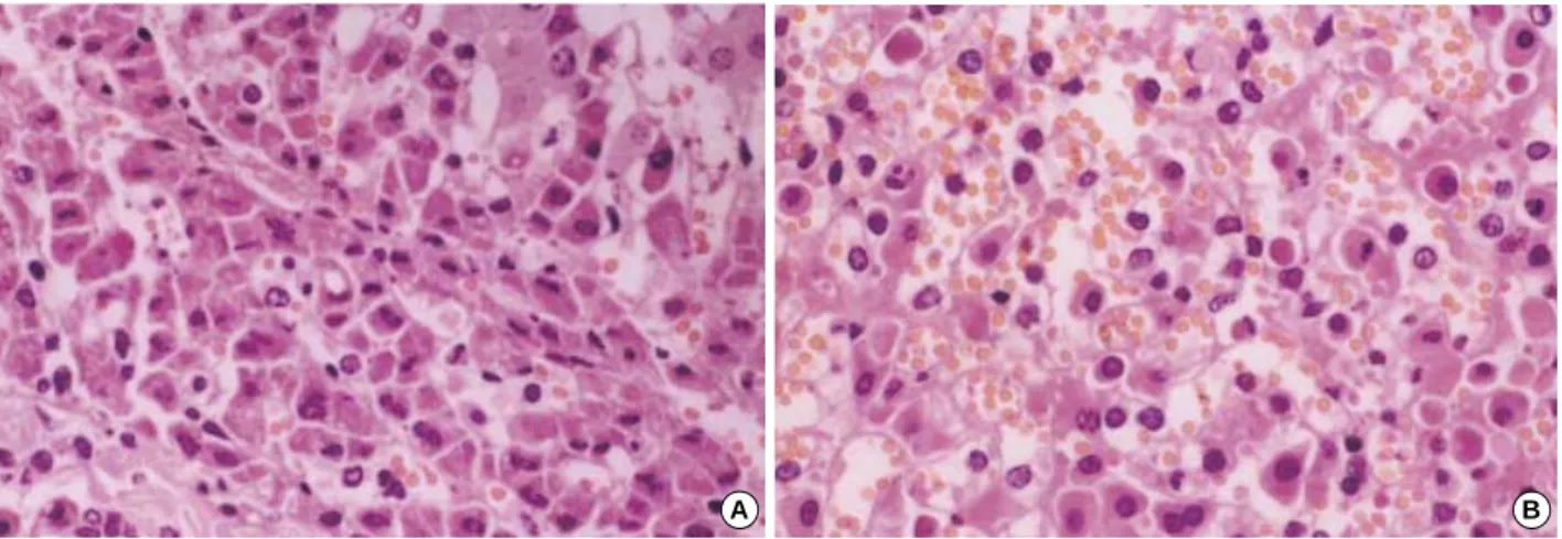
The findings obtained after 4 hr since ligating the hepatic artery (Fig. 6) were similar to those of the non-ligation group but nuclear pyknosis and cytoplasmic condensation were found in focal area. At autopsy, the histologic features mentioned above were also shown and furthermore parenchymal hem- orrhage, necrosis, nuclear pyknosis and cytoplasmic conden- sation in a multifocal area were shown (Fig. 7).
DISCUSSION
In the orthotopic liver transplantation, transient ischemia and reperfusion injury is one of the reasons of graft failure (13). Nitta et al. (16) reported that impairment of hepatic energy metabolism by intermittent portal triad cross-clamp- ing is mainly due to reinflow of pooled-portal blood to the previously ischemic liver rather than hepatic warm ischemia.
Li et al. (17) demonstrated that in the simple model of ortho- topic hepatic autotransplantation in case of dogs, the exten- sion of hepatic arterial ischemic time showed obvious ultra- microstructural changes in the liver and biliary duct, and the levels of serum bilirubin and ALT were elevated. The ischemia and reperfusion injury was represented in this model and splanchnic venous congestion was resolved by declamping the portal triad. Although several investigators verified that ischemia and reperfusion injury was the main cause of hep- atic failure in the experimental animal studies, additional hepatic artery ligation was needed to develop hepatic failure
in our model.
Terblanche et al. (4) suggested that five criteria should be met for a good animal model of hepatic failure: 1) reprodu- cibility, 2) potential reversibility, 3) the agent used to pro- duce liver failure should cause selective liver damage, 4) use of a large animal, and 5) the lack of relevant biohazard. In the present study, the surgical experimental model of liver fail- ure was reproducible. It has supported original anatomy of the portal vein and bile duct systems. Therefore liver regen- eration can be possible if a liver assist device is applied. Hep- atic coma was induced by massive hepatocytes necrosis and death could be attributed to progressive liver failure. The mor- tality rate was 100% when the hepatic artery was ligated after the intermittent clamping and the declamping of the portal triad. The period before death was long enough to allow the use of liver assist devices. Therefore, all requirements for ideal hepatic failure model except reversibility are fulfilled in this study.
In our model, the plasma level of ammonia increased, al- though it was not significant statistically. Ammonia is a neu- rotoxin which may precipitate hepatic encephalopathy. In the brain, ammonia may induce changes of blood-to-brain trans- portation of amino acids and alterations of brain energy meta- bolism. Furthermore, ammonia directly alters neuronal electric activity by inhibiting the generation of both excitatory and inhibitory post-synaptic potentials (18). Hyperammonemia is typical of fulminant hepatic failure and it indicates an impaired detoxification system.
In the ligation group, the levels of total bilirubin, AST and ALT increased progressively. Lactic acid increased abruptly and then progressively. It suggested a failure of hepatic clear- ance of peripherally generated lactic acid in the shock state (8). PT was gradually prolonged in the ligation group. Coagu- lopathy occurred as a result of decreased synthesis of clotting factors and disseminated intravascular coagulation (19). The transient increase in the level of blood glucose after clamp-
Fig. 7. Hematoxylin-eosin-stained specimens at autopsy in the ligation group (×400). The histologic features mentioned at Fig. 5B and Fig. 6 are also shown. In addition parenchymal hemorrhage, necrosis, nuclear pyknosis and cytoplasmic condensation in multifocal areas are shown.
A B
ing the portal vein and the hepatic artery in two groups was considered as a stress reaction. And it was believed for the increase to be caused by the mobilization of glycogen stores.
Hypoglycemia which is a characteristic of fulminant hepat- ic failure was seen in the ligation group. It has been shown from the result of inhibiting both gluconeogenesis and glu- cose release from the liver into the blood stream and failure of insulin metabolism (11). But hypoglycemia is not a major factor to bring on coma or death.
This model also showed features of hepatic encephalopa- thy by inducing massive necrosis of hepatocytes and releas- ing the toxic by-products into systemic circulation through the portal vessel and bile duct systems. In the ligation group, except for the first animal, the other four pigs showed the posture like spontaneously walking in the air at supine posi- tion. The animals lost pain sensation for some hours before death and even pupils did not respond to light for some hours before death. This neurological changes are similar to those of fulminant hepatic failure model in pigs induced by D- galactosamine or graded devascularization (5, 9).
In conclusion, our new model is similar to the hepatic fail- ure in human beings according to the clinical, laboratory and pathologic findings. It may be the advantage of this model that we can make it technically easily, which makes it highly reproducible. To develop fulminant hepatic failure, it is nec- essary not only to induce the ischemia and reperfusion injury to the liver parenchyma by intermittent clamping and declam- ping the portal triad, but also to ligate the hepatic artery. This model can be used to test the efficacy of liver assist devices.
ACKNOWLEDGEMENT
We are grateful to Ja June Jang M.D. for his help with the histological analysis and his many comments.
REFERENCES
1. Lee WM. Acute liver failure. Am J Med 1994; 96: S3-9.
2. Adam R, Cailliez V, Majno P, Karam V, McMaster P, Caine RY, O’Grady J, Pichlmayr R, Neuhaus P, Otte JB, Hoeckerstedt K, Bis- muth H. Normalised intrinsic mortality risk in liver transplantation:
European Liver Transplant Registry study. Lancet 2000; 356: 621-7.
3. Lee WM. Acute liver failure. N Engl J Med 1993; 329: 1862-72.
4. Terblanche J, Hickman R. Animal models of fulminant hepatic fail- ure. Dig Dis Sci 1991; 36: 770-4.
5. Benoist S, Sarkis R, Baudrimont M, Delelo R, Robert A, Vaubour- dolle M, Balladur P, Calmus Y, Capeau J, Nordlinger B. A reversible model of acute hepatic failure by temporary hepatic ischemia in the pig. J Surg Res 2000; 88: 63-9.
6. de Groot GH, Reuvers CB, Schalm SW, Boks AL, Terpstra OT, Jeekel H, ten Kate FW, Bruinvels J. A reproducible model of acute hepatic failure by transient ischemia in the pig. J Surg Res 1987; 42: 92-100.
7. Filipponi F, Fabbri LP, Marsili M, Falcini F, Benassai C, Nucera M, Romagnoli P. A new surgical model of acute liver failure in the pig:
experimental procedure and analysis of liver injury. Eur Surg Res 1991; 23: 58-64.
8. Sielaff TD, Hu MY, Rollins MD, Bloomer JR, Amiot B, Hu WS, Cerra FB. An anesthetized model of lethal canine galactosamine ful- minant hepatic failure. Hepatology 1995; 21: 796-804.
9. Kalpana K, Ong HS, Soo KC, Tan SY, Prema Raj J. An improved model of galactosamine-induced fulminant hepatic failure in the pig.
J Surg Res 1999; 82: 121-30.
10. Diaz-Buxo JA, Blumenthal S, Hayes D, Gores P, Gordon B. Galac- tosamine-induced fulminant hepatic necrosis in unanesthetized canines.
Hepatology 1997; 25: 950-7.
11. Kelly JH, Koussayer T, He DE, Chong MG, Shang TA, Whisennand HH, Sussman NL. An improved model of acetaminophen-induced fulminant hepatic failure in dogs. Hepatology 1992; 15: 329-35.
12. Mourelle M, Villlalon C, Amezcua JL. Protective effect of colchicine on acute liver damage induced by carbon tetrachloride. J Hepatol 1988; 6: 337-42.
13. Starzl TE, Demetris AJ. Liver transplantation: a 31-year perspec- tive. Part I. Curr Probl Surg 1990; 27: 49-116.
14. Nordlinger B, Douvin D, Javaudin L, Bloch P, Aranda A, Boschat M, Huguet C. An experimental study of survival after two hours of normothermic hepatic ischemia. Surg Gynecol Obstet 1980; 150:
859-64.
15. Harris KA, Wallace AC, Wall WJ. Tolerance of the liver to ischemia in the pig. J Surg Res 1982: 33: 524-30.
16. Nitta N, Yamamoto S, Ozaki N, Morimoto T, Mori K, Yamaoka Y, Ozawa K. Is the deterioration of liver viability due to hepatic warm ischemia or reinflow of pooled-portal blood in intermittent portal triad cross-clamping? Res Exp Med (Berl) 1988; 188: 341-50.
17. Li YM, Qin ZY, Kang YA, Ji ZZ, Yang WB, Xiang GA, Fang ZW.
Injurious effects of hepatic arterial ischemia on hepatic and biliary ductal tissues in canine liver autografts. Chin J Organ Transplant 1996; 17: 28-30.
18. Ferenci P. Brain dysfunction in fulminant hepatic failure. J Hepatol 1994; 21: 487-90.
19. Hillenbrand P, Parbhoo SP, Jedrychowski A, Sherlock S. Significance of intravascular coagulation and fibrinolysis in acute hepatic failure.
Gut 1974; 15: 83-8.