INTRODUCTION
Panax ginseng has the noticeable effects to protect liver and to elevate immune function. In recent years, it was proven that ginseng had anticarcinogenic effects on transferred tumors, cultured hepatoma cells, animal and human cancer (1-6). This paper describes anticarcinogenic effect of red gin- seng on the development of liver cancer induced by diethyl- nitrosamine (DEN) in rats particularly in the preventive and curative aspects.
MATERIALS AND METHODS Experiment of preventive group
33 Wistar white rats, weighing 130-180 g, were random- ly divided into experimental and control groups. In first 15 weeks, the rats in the experimental group were given 10%
DEN water solution (70 mg/kg per week) concomitantly with red ginseng fluid (Panax ginseng C.A. Meyer cultivated in JiLin, China, 3.78 g/kg per week) via gastric tube; the rats in the control group were given only 10% DEN water solu- tion (70 mg/kg per week). In the following 8 weeks, the rats
in the experimental group were continuously given red gin- seng fluid. The total amounts of DEN and red ginseng given to each rat were 180-200 mg and 17-25 g, respectively. In the 49th day and 103rd day, 5 rats each from the experimen- tal and control groups were sacrificed. In the 161st day, 7 rats in the experimental group and 6 rats in the control group were sacrificed. The liver cancer morphology and its histo- chemical changes of the rats in the two groups were studied by paraffin section, HE stain, Feulgen reaction, methyl green pyronine and periodic acid-Schiff (PAS) stain. Some fresh liver tissues were taken and processed by freezing section for SDH, 5′-NT and -GT. DNA, RNA, glycogen, -GT and SDH were detected, and measured with Univar microspec- trometer, and analyzed statistically.
The experiment of curative group
From 60 Wistar male rats, weighing 140-200 g, 40 rats were randomly taken and given 10% DEN water solution (70 mg/kg per week) via gastric tube for 15 weeks. Another 20 rats served as the control group, and were randomly divid- ed into null control group and red ginseng control group.
In the 20th week, all 60 rats were operated with laparoto- my. It was found that 20 of the 40 rats treated with DEN
Xiu-gan Wu, Da-he Zhu, Xun Li
Department of Pathology, Tongji Medical University, Wuhan, China
Address for correspondence Xiu-gan Wu, M.D.
Department of Pathology, Tongji Medical Universi- ty, Wuhan Apt. 407, Dong-cui-nan Street 7 Hai-lian Road Guangzhou 510230 P.R. China
Tel : 86(20)-3429 4545
E-mail : [email protected]
S61
Anticarcinogenic Effect of Red Ginseng on the Development of Liver Cancer Induced by Diethylnitrosamine in Rats
Anticarcinogenic effect of red ginseng (Panax ginseng C.A. Meyer cultivated in JiLin, China) on the development of liver cancer induced by diethylnitrosamine (DEN) in rats was studied, especially in preventive and curative groups. In the preventive group, the rats were given with DEN concomitantly with red ginseng fluid, and in the curative group, the rats were administered with red ginseng fluid after they developed liver cancer nodules induced by DEN. The result of the preventive group revealed that the developmental rate of liver cancer in the experimental group was 14.3%, while 100% in the control group, with the differ- ence being statistically significant. DNA, RNA, glycogen, -GT, SDH, and 5′-NT were maintained at relatively normal level in experimental group, and decreased or increased in the control group. The result of curative group showed that hep- atoma nodules of the DEN-red ginseng group I were smaller than those of con- trol group I, the structure of hepatic tissue was well preserved, the area with - GT positive was smaller, and the ultrastructure of hepatocytes was normal. The average life span the DEN-red ginseng group II and the DEN control group II were 72.8 and 42.3 days, respectively. To sum up, all findings on preventive and curative groups had clearly proved that the red ginseng had the anticarcino- genic effect on the development of liver cancer induced by DEN in rats.
Key Words : Ginseng; Diethylnitrosamine; Liver Neoplasms; Anticarcinogenic Agents
group were administered with tap water. In the 29th week, the rats in the above four groups were all sacrificed, and the body and liver were weighed. The fresh liver tissues about 1 mm3and 5×5×2 mm were taken from the low edge of middle or left lobes and fixed with 3% amyldialdehyde buffer fluid and 1:1 alcohol-acetone. The rest of liver was fixed with 10% formalin, serially cut in 2 mm thick, simulated the sec- tion surface of liver and cancer nodules with transparent paper was measured with Leitz Asm 68K Image Analysis, and the total size of the cancer nodules was calculated according to the formula of size=area×thick. The liver tissues fixed with alcohol-acetone were sectioned and stained for -GT, scanned in 4 continuous field per section with Univar-mspm Micro- spectrophotometer, and the average -GT positive area per unit area was calculated. The liver tissues fixed with amyl- dialdehyde buffer fluid were embedded in Epon812 for elec- tromicroscope analysis. The latter 19 rats with liver cancer nodules over 3 mm diameter were randomly divided into DEN-red ginseng group II with 10 rats and DEN control group II with 9 rats. At the beginning of the 21st week, the rats in the DEN-red ginseng group II were administered
The developmental rate of hepatoma
All the rats sacrificed on the 49th day and the 103rd day were found not to have liver cancer developed. But on the 161st day, all 6 rats in the control group which were admin- istered with the total average amount of DEN 200 mg per rat developed heptoma, and developmental rate was 100%.
At the meantime, only 1 of 7 rats in the experimental group which were administered with the total average amount of DEN 196 mg and ginseng 19.6 g per rat developed hep- atoma, and developmental rate was 14.3%. The difference in the developmental rates of hepatoma is highly significant between the two groups (p<0.001).
Morphologic change
The liver surface of all the rats sacrificed on the 49th day was smooth, mild in degeneration, necrosis and prolifera- tion of hepatocytes. In the 103rd day, the liver surface of the rats in the experimental group remained smooth, but the
Fig. 1.There was only one liver cancer nodule (♀4) at the later stage in the Experimental Group.
Fig. 2.Hepatic glycogen of the liver cancer cells, revealed with PAS reaction, disappeared completely (×400).
control group showed severe degeneration, necrosis, prolif- eration of hepatocytes and connective tissue in portal areas, and granular cirrhosis. On the 161st day, the liver surface of all rats in the control group was found with granular cirrho- sis and liver cancer nodules in total quantity of up to 45, and the size of the cancer nodules were 0.5-1.5 cm in diam-
eter. But only one cancer nodule with about 0.4 cm in diam- eter was found in the experimental group (Fig. 1).
Histochemical change
The details of histochemical changes are shown in Table 1
DNA PAS -GT SDH
Groups
Amount in average Nuclear area in average Amount in average Amount in average Amount in average
Nomal group 1.4 30.5 1.1 5.3 2.5
Exp. group 2.40±0.4641 3.37±6.3021 3.2±0.7742 6.8±0.8981 3.3±0.8361
Control group 6.6±0.5308 72.3±5.9641 0.1±0.0391 13.0±1.0132 0.7±0.1384
Table 2.Quantitative histochemical changes in rat liver on the 161st day
Control group was compared with normal and experimental groups, both of them with p<0.01.
Degree of histochemical changes in rats Groups (cases)
+ ++ +++ + ++ +++ - + ++ +++ + ++ +++ - + ++ +++ ± + ++ +++
DNA RNA PAS -GT SDH 5′-NT
Early stage: nomal group 1 1 1 1 1 1
exp. group 5 5 4 1 5 3 2 4 1
control group 5 5 1 3 1 1 4 3 2 2 3
middle stage: normal group 1 1 1 1 1 1
exp. group 5 5 4 1 3 2 1 4 3 2
control group 5 5 2 2 2 3 2 3 1 4
Later stage: normal group 3 3 3 3 3 3
exp. group 5 1 1 4 2 1 1 1 5 5 1 1 1 6 1 5 1
control group 6 6 6 6 3 1 4
Table 1.Hitochemical changes in the liver in rats (qualitative evaluation)
Note: “-”negative, “±”weak positive, “+”positive, “++”middle, “+++”strong positive early, middle and later stages were the lesion in the 49th, 103rd and 161st day after administration with DEN and red ginseng.
Fig. 3.Hepatic glycogen of the hepatic cells with PAS reaction is apparent (×400).
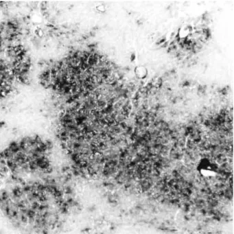
Fig. 4. -GT stain is strongly positive in the liver cancer nodule of the control group (×100).
and Table 2. Feulgen reaction and methyl green pyronine stain revealed that the stain of liver cancer tissues was stronger than that of hepatic tissues. The average amounts of DNA and area of nuclei of liver tissue in the experimental group were apparently lower than those in the control group (p<
0.01). PAS stain was negative in the cancer tissues, and pos- itive in hepatic and proliferative hepatic cells (Fig. 2, 3).
Cancer nodules were strongly positive in -GT stain, but the liver tissues of the experimental group were essentially normal (Fig. 4, 5). The activity of SDH and 5′-NT in can- cer tissues were apparently decreased or disappeared.
The result of curative group Statistics
Comparing the ratio of liver weight to body weight, there was no significant difference between ginseng control group and null control group and between DEN-ginseng group I and null control group. But the obvious increase between DEN-control group I and null control group (p<0.01), and apparent decrease between DEN-ginseng group I and DEN- control group I (p<0.01). Morphometry revealed that the total size of liver cancer nodules in each rat of DEN-ginseng group I was 5.76±2.81 L, apparently smaller than that of DEN control group I (43.81±42.46 L, p< 0.05). The result of microspectrophotometer showed that -GT positive areas per unit area of the DEN-ginseng group I was 0.10±0.12 mm2, significantly smaller than the DEN control group I (0.27±0.15 mm2, p<0.05). The life spans of the DEN- gin- seng group II were 72.80±31.40 days, significantly pro-
longed than the DEN- control group II (42.37±24.42 days, p<0.05).
Microscopic findings
Liver tissue surrounding cancer in the DEN- control group I was severely changed, and showed fat degeneration and focal hydropic degeneration, but they were maintained essen- tially normal and seldom saw fat degeneration and hydropic degeneration in the DEN-ginseng group I. In addition, the hepatic cells around cancer in both two groups showed sim- ple or atypical proliferation, and the proliferations were dif- ferent in morphology, but both were positive in -GT stain.
The ultrastructural changes
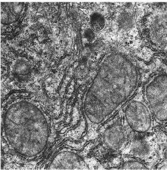
On the 20th week, the nuclear membrane of hepatocytes was not smooth, nucleoli were larger and increased in quan- tity, mitochondria increased and were different in size and form, fat drop increased, and the contact surface of hepato- cytes was widened. On the 29th week, the hepatocytes of the DEN-control group I revealed severe atypia, large nucleus and dental nuclear membrane, chromatin increased and coag- ulated pieces, mitochondria degeneration, rough reticuloen- doplasma decreased, microvilli in sinus surface of hepatocytes decreased and endothelium was defective. But on the 29th week, the form of hepatic nuclei of the DEN-ginseng group I was maintained regular and intact, chromatin increased but not prominent, mitochondria increased and became larger with the intact structure, rough reticuloendoplasmic reticu-
Fig. 5. -GT stain was focally weak positive in the hepatic tissue of the experimental group (×100).
Fig. 6.Mitochondria increased and were large in size and rough endoplasmic reticula and Golgi apparatus well devel- oped, under electron microscopy (×10,000).
lum developed, and Golgi apparatus was normal (Fig. 6).
DISCUSSION
The results of the preventive group revealed that red gin- seng had significantly inhibited the development of liver cancer induced by DEN in rats. In the early stage, degener- ation, necrosis, and proliferation of hepatocytes and cirrhosis of the experimental group were less than those of the control group. In the later stage, the developmental rate of liver cancer was 14.3% in the experimental group, and 100% in the control group. The difference between the two groups was statistically significant. DNA, RNA, glycogen, -GT, SDH, 5′-NT were maintained at relatively normal level in the experimental group, and decreased or increased in the control group. These findings suggest that the ginseng had a role of protecting the hepatocytes from injury by DEN, thereby inhibiting the development of liver cancer induced by DEN.
The result of the curative group displayed that the hepatoma nodules of the DEN- red ginseng group I were smaller than those of the DEN control group I. The structure of hepatic tissue was normal, the -GT positive area was smaller, and the ultrastructure of hepatocytes was essentially normal. These findings suggest that the red ginseng could decrease the developmental speed of precancerous lesions and cancer, pro- tect the ultrastructure of hepatocytes, therefore prolonging
the life spans of the rats with cancer.
To sum up, all findings on preventive and curative groups had proved clearly that the red ginseng has anticarcinogenic effect on the development of liver cancer induced by DEN in rats.
REFERENCE
1. Yun TK, Yun YS, Han IW. Anticarcinogenic effect of long-term oral administration of red ginseng on newborn mice exposed to various chemical carcinogens. Cancer Detect Prev 1983; 6: 515-25.
2. Hikino H, Kiso Y, Kinouchi J, Sanada S, Shoji J. Antihepatotoxic actions of ginsenosides from Panax ginseng roots. Planta Med 1985;
51: 62-4.
3. Wang BX. Pharmacological basis of ginseng treating the liver dis- eases. Jilin Medicine 1985; 6: 48-50 (Chinese).
4. Lie SG. Antitumorous effects of ginseng. J Trad Chinese Med 1982;
11: 1(Chinese).
5. Abe H, Arichi S, Hayashi T, Odushima S. Ultrastructural studies of Morris hepatoma cells reversely transformed by ginsenosides. Experi- entia 1979; 35: 1647-9.
6. Odashima S, Nakayabu Y, Honjo N, Abe H, Arich S. Induction of phenotypic reverse transformation by ginsenosides in cultured Mor- ris hepatoma cells. Eur J Cancer 1979; 15: 885-92.
7. Wu XG, Zhu DH. Influence of ginseng upon the development of liver cancer induced by diethylnitrosamine in rats. J Tongji Med Univ 1990; 10: 141-5.