INTRODUCTION
Antioxidant enzymes such as superoxide dismutase, cata- lase, glutathione peroxidase, heat shock protein and heme oxygenase-1 (HO-1) are induced by various stimuli, and they represent a powerful endogenous protective mechanism that act within the pancreatic islets against free radicals (1, 2).
HO-1 catalyzes the rate-limiting step in heme catabolism and it generates CO and bilirubin, and bilirubin has been demon- strated as a potent antioxidant (3). In addition, HO-1 has been described as an inducible protein that is capable of cyto- protection via radical scavenging and preventing apopotosis.
Glucose toxicity is defined as nonphysiological and poten- tially irreversible cellular damage that results in defective insulin gene expression, and this is caused by chronic expo- sure to supraphysiologic glucose concentrations (4-9). The beta cell in type 2 diabetes is also adversely affected by chronic hyperglycemia and, in this sense, is also a target for secondary complications. As hyperglycemia worsens, the beta cell steadi- ly undergoes deterioration, secretes less and less insulin, and becomes a participant in a downward spiral of loss of func- tion (9). Recently, the overexpression of antioxidant gene
products has been induced in the islets, and along with using antioxidant drugs, this helps to protect against oxidative stress (10-14). However, the relation between glucose toxicity and HO-1 in the islets is still not known completely.
Thus, we would like to determine whether prolonged expo- sure of the pancreatic islets to a supraphysiologic glucose con- centration disrupts the intracellular balance between reactive oxygen species (ROS) and HO-1, and if this causes defective insulin secretion; we also wanted to evaluate a protective role for HO-1 against high glucose concentrations in the pancre- atic islets.
MATERIALS AND METHODS INS-1 cell culture
The INS-1 cells (15) were grown in 5% CO2-95% air at 37℃in RPMI-1640 medium containing 11.1 mM pyru- vate, 10 mM HEPES, 50 M 2-mercaptoethanol, 100 U penicillin/mL and 100 g streptomycin/mL. The RPMI-1640 medium used in all the experiments contained the supple-
Kyu Chang Won, Jun Sung Moon, Mi Jung Eun, Ji Sung Yoon, Kyung Ah Chun*, Ihn Ho Cho*, Yong Woon Kim�, Hyoung Woo Lee
Department of Internal Medicine, Department of Nuclear Medicine*, Department of Physiology�, College of Medicine, Yeungnam University, Daegu, Korea
Address for correspondence Kyu Chang Won, M.D.
Department of Internal Medicine, College of Medicine, Yeungnam University, 317-1 Daemyung-dong, Nam-gu, Daegu 705-717, Korea
Tel : +82.53-620-3846, Fax : +82.53-654-8386 E-mail : [email protected]
*This study was supported by a Grants of Yeungnam University Medical Center.
418
A Protective Role for Heme Oxygenase-1 in INS-1 Cells and Rat Islets that are Exposed to High Glucose Conditions
Heme oxygenase-1 (HO-1) has been described as an inducible protein that is capa- ble of cytoprotection via radical scavenging and the prevention of apoptosis. Chronic exposure to hyperglycemia can lead to cellular dysfunction that may become irre- versible over time, and this process has been termed glucose toxicity. Yet little is known about the relation between glucose toxicity and HO-1 in the islets. The pur- poses of the present study were to determine whether prolonged exposure of pan- creatic islets to a supraphysiologic glucose concentration disrupts the intracellular balance between reactive oxygen species (ROS) and HO-1, and so this causes defective insulin secretion; we also wanted to evaluate a protective role for HO-1 in pancreatic islets against high glucose levels. The intracellular peroxide levels of the pancreatic islets (INS-1 cell, rat islet) were increased in the high glucose media (30 mM glucose or 50 mM ribose). The HO-1 expression was induced in the INS-1 cells by the high glucose levels. Both the HO-1 expression and glucose stimulated insulin secretion (GSIS) was decreased simultaneously in the islets by treatment of the HO-1 antisense. The HO-1 was upregulated in the INS-1 cells by hemin, an inducer of HO-1. And, HO-1 upregulation induced by hemin reversed the GSIS in the islets at a high glucose condition. These results suggest HO-1 seems to medi- ate the protective response of pancreatic islets against the oxidative stress that is due to high glucose conditions.
Key Words : HMOX1 protein, human; HO-1; Glucose Toxicity; Oxidative Stress; Islets of Langerhans
Received : 5 August 2005 Accepted : 4 November 2005
ments described above. The cells were passaged weekly after they has been detached with trypsin-EDTA. All the studies were performed on INS-1 cells that were between passages 21 and 29.
Pancreatic islet isolation
The pancreata from male Wistar rats were infused with 10 mL of 1.5 mg/mL collagenase type XI (Sigma, St. Louis, MO, U.S.A.)/1% fetal bovine serum/2 units/mL RQ1 DNase (Promega, U.S.A.) solution in Medium 199 (Sigma). After it was surgically excised, the pancreas was incubated in the collagenase solution at 37℃. The undigested tissue was re- moved by using a 500 m screen, and the recovered tissue was washed twice with ice-cold Hanks’ balanced salt solution containing 0.1% bovine serum albumin; this was followed by centrifugation at 250×g for 4 min. The islets in the pellet were separated by using a Histopaque-1077 gradient (Sigma), and then islets were hand-picked and cultured in RPMI medi- um 1640 containing 10% fetal bovine serum, 11.1 mM glu- cose and penicillin/streptomycin/amphotericin B before the experimentation.
In vitro induction of HO-1
Hemin (Sigma) was dissolved in 0.1N NaOH and it was diluted 1:1 in phophate-buffered saline (PBS); the pH was adjusted to 7.4 and the solution was sterilized by filtration.
The INS-1 cell or islets were incubated at 37℃for 24 hr with the selected concentration of hemin.
Evaluation of ROS with flow cytometry
The intracellular peroxide levels (16) were detected by flow cytometric analysis with using an oxidation-sensitive fluo- rescein-labeled dye, carboxylated dichlorodi-hydrofluores- cein diacetate (carboxy-H2DCFDA, Molecular Probes, Carls- bad, CA, U.S.A.). Upon oxidation by intracellular ROS, the non-fluorescent dye is converted into its fluorescent form.
The islets and INS-1 cells were labeled with 100 M car- boxy-H2DCFDA for 1 hr at 37℃. Following the cell load- ing of the dye, the islets were washed twice with PBS and then put back into culture conditions for 2 hr. The islets and INS-1 cells were then harvested, washed twice with PBS and resuspended in trypsin-EDTA (0.25% trypsin, 2 mM Na4-EDTA, Invitrogen) for 5 min at 37℃. To disperse the islets into a single cell suspension, the islets and INS-1 cells were gently passed 20 times in and out of a 200-1,000- L tip. The cells were then washed twice with ice-cold PBS. The pellet was then resuspended in ice-cold PBS, and 2 g/mL propidium iodide was added. The cells were analyzed using a 488 nm argon laser EPICS XL-MCL flow cytometer that was controlled by EXPO 32-ADC software (Beckman Coul- ter, Fullerton, CA, U.S.A.). The ROS values were analyzed
using a histogram plot of carboxy-H2DCFDA (the log of the fluorescence). The results were calculated as the fold differ- ence from the untreated control cells.
Glucose stimulated insulin secretion (GSIS)
Static incubation of the islets in Krebs-Ringer buffer that contained either non-stimulatory or stimulatory concentrations of glucose was performed for 1 hr (4). The insulin levels in the Krebs-Ringer buffer samples collected from the static incuba- tions (5.6 mM and 30 mM glucose conditions) from the islets and INS-1 cells by using a 95.5 ethanol:hydrochloric acid solu- tion were measured by using a sensitive rat insulin radioimmu- noassay kit (Linco Research Immunoassay, St. Charles, MO, U.S.A.).
HO activity assay
The HO activity was assayed by preparing a cell homogenate from the INS-1 cells that were treated with hemin. The cell homogenate was incubated with 50 M heme, 2 mg/mL rat liver cytosol (the source of the biliverdin reductase), 1 mM MgCl2, 3 U of glucose 6-phosphate dehydrogenase, 1 mM glucose 6-phosphate, and 2 mM NADP+in 0.5 mL of 0.1 M potassium phosphate buffer, (pH 7.4) for 30 min at 37℃. Placing the tubes on ice terminated the reaction, and the bilirubin was extracted with chloroform as described by Wa- gener et al. (2). The amount of bilirubin generated was deter- mined by using a scanning spectrophotometer (Lambda 17 UV/VIS, Perkin-Elmer Cetus Instruments, Norwalk, CT, U.S.A.) and this was defined as the difference between the wavelengths 464 and 530 nm (the extinction coefficient for bilirubin was 40/mM/cm). The HO activity was expressed as the picomoles of bilirubin formed per milligram of INS-1 cell protein per hour.
HO-1 sense and antisense oligodeoxynucleotide (ODN) treatment
For inhibition of HO-1, pretreated HO-1 antisense ODNs (2.5 mg/mL) were transfected using a lipofection reagent to the INS-1 cells (35). The INS-1 cells at 50% confluence were washed three times with PBS and then they were cultured in medium containing 10% Nu-serum, which lacks nuclease activity (Collaborative Research Inc., Waltham, MA, U.S.A.), for 5 hr before the addition of the ODNs. N-[-(2,3-dioleoy- loxy)propyl]-N,N,N-trimethylammonium methylsulfate (DOTAP; Boehringer Manngeim Diagnostics, Indianapolis, IN, U.S.A.) was used as a vehicle for transfecting the cells with the ODNs. The proportions used were 2 g ODN/1 g DOTAP/mL of medium and the protocol used was described by the manufacturer. The phosphorothioated oligonucleotides derived from the rat HO-1 sequence were synthesized at the Life technologies, U.S.A.. The sense/antisense ODNs for the
HO-1 were directed against the flanking translation initia- tion codon in the human HO-1 cDNA. The antisense se- quence for the HO-1 was 5′-GCGCTCCATCGCGGG-3′, whereas the sense sequence for the HO-1 was 5′-CCCGC- GATGGAGCGC-3′and for HO-1 scramble, it was 5′-GG- CCCTCTACGGGCG-3′. Each ODN was phospho-rothioat- ed on the first three bases on the 3′end and then it was puri- fied by HPLC. The cells were incubated for 24 hr with the ODNs, and then the medium was replaced with fresh medi- um containing 10% fetal bovine serum; the cells were then incubated for 24 hr in the absence or presence of heme.
Western blot analyses
The pancreatic islets and the INS-1 cells are washed once in ice-cold PBS buffer. The cells were lysed in buffer (140 mM NaCL, 10 mM Tris pH 7.4, 1 mM CaCl2, 1 mM MgCl2, 10% glycerol, 1% Nonidet P-40 and 1×complete protease
inhibitors [Roche, Indianapolis, ID, U.S.A.]) and then homo- genized by sonication for 2 to 20 sec on ice. The cell homoge- nates are centrifuged at 12,000 rpm at 4℃for 10 min to pellet the insoluble material. The supernatant is used for Western blot analyses. The protein was determined by the bicinchonic acid (BCA) assay (Pierce, Rockford, IL, U.S.A.).
The protein was fractionated by SDS-polyacrylamide gel elec- trophoresis and then it was electroblotted to a nitrocellulose membrane. Nonspecific binding sites are blocked by nonfat dry milk for 1 hr at room temperature. The blots are then incubated with the specific primary antibodies against HO-1 at a dilution of 1:2,000-1:5,000 for 2 hr at room temperature;
this was followed by a 1 hr incubation period with the peroxi- dase-labeled secondary antibody at a dilution of 1:10,000.
The protein bands are visualized by chemiluminescence with using the NEN detection system.
Data analysis
All the values are expressed as means±SE. Analysis of variance and Student’s t-test were used for the statistical anal- ysis, and the differences between groups were considered to be significant at p values <0.05.
RESULTS
Intracellular peroxide levels in the islets at the high glucose condition
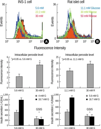
The INS-1 cells cultured for 3 days in glucose concentra- tions ranging from 5.6 to 30 mM had progressively greater peroxide levels with the higher glucose concentrations (Fig.
1A, p<0.05). The rat islets cultured for 3 days in 30 mM or 50 mM ribose had greater peroxide levels than that in the rat islets cultured in 11.1 mM glucose (Fig. 2B, p<0.05). In addition, the cells at higher glucose or ribose concentrations displayed a decreased GSIS (p<0.05).
HO-1 was induced in the INS-1 cells by the high glucose levels
The INS-1 cells were cultured for 3 days in 5.6 mM or 30 mM glucose concentrations. Compared with 5.6 mM glu- cose, 30 mM glucose caused an increase of the HO-1 expres- sion and activity in the INS-1 cells (Fig. 2, p<0.05).
HO-1 downregulation in the INS-1 cells by the HO-1 anti- sense
After 3 days culture (5 hrs exposure of the ODN) of the INS-1 cells at 5.6 mM or 30 mM glucose concentrations, the intracellular peroxide level, the HO-1 expression and the GSIS were measured. HO-1 and GSIS were decreased
90
100 101 102 103
INS-1 cell
5.6 mM 22.2 mM 30 mM
A
Events
36
0
100 101 102 103 104
Fluorescence Intensity
Rat islet cell
11.1 mM Glucose 30 mM Ribose 50 mM Ribose
B
Events
Fluorescence intensity
6 5 4 3 2 1 0
5.6 mM G 30 mM G Intracellular peroxide level
*p<0.05 vs. 5.6 mM G
Fig. 1.The effects of high glucose on the intracellular peroxide level and Glucose stimulating insulin secretion (GSIS) in the INS- 1 cells and rat islets. (A) INS-1 cells were incubated at 5.6, 22.2 or 30 mM glucose for 3 days. INS-1 cells incubated at 30 mM glu- cose increased levels of intracellular peroxides compared with the 5.6 mM concentration of glucose. (B) Isolated rat islets were incubated with 11.1 mM glucose or 30 mM ribose for 3 days. 30 mM ribose caused an increase of intracellular peroxide levels compared with the 11.1 mM glucose. Each cell at the high glucose or ribose concentrations showed decreased GSIS (p<0.05). Data are means±SD from 3 separate experiments.
Insulin secretion (U/mL)
1,200 1,000 800 600 400 200 0
5.6 mM G 30 mM G GSIS
4 mM G
Fluorescence intensity
25 20 15 10 5 0
11.1 mM G 30 mM R Intracellular peroxide level
*p<0.05 vs. 11.1 mM G
16.7 mM G
Insulin secretion (U/Islet)
1,000
800
600
400
200
0
11.1 mM G 30 mM G GSIS
5.6 mM G 16.7 mM G
*
*
*
simultaneously by treatment of the HO-1 antisense (Fig. 3, p<0.05), suggesting GSIS is associated with HO-1.
HO-1 upregulation in the islets by hemin
The INS-1 cells cultured for 3 days (with 1day pre-expo- sure of the hemin) in hemin concentrations ranging from 0.1 mM to 10 mM had progressively greater HO-1 levels with the higher hemin concentrations, and the cells had pro- gressively smaller peroxide levels with the higher hemin con- centrations (Fig. 4A, p<0.05). Similar results were also ob- tained in the rat islets (Fig. 4B, p<0.05).
HO-1 upregulation induced by hemin increased GSIS in the INS-1 cells at high glucose conditions
The GSIS in both the INS-1 cells and rat islets after 3 days subculture with high glucose concentration was increased in a dose-dependent manner by additional treatment of hemin for 1 day, which was associated with HO-1 upregulation induced by hemin (Fig. 5, p<0.05).
DISCUSSION
Glucose toxicity is defined as the nonphysiological and potentially irreversible cellular damage that results in defec- tive insulin gene expression, and this is caused by chronic exposure to supraphysiologic glucose concentrations (4-9).
With using HIT-T15 cells, Robertson et al. (4) observed that cells chronically cultured for 6 months in media containing 11.1 mM glucose, a concentration exceeding what is neces- sary to elicit maximal insulin responses, caused a marked loss of insulin mRNA, greatly diminished levels of the insulin content and almost a complete disappearance of insulin secre- tion. In contrast, the HIT-T15 cells of the same passage that were serially cultured in media containing 0.8 mM glucose for 6 months retained their insulin mRNA, insulin content and their glucose-induced insulin secretion (4). The concept of glucose autoxidation along with the consequent excess generation of ROS in relation to diabetes mellitus has been proposed as early as 1987 by Wolff and Dean (17). Hunt et al. (18) demonstrated that glucose autoxidation produces hydroxyl radicals and that the hydroxyl radical scavengers protected against the glucose-induced fragmentation of pro- tein. Earlier work by Grankvist et al. (19) demonstrated that the pancreatic islets contained relatively small amounts of
Fig. 2.The HO-1 expression and activity after 3 days subculture of the INS-1 cells. Compared with the 5.6 mM glucose concen- tration, 30 mM glucose caused an increase in the HO-1 expres- sion and activity in the INS-1 cells (p<0.05). Data are means±SD from 3 separate experiments.
Glucose concentrations
5.6 mM 5.6 mM 5.6 mM 30 mM 30 mM 30 mM
6 5 4 3 2 1 0
5.6 mM G 30 mM G
* HO-1 expression
6 5 4 3 2 1 0
5.6 mM G 30 mM G
* HO-1 activity
p<0.05 vs. 5.6 mM G
nmol bilirubin/mg protein/hrIntensity Fluorescence intensity
6 5 4 3 2 1
0 5.6 mM G 30 mM G Scramble Sense Antisense HO-1 expresion
*p<0.05 vs. 30 mM G
*p<0.05 vs. 30 mM G
Fig. 3.The intracellular peroxide level, HO-1 expression and GSIS after 3 days culture (5 hrs exposure to the ODNs) in the INS-1 cells.
HO-1 was downregulated in the INS-1 cells by the HO-1 antisense ODNs (p<0.05). Data are means±SD from 3 separate experi- ments.
*
6 5 4 3 2 1 0
5.6 mM G 30 mM G Scramble Sense Antisense 30 mM G Intracellular peroxide level *
30 mM G
Insulin secretion (mU/mL)
1,200 1,000 800 600 400 200
0 5.6 mM G 30 mM G Scramble Sense Antisense 4 mM G 16.7 mM G
*
antioxidant enzymes such as Cu, Zn-superoxide dismutase, Mn-superoxide dismutase, catalase and glutathione peroxi- dase. Oliveira et al. (20) reported that superoxide dismutase
activity increases with the increasing glucose concentration.
These observations set the stage for the increased risk for ROS- induced damage. We also found that the intracellular perox-
Fluorescense intensity
7 6 5 4 3 2 1 0
5.6 mM G 30 mM G 0.1 M 1 M 10 M Hemin INS-1 cells
HO-1 expression
Fig. 4.The intracellular peroxide level and the HO-1 expression and activity after 3 days subculture (1 day pre-exposure of Hemin) in the INS-1 cells (A) and rat islets (B). HO-1 was upregulated in the INS-1 cells and rat islets by Hemin (p<0.05). Data are means±SD from 3 separate experiments.
A
Fluorescense intensity
12 10 8 6 4 2 0
11.1 mM G 50 mM R 0.1 M 1 M 10 M Hemin Rat Islets
B
*
nmol bilirubin/mg protein/hr
9 8 7 6 5 4 3 2 1 0
5.6 mM G 30 mM G 0.1 M 1 M 10 M Hemin
HO-1 activity *
Fluorescense intensity
6 5 4 3 2 1
0 5.6 mM G 30 mM G 0.1 M 1 M 10 M Hemin HO-1 activity
*
*
Fluorescense intensity
25 20 15 10 5 0
11.1 mM G 50 mM R 0.1 M 1 M 10 M Hemin
*
*p<0.05 vs. 30 mM G Glucose
*p<0.05 vs. 50 mM G Ribose
30 mM G 50 mM R
Insulin secretion (mU/mL)
1,000
800
600
400
200
0
5.6 mM G 30 mM G 0.1 M 1 M 10 M Hemin
30 mM G A
* 4 mM G 16.7 mM G
*p<0.05
INS-1 Cell
Insulin secretion (mU/mL)
1,000
800
600
400
200
0
11.1 mM G 50 mM R 0.1 M 1 M 10 M Hemin
50 mM R B
*
5.6 mM G 16.7 mM G
*p<0.05
Rat Islets
Fig. 5.GSIS after 3 days subculture (1 day pre-exposure of Hemin) in the INS-1 cells (A) and rat islets (B). Hemin induced HO-1 upregula- tion and it reserved the GSIS in the islets at the high glucose condition (p<0.05). Data are means±SD from 3 separate experiments.
ide level was increased by a chronic exposure of high glucose conditions in both the INS-1 cells as well as rat islets, which was accompanying with a decreased GSIS (Fig. 1).
The heme oxygenases play critical roles in physiological iron homeostasis, antioxidant defense, and, as has been shown from the accumulating evidences, in the signaling pathways that employ CO as a messenger (21). Three mammalian iso- forms of HO have been identified: HO-1, an inducible en- zyme that is most highly concentrated in the tissues that are heavily involved in the catabolism of heme proteins (22);
HO-2, a non-inducible isoform that is present at the high- est concentrations in the brain and testes, and it is thought to be particularly involved in signaling pathways (23); and HO-3, an isoform with low catalytic activity and an uncer- tain physiological role (24). HO uses dioxygen and nicoti- namide-adenine dinucleotide phosphate as cofactors, and the resulting products of the reaction are carbon monoxide, iron and biliverdin (21). Biliverdin is converted to bilirubin by a ubiquitous cytosolic enzyme biliverdin reductase (22).
Both HO-2 and an inducible HO-1 have been identified in rat pancreatic islets (23-25), as well as in other tissues (26).
Pancreatic islets respond to stress through the induction and activation of several stress-activated proteins. Interleukin-1 (IL-1 ) induces an inflammatory response in pancreatic islets that is characterized by increased levels of inducible nitric oxide synthase (iNOS) and increased nitric oxide (NO)/nitrite levels (27-30). IL-1 and heat shock protein increase the expression of hsp70 (31, 32), as well as HO-1 (23, 33). The protective effect of heat shock protein (HSP) on the islet cells may be associated with the reduced lysis from NO, reactive oxygen intermediates and streptozotocin (32); but the response is nonspecific because many HSPs respond to these stimuli.
On the other hand, liposomal delivery of hsp70 into islet cells protected the cells from the IL-1 effects on insulin secretion (34), suggesting that heightened levels of specific HSP can protect cells from the inhibitory effects of the cytokine. Hemin induces the synthesis of HO-1 and so it partly counteracts the IL-1 induced inhibition of aconitase activity and glucose oxidation (33), and perhaps this occurs through antioxidant mechanisms. However, hemin has also been reported to increase insulin and glucagon secretion from normal rat islets (24). Ye and Laychock (1) reported that HO, which is also known as a heat shock protein (hsp 32), is an enzyme that may protect cells by reducing the heme levels that catalyze the oxygen radical reactions, and it appears to be a protective agent for pancreatic islets against interleukin- 1 . Similarly, our study has shown that compared with 5.6 mM glucose, 30 mM glucose caused an induction of HO-1 expression and activity in the INS-1 cells (Fig. 2, p<0.05).
The HO-1 expression increases in response to heme and to such stressors as UV radiation and oxidative stress, as well as to endotoxin, hormones and heavy metals (26). HO-1 induc- tion may protect cells by reducing the heme levels that cat- alyze oxygen radical reactions and by elevating bilirubin,
which has antioxidant properties (21). Bilirubin inhibits the autoxidation or peroxyl-radical-induced oxidation of the unsaturated fatty acids, and it apparently does so through its peroxyl radical-trapping antioxidant abilities (35-37). In the present study, we hypothesized that HO-1 can protect the suppression of GSIS resulted from glucose toxicity.
To evaluate our hypothesis, we measured GSIS after cul- tured with high glucose conditions in both HO-1 downreg- ulated state and upregulated state. We observed that the GSIS was decreased by treatment of HO-1 antisense ODNs, which was accompanying with a downregulation of HO-1 expression (Fig. 3, p<0.05). Moreover, the GSIS was increased incompletely by hemin administration associating with the upregulation of HO-1 expression and activity (Fig. 4, 5).
These results in this study supported our hypothesis. Thus, our results suggest that HO-1 seems to mediate the protec- tive responses of the pancreatic islets against the oxidative stress that is due to high glucose conditions. Also, we sug- gested that HO-1 is one of the targets for preserving islets from glucose toxicity in diabetic state.
REFERENCES
1. Ye J, Laychock SG. A protective role for heme oxygenase expres- sion in pancreatic islets exposed to interleukin-1 . Endocrinology 1998; 139: 4155-63.
2. Wagener FA, da Silva JL, Farley T, de Witte T, Kappas A, Abraham NG. Differential effects of heme oxygenase isoforms on heme medi- ation of endothelial intracellular adhesion molecule 1 expression. J Pharmacol Exp Ther 1999; 291: 416-23.
3. Pileggi A, Molano RD, Berney T, Cattan P, Vizzardelli C, Oliver R, Fraker C, Ricordi C, Pastori RL, Bach FH, Inverardi L. Heme oxy- genase-1 induction in islet cells results in protection from apoptosis and improved in vivo function after transplantation. Diabetes 2001;
50: 1983-91.
4. Robertson RP, Zhang HJ, Pyzdrowski KL, Walseth TF. Preservation of insulin mRNA levels and insulin secretion in HIT cells by avoid- ance of chronic exposure to high glucose concentration. J Clin Invest 1992; 90: 320-5.
5. Won KC. Oxidative stress in pancreatic islet beta-cells exposed to high glucose concentration. J Korean Diabetes Assoc 2004; 28: 250- 54.
6. Harmon JS, Stein R, Robertson RP. Oxidative stress-mediated, post- translational loss of MafA protein as a contributing mechanism to loss of insulin gene expression in glucotoxic beta cells. J Biol Chem 2005; 280: 11107-13.
7. Sharma A, Olson LK, Robertson RP, Stein R. The reduction of insulin gene transcription in HIT-T15 beta cells chronically exposed to high glucose concentration is associated with the loss of RIPE3b1 and STF-1 transcription factor expression. Mol Endocrinol 1995; 9: 1127- 34.
8. Harmon JS, Tanaka Y, Olson LK, Robertson RP. Reconstitution of glucotoxic HIT-T15 cells with somatostatin transcription factor-1
partially restores insulin promoter activity. Diabetes 1998; 47: 900-4.
9. Robertson RP. Chronic oxidative stress as a central mechanism for glucose toxicity in pancreatic islet beta cells in diabetes. J Biol Chem 2004; 279: 42351-4.
10. Tiedge M, Lortz S, Munday R, Lenzen S. Complementary action of antioxidant enzymes in the protection of bioengineered insulin-pro- ducing RINm5F cells against the toxicity of reactive oxygen species.
Diabetes 1998; 47: 1578-85.
11. Kralik PM, Xu B, Epstein PN. Catalase transfection decreases hydro- gen peroxide toxicity in a pancreatic beta cell line. Endocr Res 1998;
24: 79-87.
12. Tiedge M, Lortz S, Munday R, Lenzen S. Protection against the co- operative toxicity of nitric oxide and oxygen free radicals by overex- pression of antioxidant enzymes in bioengineered insulin-producing RINm5F cells. Diabetologia 1999; 42: 849-55.
13. Hohmeier HE, Thigpen A, Tran VV, Davis R, Newgard CB. Stable expression of manganese superoxide dismutase (MnSOD) in insuli- noma cells prevents IL-1 -induced cytotoxicity and reduces nitric oxide production. J Clin Invest 1998; 101: 1811-20.
14. Moriscot C, Pattou F, Kerr-Conte J, Richard MJ, Lemarchand P, Benhamou PY. Contribution of adenoviral-mediated superoxide dismutase gene transfer to the reduction in nitric oxide-induced cy- totoxicity on human islets and INS-1 insulin-secreting cells. Diabe- tologia 2000; 43: 625-31.
15. Asfari M, Janjic D, Meda P, Li G, Halban PA, Wollheim CB. Estab- lishment of 2-mercaptoethanol-dependent differentiated insulin-secret- ing cell lines. Endocrinology 1992; 130: 167-78.
16. Tanaka Y, Tran PO, Harmon J, Robertson RP. A role for glutathione peroxidase in protecting pancreatic beta cells against oxidative stress in a model of glucose toxicity. Proc Natl Acad Sci USA 2002; 99:
12363-8.
17. Wolff SP, Dean RT. Glucose autoxidation and protein modification.
The potential role of autoxidative glycosylation in diabetes. Biochem J 1987; 245: 243-50.
18. Hunt JV, Dean RT, Wolff SP. Hydroxyl radical production and autox- idative glycosylation. Glucose autoxidation as the cause of protein damage in the experimental glycation model of diabetes mellitus and ageing. Biochem J 1988; 256: 205-12.
19. Grankvist K, Marklund SL, Taljedal IB. CuZn-superoxide dismutase, Mn-superoxide dismutase, catalase and glutathione peroxidase in pancreatic islets and other tissues in the mouse. Biochem J 1981;
199: 393-8.
20. Oliveira HR, Curi R, Carpinelli AR. Glucose induces an acute increase of superoxide dismutase activity in incubated rat pancreatic islets.
Am J Physiol Cell Physiol 1999; 276: 507-10.
21. McDonagh AF. Is bilirubin good for you? Clin Perinatol 1990; 17:
359-69.
22. George JW, Nulk K, Weiss A, Bruss ML, Cornelius CE. Biliverdin reductase activity in cattle, sheep, rabbits and rats. Int J Biochem 1989; 21: 477-81.
23. Helqvist S, Polla BS, Johannesen J, Nerup J. Heat shock protein induc- tion in rat pancreatic islets by recombinant human interleukin 1 . Diabetologia 1991; 34: 150-6.
24. Henningsson R, Alm P, Lundquist I. Occurrence and putative hor- mone regulatory function of a constitutive heme oxygenase in rat pancreatic islets. Am J Physiol Cell Physiol 1997; 273: 703-9.
25. Welsh N, Margulis B, Borg LA, Wiklund HJ, Saldeen J, Flodstrom M, Mello M, Andersson A, Pipeleers DG, Hellerstrom C, Eizirid DL.
Differences in expression of heat-shock proteins and antioxidant emzymes between human and rodent pancreatic islets: implications for the pathogenesis of insulin-dependent diabetes mellitus. Mol Med 1995; 1: 806-20.
26. Abraham NG, Lin JH, Schwartzman ML, Levere RD, Shibahara S.
The physiological significance of heme oxygenase. Int J Biochem 1998; 20: 543-58.
27. Southern C, Schulster D, Green IC. Inhibition of insulin secretion by interleukin-1 and tumor necrosis factor necrosis factor- via an L-arginine-dependent nitric oxide generating mechanism. FEBS Lett 1990; 276: 42-4.
28. Welsh N, Eizirik DL, Bendtzen K, Sandler S. Interleukin-1 -induced nitric oxide production in isolated rat pancreatic islets requires gene transcription and may lead to inhibition in isolated of the Krebs cycle enzyme aconitase. Endocrinology 1991; 129: 3167-73.
29. Corbett JA, Wang JL, Hughes JH, Wolf BA, Sweetland MA, Lan- caster JR Jr, McDaniel ML. Nitric oxide and cyclic GMP formation induced by interleukin 1 in islets of Langerhans: evidence for a role of nitric oxide in islet dysfunction. Biochem J 1992; 287: 229-35.
30. Corbett JA, Sweetland MA, Wang JL, Lancaster JR, McDaniel ML.
Nitric oxide mediates cytokine-induced inhibition of insulin secretion by human islets of Langerhans. Proc Natl Acad Sci USA 1993; 90:
1731-5.
31. Eizirik DL, Welsh M, Strandell E, Welsh N, Sandler S. Interleukin-1 beta depletes insulin messenger ribonucleic acid and increases the heat shock protein hsp 70 in mouse pancreatic islets without impair- ing the glucose metabolism. Endocrinology 1990; 127: 2290-7.
32. Bellmann K, Wenz A, Radons J, Burkhart V, Kleemann R, Kolb H.
Heat shock induces resistance in rat pancreatic islet cells against nitric oxide, oxygen radicals and streptozotocin toxicity in vitro. J Clin Invest 1995; 95: 2840-5.
33. Welsh N, Sandler S. Protective action by hemin against interleukin- 1 induced inhibition of rat pancreatic islet function. Mol Cell En- docrinol 1994; 103: 109-14.
34. Margulis BA, Sandler S, Eizirik DL, Welsh N, Welsh N, Welsh M.
Liposomal delivery of purified heat shock protein hsp 70 into rat pancreatic islets as protection against interleukin-1 -impaired - cell function. Diabetes 1991; 40: 1418-22.
35. Polte T, Abate A, Dennery PA, Achroder H. Heme oxygenase-1 is a cGMP-inducible endothelial protein and mediates the cytoprotective action of nitric oxide. Arterioscler Thromb Vasc Biol 2000; 20: 1209- 15.
36. Farrera JA, Jauma A, Ribo JM, Peire MA, Parallada PP, Roques- Choua S, Bienvenue E, Seta P. The antioxidant role of bile pigments evaluated by chemical tests. Bioorg Med Chem 1994; 2: 181-5.
37. Choi MK, Alam J. Heme oxygenase-1: function, regulation and implication of a novel stress-inducible protein in oxidant-induced lung injury. Am J Respir Cell Mol Biol 1996; 15: 9-19.