DOI:10.4078/jkra.2009.16.2.74
<접수일:2009년 6월 5일, 심사통과일:2009년 6월 15일>
※통신저자:윤 종 현
경기도 의정부시 금오동 65-1번지
가톨릭대학교 의과대학 류마티스내과학교실
Tel:031) 820-3653, Fax:031) 847-2719, E-mail:[email protected] 본 연구는 일부 보건복지가족부 보건의료기술연구개발사업의 지원에 의해 이루어진 것임(A084794).
류마티스관절염에서 초음파의 이용
건국대학교 의학전문대학원 류마티스내과학교실1, 가톨릭대학교 의과대학 류마티스내과학교실2
김 해 림
1ㆍ윤 종 현
2= Abstract =
Ultrasonographic Assessment in Rheumatoid Arthritis
Hae-Rim Kim1, Chong-Hyun Yoon2
Division of Rheumatology, Department of Internal Medicine, Konkuk University School of Medicine1, The Catholic University of Korea, School of Medicine2, Seoul, Korea
The administration of disease-modifying antirheumatic drugs (DMARDs) in the early period of rheumatoid arthritis (RA) is critical for protecting against joint damage and inducing remission.
Physicians need to identify patients at risk of progression to RA at the early stages of arthritis.
Musculoskeletal ultrasonography (MSUS) allows the direct visualization of synovitis and bone erosion in the early phase, and may be useful for differentiating early rheumatoid arthritis from other inflammatory arthritis. Power Doppler sonography is a promising tool for assessing the disease activity and monitoring the effects of DMARDs. This article reviews the current status and recent advances in MSUS imaging in RA.
Key Words: Rheumatoid arthritis, Ultrasonography, Power doppler, Synovitis, Bone erosion
서 론
류마티스관절염은 만성 염증성 관절염으로 윤활막 의 증식과 신생혈관 생성이 특징적이다. 만성적으로
진행하는 관절내 염증은 골미란과 연골 손상을 통해 관절을 파괴하여 장애를 남긴다. 현재까지 정확한 발 병기전이 규명되지 않았지만 1990년대 후반부터 류 마티스관절염의 발병기전에 관여하는 시토카인이나 면역세포의 기능을 조절할 수 있는 생물학적 제재가
도입되면서 치료 효과가 현저히 개선되고 있다. 이 러한 치료 효과의 개선으로 관절의 파괴를 예방하거 나 최소화하기 위해 조기진단에 대한 관심이 높아지 고 류마티스관절염이 치료 가능 할 수 있다는 기대 를 갖게 되었다.
현재 널리 사용되고 있는 미국류마티스학회에서 제시한 류마티스관절염의 분류기준은 새로 발병하는 류마티스관절염을 진단하는데 민감도와 특이도가 매 우 낮아서 진단이 늦어지는 단점이 있다 (1,2). 이 분류기준에 따라 새롭게 진단된 류마티스관절염 환 자에서 이미 관절의 파괴가 진행된 경우가 많아 분 류기준을 만족시키지 못 하는 질병의 초기단계에서 류마티스 관절염을 조기 진단하고 치료함으로써 관 절의 파괴를 예방하려는 시도가 활발히 진행되고 있 다 (3). 염증성 활막염에서 류마티스관절염으로의 진 행을 예상할 수 있는 지표나 조기진단 기준이 아직 까지 정립되지 않았으나, 자기공명영상이나 초음파 영상 등을 이용한 방법들이 제시되고 있다. 현재의 분류 기준에는 단순 방사선 영상만이 포함되어 있는 데, 단순 방사선 영상은 연부조직의 변화를 직접적 으로 관찰할 수 없기 때문에 활막염의 중등도를 평 가할 수 없고 골미란을 조기에 검출할 수 없다는 한 계가 있다 (4,5). 반면 자기공명영상이나 초음파 영 상을 이용하면 관절내 윤활막의 증식, 신생혈관 증 식, 힘줄의 침범, 연골 손상 및 작은 골미란을 조기 에 민감하게 관찰할 수 있다는 장점이 있다. 따라서 단순 방사선 영상에서 관절의 변화를 발견하기 수개 월에서 수년 전에 미리 관절의 염증 상태 및 뼈의 손상을 검출할 수 있어 류마티스관절염의 조기 진단 에 유리하다 (6-8). 또한 류마티스관절염의 진단과 활성도 평가에 필수적인 침범된 관절의 개수를 평가 하는데 있어 기존에 사용해 왔던 진찰에 비해서 초 음파 영상을 이용하는 경우가 민감도가 높고 재현성 도 좋아 조기 진단과 정확한 활성도 평가에 더 유리 하다 (9-11).
과학 기술의 발전에 따라 15 MHz 이상의 고해상 도 초음파 영상이 이용 가능해 지면서 영상의 해상 도가 높아지고, 파워도플러를 이용하여 윤활막염의 혈관 증식을 발견할 수 있는 민감도가 개선되어 류 마티스관절염 진료에 초음파의 사용이 점점 확대되 는 추세이다(12,13). 치료 효과의 객관적이고 정확한
판정은 매일의 진료 과정뿐 아니라 임상시험에서도 필수적인 요소이다. 치료에 대한 질병 활성도의 변 화를 평가하기 위해서 주로 사용되는 미국류마티스 학회 또는 유럽류마티스학회의 지표에 임상 자료와 검사실 소견 등은 활막염의 정도를 간접적으로 반영 한다. 이에 비해 자기공명영상이나 초음파 영상을 이용하면 관절내 활막염의 변화를 직접 관찰함으로 써 치료 효과를 판정하는데 좀더 정확하고 객관적인 정보를 얻을 수 있다 (9,14-16). 초음파 영상은 자기 공명영상에 비해서 검사자의 능력에 의존적인 부분 이 많으며 골부종(bone edema)를 관찰 할 수 없지만, 상대적으로 가격이 저렴하며 진료의가 직접 검사할 수 있어 이용이 편리하고 동시에 여러 부위의 관절 을 관찰 할 수 있어 침범 관절의 개수를 평가하기 유리하다는 장점이 있다. 본문에서는 초음파를 이용 한 류마티스관절염의 조기 진단, 감별 진단, 그리고 활성도 평가에 있어 최신 연구 결과를 통해 입증된 유용성을 다루고자 한다.
본 론
1. 조기 류마티스관절염의 진단 1) 윤활막염(Synovitis)
류마티스관절염의 대표적인 병변은 크게 윤활막염 과 골미란이다. 여러 임상연구를 통해 활성 관절 촉 진 등 임상적 진찰에 비해 초음파 검사가 윤활막염 을 진단하는데 민감하고 유용한 검사임이 입증되었 다 (5,17-19). 몇 가지 대표적인 연구를 살펴보면 발 허리발가락(metatarsophalangeal, MTP) 관절의 윤활막 염을 조사한 연구에서 자기공명영상을 기준 방법으 로 설정했을 때 임상적 진찰로 윤활막염을 진단하는 경우 민감도, 특이도, 정확도가 각각 0.43, 0.89, 0.71 인 반면, 초음파의 경우 각각 0.87, 0.74, 0.79로 임상 적 진찰에 비해 초음파 검사의 민감도가 특히 우수 하였고, 자기공명영상에서 측정한 윤활막염의 정도 와 잘 비례하였다 (5). 다른 연구에서 류마티스관절 염 환자에서 무릎 관절의 삼출을 임상적 진찰로 진 단한 경우가 36%인데 반해 초음파로 검출한 경우가 61%이었으며, 베이커씨 낭종이나 활막염의 진단에 도 초음파 검사가 훨씬 민감한 것으로 나타났다 (10).
또 한 연구에서는 통증과 종창 등 윤활막염의 임상
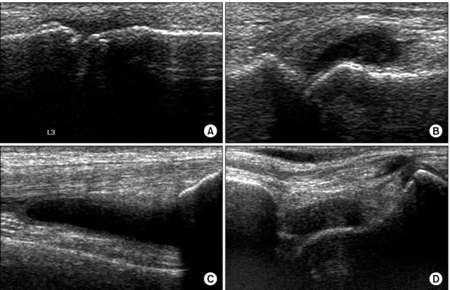
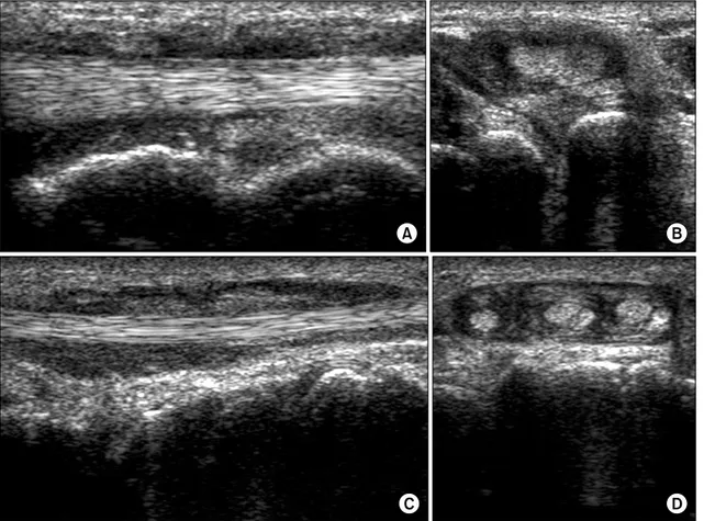
Fig. 1. Ultrasonographic findings of joint effusion. (A) Metacarpophalangeal joint, (B) metatarsophalangeal joint, (C) suprapatellar recess of the knee and (D) anterior talotibial joint.
증상이 전혀 없는 관절의 13%와, 종창 없이 통증만 있던 33%의 관절에서도 초음파로 윤활막염을 검출 하여, 임상 증상이 없는 관절에서도 윤활막염을 객 관적인 영상으로 진단하여 적절한 치료 방향을 제시 할 수 있음을 시사하였다 (11). 더욱이 임상적인 관 해가 이루어진 류마티스관절염 환자의 73% 가량에서 초음파로 윤활막염이 남아있음을 확인할 수 있어 (20) 류마티스관절염의 분류 기준이나 관해 기준에 단순 방사선촬영 외 초음파 소견을 추가해야 한다는 의견 이 제시되고 있다.
윤활막염의 특징적 초음파 소견은 관절내 삼출(effu- sion)의 존재, 윤활막 증식(synovial hypertrophy) 및 도플러 초음파상 혈관음영의 증가 등이며, 파누스와 골미란이 관찰될 수 있다 (21).
먼저 삼출의 초음파적 소견은 피하 지방층과 비교 하여 비정상적인 저음영(hypoechoic) 또는 무음영(ane- choic)의 물질이 관절내 존재하는 것이다 (22) (그림 1). 삼출의 원인이나 성상, 급만성도, 감염이나 출혈
등의 동반 여부에 따라 음영에 차이가 있을 수 있 다. 이 병변은 이동성이며, 압박 가능하지만 도플러 음영이 보이지 않는 특징을 보인다 (22). 윤활막염의 가장 초기 소견은 삼출이기 때문에 무증상의 초기 윤활막염을 시사하는데 중요한 근거가 된다. 손가락 이나 발가락 관절에서 촉진 등 임상적 진찰로 진단 되지 않는 소량의 삼출액을 검출하는데 초음파는 매 우 유용하며, 또한 삼출의 정확한 위치를 파악할 수 있어 관절 주사 주입의 정확도를 단순 촉진 시 59%
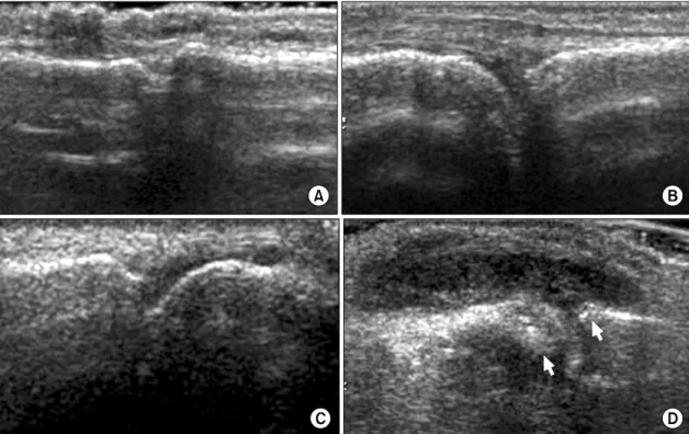
에서 초음파 이용 시 96%까지 높일 수 있다는 치료 적인 장점까지 가지고 있다 (23). 아직까지 초음파를 이용한 삼출액의 부피에 대한 정량적인 측정은 쉽지 않고 표준화되어 있지 않으나, 그 양에 따른 반정량 적인 분류로, grade 0=삼출액이 없는 경우, grade 1=
최소량의 삼출액, grade 2=중등도의 삼출액이 존재하 나 관절주머니(joint capsule)가 팽창되지 않는 경우, grade 3=과량의 삼출액이 존재하여 관절주머니가 팽 창된 경우로 나누는 방법이 제안되었다(그림 2) (17).
Fig. 2. Ultrasonographic findings of joint effusion can be graded according to the amount. (A) Grade 0 (no effusion), (B) grade 1 (minimal amount of fluid), (C) grade 2 (moderate amount of fluid without distension of the joint capsule), and (D) grade 3: extensive amount of fluid with distension of the joint capsule.
그러나 정상인에서도 관절에 따라서 소량의 삼출이 존재할 수 있으므로 무증상의 반대쪽과 비교하고, 결과 해석에 주의해야 한다.
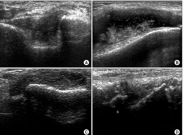
윤활막 증식은 초음파에서 피하 지방층과 비교하 여 비정상적인 저음영의 관절내 조직으로 관찰되며, 때로는 초음파적 음영이 피하 지방층과 동일 음영이 나 고음영으로 보일 수 있다 (22) (그림 3). 삼출과는 달리 이동되지 않고 쉽게 압박되지 않으며 도플러 신 호가 보일 수 있다는 특징을 보인다. 정상적인 윤활 막은 초음파로 관찰이 되지 않으나 윤활막염 발생 시 활막 두께의 증가로 인해 관찰이 가능해 지며, 윤활막의 부종에 의해 윤활막 자체가 저음영 또는 무음영으로 보이는 경우에는 삼출액과의 감별이 필 요하다. 두 병변을 감별하는 방법으로 윤활막 증식 의 경우 압박에도 병변이 이동하지 않는다는 점과 컬러 또는 파워도플러 초음파상 이상 혈관 음영의 증가가 관찰된다는 점이다 (21). 윤활막 증식에 대한 반정량적 분류는 여러 연구자에 의해 몇 가지 방법
이 제안되었으며 표 1과 같다(표 1). Scheel AK 등은 손허리손가락(metacarpophalangeal, MCP) 관절과 몸쪽 손가락뼈사이(proximal interphalangeal, PIP) 관절을 초 음파와 자기공명영상으로 관찰하였을 때 윤활막염은 손바닥 몸쪽(palmar proximal)에서 가장 잘 관찰되었 고(86%), 반정량적인 분류와 정량적인 수치 측정에 는 큰 차이가 없었으며, 자기공명영상 소견과도 잘 일치한다고 보고하였다. 이에 류마티스관절염의 평 가에 “세 손가락의 합(sum of three fingers)”-두 번째 부터 네 번째 MCP관절과 PIP 관절을 분석-을 제안 하기도 하였다. 류마티스관절염 환자의 손과 손목의 소견을 비교한 최근 두 연구에서 자기공명영상이 골 미란을 더 민감하게 진단하지만 초음파가 삼출액 등 을 더 민감하게 검출한다는 결과와 (24), 초음파가 이 두 관절의 골미란을 진단하는데 있어 자기공명영 상과 동등한 민감도를 보였다는 결과가 있다 (25).
윤활막염을 진단하고 다른 병변과 감별하며 활성 도를 파악하는데 있어 일반적인 회색조(gray-scale) 초
Fig. 3. Ultrasonographic findings of synovial proliferation. (A) Olecrenon fossa of the elbow, (B) lateral recess of the knee, (C) metacarpophalangeal joint, and (D) dorsal carpal joint.
음파 검사에 비해 혈류 신호를 감지할 수 있는 도플 러 방법은 매우 유용하게 사용된다. 도플러 초음파는 색도플러(color Doppler) 초음파와 강화도플러(power Doppler) 초음파 방법으로 나뉘어 지는데, 이중 윤활 막염을 평가하는데 있어 강화도플러가 중요한 역할 을 한다. 평균 도플러 주파수변위를 부호화하여 신 호의 속도를 측정하는 색도플러와 달리 강화도플러는 도플러 신호의 강화 분광밀도의 진폭을 부호화 하기 때문에 총신호의 강도를 측정하여 작은 혈관의 혈류 를 검출하는데 더 민감하다. 또한 둘러겹침 허상(alia- sing artifact)이 없고 각도 의존적(angle dependency)이 지 않다는 것이 장점이다 (26-29). 무릎과 엉덩이 등 큰 관절의 조직학적 소견과의 비교에서 강화도플러 의 혈관 신호가 윤활막 조직내 신생 혈관의 존재 정 도와 잘 비례하며 (28,30,31), 적혈구침강속도, C반응
단백, DAS28 등 질병 활성도를 정확히 반영하는 것 으로 알려져 있다 (32,33). 또한 치료 효과를 판정하 는 도구로 사용될 수 있는데, 관절내 스테로이드 주 입이나 생물학적 제제 치료 전후로 비교하였을 때 임상 증상이 좋아지는 것에 비례하여 강화도플러의 혈관신호가 감소하는 것을 알 수 있다 (33-39).
도플러 초음파는 장비의 질적 수준, 기술적 조건, 검사자의 숙련도에 의해 촬영과 해석이 좌우될 수 있다 (39,40). 검사 시 주의할 점으로는 대상 관절이 나 힘줄의 긴장이 최소화 된 자세로 관찰하고, 젤리 를 충분히 이용하여 탐색자의 압력을 최소화 함으로 써 혈관이 눌려 신호가 감소하는 것을 피하고, 탐색 자를 정지된 상태에서 관찰하고, 주변 온도를 일정 하게 유지해야 하며, 무증상의 반대 쪽과 비교하여 관찰해야 한다 (21). 그리고 펄스반복주파수(pulse repeti-
Table 2. Scoring systems of the vascularity of synovitis using power Doppler sonography
Grade 0 Grade 1 Grade 2 Grade 3
Naredo E (33)
Absence, No synovial flow
Mild, ≤3 isolated signals Moderate, >3 isolated signals or confluent signal in less than half of the synovial area
Marked, Signals in more than half of the synovial area
Kubassova O (41)
No Doppler activity in the synovium
One or two single spots Confluent areas covering up to half of the synovial area
Confluent areas covering more than half of the synovial area Newman JS (34) No flow Mild or moderate flow Intense flow Strunk J (61) Normal or minimal perfusion Mild and moderate flow
Szkudlarek M (17)
No flow in the synovium
Single vessel signal Confluent vessel signals in less than half of the area of the synovium
Vessel signals in more than half of the area of the synovium
Weidekamm C (60) No vascularization Small vascularization Moderate vascularization Strong vascularization Table 1. Scoring systems of the ultrasonographic signs of synovitis
Grade 0 Grade 1 Grade 2 Grade 3 Grade 4
Szkudlarek M (17)
No synovial thickening
Minimal synovial thickening (filling the angle between the periarticular bone, without bulging over the line linking the tops of the bones)
Synovial thickening (bulging over the line linking the tops of the bones but without extension along the bone diaphyses)
Synovial thickening (bulging over the line linking the tops of the bones and with extension to at least one of the bone diaphyses) Scheel AK
(58)
No effusion/
hypertrophy
Minimal effusion/
hypertrophy
Moderate effusion/
hypertrophy
Extensive effusion/
hypertrophy Ostergaard
M (50)
No synovial thickening
Minimal synovial thickening
Synovial thickening (bulging over the line linking the tops of the bones forming the joint without ex- tension along the bone diaphyses)
Synovial thickening (bulging over the line linking the tops of the bones forming the joint with exten- sion to one of the diaphyses)
Synovial thickening (bulging over the line linking the tops of the bones forming the joint with exten- sion to one of the diaphyses) Naredo E (14) Absence Mild Moderate Marked
Weidekamm C (60)
No change Slight change Moderate change Strong changes
tion frequency)는 민감도가 최대화 되는 최소 값으로 조절하며, 근골격계 영역에서는 대개 500∼750 Hz 범위가 적당하다 (33). 일반적으로 관절이나 힘줄 내 부에서 혈관 신호가 보이지 않는 것이 정상이다. 혈 관 신호의 정도에 따라 등급을 분류하기도 하는데, 대상 관절의 윤활막에서 도플러 초음파에서 혈관 신
호가 차지하는 범위에 따라 반정량적으로 분류하는 것이 일반적이며(표 2), 정량적인 방법으로는 색분율 (color fraction)을 계산하는 방법이 있다 (4,28,32,41).
이는 윤활막을 투사(trace)한 후 소프트웨어 프로그 램에 의해 총화소(total pixel) 중 색화소(color pixel) 의 분율을 계산한 값이다. 윤활막의 관류를 보다 객
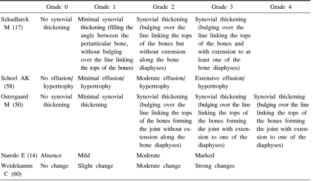
Fig. 4. Ultrasonographic findings of bone erosion. (A) Small erosion in the talus, (B) irregular and moderate-sized erosions of the metacarpal head, (C) large and deep erosion of the metacarpal head, and (D) inflammatory pannus with increased vascularity on power Doppler ultrasound in the metatarsal joint.
관적이며 정량적으로 파악할 수 있다는 이론적인 장 점이 있으나, 류마티스관절염의 경우 윤활막염이 호 전되어 두께와 부피가 감소하면 상대적으로 색분율 이 높은 값으로 나타날 수 있다는 문제점이 있어 질 병 활성도의 변화를 반영하는데 반정량적인 방법보 다 우수하거나 유용하지 않다 (4,28,32,41).
2) 골미란
류마티스관절염에서 발생하는 골미란의 초음파 소 견은 관절내부 골표면의 불연속성이며, 이는 두 개 의 수직면에서 모두 관찰되어야 한다(그림 4) (22).
골미란의 내부에 고음영의 조직이나 도플러상 혈관 신호가 증가된 조직이 보이는 경우 증식성, 혈관성 파누스가 존재함을 시사한다 (42). 처음 진단 당시 단순방사선 촬영에서 골미란이 관찰되는 경우는 약 40% 가량밖에 되지 않으나 (41) 실제 골미란이 존재
하는 경우가 96∼97%에 달하기 때문에 (43,44), 단순 방사선 검사 외 조기진단에 민감한 영상 방법이 필 요하게 되는데 자기공명영상이나 초음파가 적합하 다. 류마티스관절염 환자의 MCP 관절에서 초음파 검사와 단순방사선 촬영의 비교연구를 보면, 골미란 을 검출하는데 있어 초음파 검사가 조기 관절염에서 6.6배, 진행된 관절염에서 3.4배의 검출력을 보였다 (6). 또한 MTP 관절의 골미란 검출 시 자기공명영상 소견을 기준으로 보았을 때 단순방사선 촬영이 0.32 의 민감도를 보이는 것에 비해 초음파 검사는 0.79 의 높은 민감도를 보였다 (5). 손목 관절의 골미란을 검출하는데 자기공명영상과 초음파 검사의 차이가 없었으며, MCP 관절의 골미란을 검출하는데 초음파 가 더 많은 관절을 검출했다는 연구가 있으나 (19,25), 일반적인 비교 연구에서 골미란을 검출하는 데 있어
Fig. 5. Ultrasonographic findings of tenosynovitis. (A, B) Anechoic fluid accumulation in the sheath of posterior tibial tendon (longitudinal and transverse scans). (C, D) Hyperechoic synovial proliferation in the sheath of the extensor digitorum tendons (longitudinal and transverse scans).
자기공명영상이 더 예민한 방법인 것으로 보인다 (5,24). 아직까지 류마티스관절염의 골미란의 등급을 평가하는 방법이 표준화 되어있지 않으나, 한 연구 에서 grade 0=균질한 골표면, grade 1=두 면에서 보 았을 때 소실을 형성하지 않은 불균질한 골표면, grade 2=두 면에서 보았을 때 골표면의 소실이 보이 는 경우, grade 3=광범위한 골파괴를 동반한 골소실 로 분류하였다 (17).
3) 연골의 변화
초음파에서 관절연골은 주변과 경계가 명확한 저 음영 또는 무음영의 구조물로 보이지만, 자기공명영 상과는 달리 뼈를 투과하지 못해 연골의 모든 부분 이 관찰되지 않는다는 한계가 있다 (40). 류마티스관 절염에서 연골의 두께가 부분적 또는 전체적으로 얇
아지고 주변과의 경계가 불분명해지는 소견을 보이 며 (45,46), MCP와 PIP 관절의 연골 두께가 질병이 조절되지 않았던 기간과 반비례한다는 연구결과가 있다 (46). 류마티스관절염의 골파괴 정도를 파악하 는데 일반적으로 사용되는 modified Sharp score의 항 목 중 관절간격 감소는 결국 연골 두께의 감소를 의 미하므로 단순 방사선촬영에 비해 초음파가 훨씬 민 감하게 검출할 수 있어 조기 골파괴를 시사하는 주 요 소견이 될 수 있을 것이다. 아직까지의 류마티스 관절염에서 연골에 대한 초음파 연구는 그 두께의 변화에 국한되었으나, 두께의 감소뿐 아니라 그 경 계의 불명확성이나 연골내 음영의 변화가 연골의 초 기 변화로 발생할 수 있다. 이러한 연골 손상은 관 절 장애를 예견하는 중요한 변화이므로 조기 진단과
적절한 치료로 이어질 수 있어 추후 자세한 연구가 필요하다.
4) 힘줄의 변화
힘줄윤활막염(tenosynovitis)은 조기 류마티스관절염 에서 흔하게 발생하며, 분류되지 않은 염증성 관절 염에서 항CCP 항체와 더불어 류마티스관절염으로의 진행을 시사하는 조기 표지자이기 때문에 중요한 병 변이다 (47).
초음파 소견은 힘줄윤활막내 액체의 저류가 존재 하거나 저음영의 또는 무음영의 두꺼운 조직이 존재 하는 것이며, 이는 두 수직면에서 관찰되고 도플러 상 혈관 음영이 보일 수 있다(그림 5) (22). 이 때 힘줄 자체의 염증이 동반되기도 하는데 초음파상 힘 줄의 경계가 불분명해지면서 힘줄내 저음영 부위가 존재하여 불균질한 음영을 보이게 되고, 이러한 힘 줄내 변화는 자기공명영상보다 초음파에서 더 잘 관 찰되므로 매우 유용하다 (42,48).
2. 조기 류마티스관절염의 감별 진단
이상에서 조기 류마티스관절염의 초음파 소견으로 윤활막염, 힘줄윤활막염, 윤활낭염, 골미란 등의 소 견을 언급했다.
조기 류마티스관절염과 감별해야 할 만성 염증성 관절염으로 건선성 관절염이 대표적이다. 건선성 관 절염은 윤활막염, 힘줄윤활막염, 골미란 등 류마티스 관절염과 비슷한 초음파 소견을 보인다. 감별점으로 는 건선성 관절염의 경우 주로 먼쪽가락사이 관절(distal interphalangeal joint)을 침범하고, 굽힘근(flexor) 침범 이 더 흔하며, 피하 연부조직의 염증이나 부종을 동 반하는 경우가 많다 (19,42). 류마티스관절염은 관절 내 염증이 힘줄 부착부보다 더 흔하게 발생하는데 반해 건선성 관절염은 힘줄 부착부병증이 더 흔하게 발생한다. 이렇게 건선성 관절염에서 말초 부착부병 증이 발생하는 경우 작은 힘줄의 경우에도 자기공명 영상보다 초음파로 민감한 검출이 가능하다. 또한 골 미란 뿐 아니라 골증식이나 뼈막염이 발생하는 경우 가 있고 초음파가 작은 골증식을 검출하는데 유용하 다 (19).
3. 류마티스관절염의 활성도 평가와 치료 효과 판정 류마티스관절염의 활성도를 대표하는 적혈구침강
속도, C반응 단백, DAS28 등이 초음파적인 활막염 의 정도와 잘 비례하고 (32,33,49) 초음파 검사가 임 상 진찰보다 민감하게 활성 관절을 파악할 수 있기 때문에 (11,20) 최근 치료 후 초음파로 질병 활성도 의 변화를 초음파로 확인하는 연구가 많이 이루어 지는 추세이다. 한 연구에서 초음파로 윤활막염, 삼 출액, 골미란, 힘줄윤활막염과 윤활낭염에 대해 각각 0∼3점으로 반정량적 등급을 주어 생물학적 제제 투 여 전후의 변화를 관찰한 결과 치료 시작 전에 비해 치료 후 총점이 감소하였고 이는 DAS28의 변화와 일치했음을 발표하였다 (16). 그러나 아직 자기공명 영상이나 단순 방사선 검사처럼 질병 활성도나 관절 파괴 정도에 따른 등급의 분류가 없고, 검사 방법에 대한 표준화가 이루어지지 않았으며, 검사자나 장비 의 수준에 따라 결과 판정에 차이가 있을 수 있다는 것은 문제점이며 남은 과제이다 (50).
4. 3차원 초음파
현재 주로 사용되고 있는 2차원 초음파는 검사자 가 탐촉자를 움직여서 여러 개의 영상을 얻어 머리 속에서 3차원 영상으로 통합하는 과정을 거쳐 해부 학적인 이상을 판단하기 때문에 검사자의 능력에 따 라 결과에 많은 차이를 보인다. 따라서 정확한 술기 를 습득하는데 많은 시간을 투자해야 하며 술기에 대한 표준화가 어려워 관찰자내 가변성(intra-observer variability)과 관찰자간 가변성(inter-observer variability) 이 크고 검사에 소요되는 시간이 다른 영상 검사에 비해서 상대적으로 길다. 이러한 한계를 극복할 수 있는 방법이 3차원 초음파이다.
부피를 측정할 수 있는 탐촉자(volumetric probe)를 사용하면 검사자의 능력에 관계없이 자동화된 기계 적인 방법으로 연속된 초음파 영상을 얻을 수 있다.
얻은 영상을 3차원 영상으로 재조합하면 2차원에서 얻을 수 없는 관상영상(coronal plane)도 볼 수 있어 더 많은 정보를 얻을 수 있다 (51). 또한 넓은 해부 학적 부위의 연속된 영상을 얻을 수 있기 때문에 중 요한 해부학적인 지점들에 대한 정확한 이해가 없어 도 검사를 시행할 수 있어 술기 습득을 위한 시간이 단축될 뿐 아니라 검사에 소요되는 시간도 몇 초면 충분하기 때문에 2차원 초음파에 비해서 현저히 단 축되고 표준화된 영상을 얻기가 용이하다. 기술의
발전으로 인해 고주파수의 3차원 초음파 탐촉자를 이용하여 관절의 3차원 영상을 선명하게 관찰하는 것이 가능해졌으며 파워도플러를 이용하여 혈관의 주행까지 확인할 수 있게 되었다 (52). 특히 검사 술 기를 습득하는데 많은 시간을 필요로 하는 강화도플 러 영상의 경우에도 3차원 초음파를 이용하면 허상 이 거의 없는 영상을 얻을 수 있어 관찰자간 일치도 (inter-observer agreement)가 2차원 초음파에 비해서 매우 높다 (53). 이러한 장점에도 불구하고 3차원도 단점이 있는데 움직이는영상(dynamic image)을 얻기 어렵고 탐촉자의 크기가 상대적으로 커서 특정 해부 학적 부위를 검사하는데 제한이 있고 관절을 관찰하 기 위해 고주파수의 장비가 필요하기 때문에 초기 투자비용이 크다.
류마티스관절염에 3차원 초음파를 이용한 보고들 에 의하면 윤활막 두께 및 관절내 혈관의 개수에 대 한 관찰자 사이의 동의가 매우 높았으며 치료에 대 한 반응으로 활막내 혈류의 감소도 쉽게 관찰할 수 있다 (53,54). 류마티스관절염의 진단과 경과 관찰에 초음파 이용에 대해 부정적인 의견이 도출되는 경우 의 대부분이 2차원 초음파 영상의 한계에 기인한 바 가 크기 때문에 3차원 초음파의 도입으로 인해 신뢰 도와 재현성이 높아져 임상에서 이용이 확대될 것으 로 기대된다.
결 론
현재까지는 초음파 영상을 이용하여 류마티스관절 염의 활성도와 관절 손상을 평가하는데 있어서 합의 된 지침이나 표준화된 척도는 없다. OMERACT 산 하의 연구집단이 관절의 염증에 대한 초음파 영상의 정의를 제안하였고 후속 연구들을 통해서 재현성이 높다고 보고 한 정도이다 (22,55,56). Ellegaard 등은 정상인의 손가락 관절들을 대상으로 여러 위치에서 초음파 영상을 얻어서 다양한 평가 체계를 시도해 보았는데 관절에 염증이 의심되는 높은 점수를 받은 경우가 예상보다 많았다고 보고하여 정상인을 류마 티스관절염으로 오진할 수 있는 문제점이 제시하였 고 (53), Terslev 등은 류마티스관절염 환자들의 손목과 손관절에서 얻은 초음파 영상의 염증 정도와 MRI의 강조영상 결과가 높은 상관관계를 보였지만 적혈구
침강 속도나 C-반응단백과 같이 염증을 반영하는 임 상자료와 관련이 없었다고 보고한 것으로 보면 (57), 초음파 소견이 임상적 판단에 있어 절대적인 결정적 인 요소는 아니며 다른 활성도 표지자 들과 더불어 고려해야 할 것으로 보인다.
류마티스관절염을 정확히 관찰하기 위해서 어떤 관절들을 대상으로 초음파 검사를 시행해야 하는지 에 대한 합의는 아직 없으며 평가 체계가 몇 가지 제안되어 있으나 추가적인 연구가 필요한 상태이다 (33,58,59). 특히 Naredo 등은 12개 관절(양측의 팔꿈 치, 손목, 두 번째와 세 번째 손허리손가락 관절, 무 릎, 발목)의 파워도플러 평가 체계가 44개 관절을 평가한 체계와 상관관계가 높았으며 재현성과 민감 도가 높았다고 제시하여 시간이 많이 소요되는 관절 초음파 검사의 단점을 극복하여 환자와 의사가 효율 적으로 이용할 수 있는 가능성을 보여주었다 (33).
현재까지 초음파 영상을 이용한 류마티스관절염의 평가에서 신뢰할 수 있는 표준 지침이 마련되어 있 지 않음에도 불구하고 관절내 염증의 중등도 평가와 관절내 주사에 관절초음파 이용은 널리 확산되고 있 다. 고주파수 3차원 초음파의 이용이 가능해 지면서 초음파 영상에 대한 신뢰도와 재현성이 높아질 것으 로 예상되며 검사 방법에 대한 표준 지침을 마련할 수 있는 기술적인 기반이 제공되었다고 할 수 있다.
앞으로 더 많은 연구와 자료의 축적을 통해서 초음 파 영상을 이용한 평가 체계가 마련됨으로써 류마티 스관절염을 최대한 조기에 발견하고 정확한 치료 효 과 판정을 통해 관절의 파괴를 최소화하려는 시도에 커다란 도움이 되길 기대한다.
참고문헌
1) Harrison BJ, Symmons DP, Barrett EM, Silman AJ.
The performance of the 1987 ARA classification criteria for rheumatoid arthritis in a population based cohort of patients with early inflammatory polyarthritis. Ame- rican Rheumatism Association. J Rheumatol 1998;
25:2324-30.
2) Chan KW, Felson DT, Yood RA, Walker AM. The lag time between onset of symptoms and diagnosis of rheumatoid arthritis. Arthritis Rheum 1994;37:814-20.
3) van Dongen H, van Aken J, Lard LR, Visser K, Ronday HK, Hulsmans HM, et al. Efficacy of metho-
trexate treatment in patients with probable rheumatoid arthritis: a double-blind, randomized, placebo-controlled trial. Arthritis Rheum 2007;56:1424-32.
4) Taylor PC, Steuer A, Gruber J, Cosgrove DO, Blom- ley MJ, Marsters PA, et al. Comparison of ultrasono- graphic assessment of synovitis and joint vascularity with radiographic evaluation in a randomized, placebo- controlled study of infliximab therapy in early rheu- matoid arthritis. Arthritis Rheum 2004;50:1107-16.
5) Szkudlarek M, Narvestad E, Klarlund M, Court-Payen M, Thomsen HS, Ostergaard M. Ultrasonography of the metatarsophalangeal joints in rheumatoid arthritis:
comparison with magnetic resonance imaging, con- ventional radiography, and clinical examination. Arthritis Rheum 2004;50:2103-12.
6) Wakefield RJ, Gibbon WW, Conaghan PG, O'Connor P, McGonagle D, Pease C, et al. The value of sono- graphy in the detection of bone erosions in patients with rheumatoid arthritis: a comparison with con- ventional radiography. Arthritis Rheum 2000;43:2762- 70.
7) Schmidt WA. Value of sonography in diagnosis of rheumatoid arthritis. Lancet 2001;357:1056-7.
8) Filippucci E, Iagnocco A, Meenagh G, Riente L, Delle Sedie A, Bombardieri S, et al. Ultrasound ima- ging for the rheumatologist VII. Ultrasound imaging in rheumatoid arthritis. Clin Exp Rheumatol 2007;25:
5-10.
9) Backhaus M, Kamradt T, Sandrock D, Loreck D, Fritz J, Wolf KJ, et al. Arthritis of the finger joints: a comprehensive approach comparing conventional ra- diography, scintigraphy, ultrasound, and contrast- enhanced magnetic resonance imaging. Arthritis Rheum 1999;42:1232-45.
10) Kane D, Balint PV, Sturrock RD. Ultrasonography is superior to clinical examination in the detection and localization of knee joint effusion in rheumatoid arthritis. J Rheumatol 2003;30:966-71.
11) Wakefield RJ, Green MJ, Marzo-Ortega H, Conaghan PG, Gibbon WW, McGonagle D, et al. Should oligoar- thritis be reclassified? Ultrasound reveals a high pre- valence of subclinical disease. Ann Rheum Dis 2004;
63:382-5.
12) Joshua F, Edmonds J, Lassere M. Power Doppler ultrasound in musculoskeletal disease: a systematic review. Semin Arthritis Rheum 2006;36:99-108.
13) Naredo E, Moller I, Cruz A, Carmona L, Garrido J.
Power Doppler ultrasonographic monitoring of re- sponse to anti-tumor necrosis factor therapy in pa-
tients with rheumatoid arthritis. Arthritis Rheum 2008;58:2248-56.
14) Naredo E, Bonilla G, Gamero F, Uson J, Carmona L, Laffon A. Assessment of inflammatory activity in rheumatoid arthritis: a comparative study of clinical evaluation with grey scale and power Doppler ultra- sonography. Ann Rheum Dis 2005;64:375-81.
15) Hau M, Kneitz C, Tony HP, Keberle M, Jahns R, Jenett M. High resolution ultrasound detects a de- crease in pannus vascularisation of small finger joints in patients with rheumatoid arthritis receiving treat- ment with soluble tumour necrosis factor alpha receptor (etanercept). Ann Rheum Dis 2002;61:55-8.
16) Iagnocco A, Filippucci E, Perella C, Ceccarelli F, Cassara E, Alessandri C, et al. Clinical and ultrasono- graphic monitoring of response to adalimumab treat- ment in rheumatoid arthritis. J Rheumatol 2008;35:
35-40.
17) Szkudlarek M, Court-Payen M, Jacobsen S, Klarlund M, Thomsen HS, Ostergaard M. Interobserver agree- ment in ultrasonography of the finger and toe joints in rheumatoid arthritis. Arthritis Rheum 2003;48:
955-62.
18) Pincus T. Limitations of a quantitative swollen and tender joint count to assess and monitor patients with rheumatoid arthritis. Bull NYU Hosp Jt Dis 2008;66:
216-23.
19) Wiell C, Szkudlarek M, Hasselquist M, Moller JM, Vestergaard A, Norregaard J, et al. Ultrasonography, magnetic resonance imaging, radiography, and clinical assessment of inflammatory and destructive changes in fingers and toes of patients with psoriatic arthritis.
Arthritis Res Ther 2007;9:119.
20) Brown AK, Quinn MA, Karim Z, Conaghan PG, Peterfy CG, Hensor E, et al. Presence of significant synovitis in rheumatoid arthritis patients with disease- modifying antirheumatic drug-induced clinical remi- ssion: evidence from an imaging study may explain structural progression. Arthritis Rheum 2006;54:3761- 73.
21) Farrant JM, O'Connor PJ, Grainger AJ. Advanced imaging in rheumatoid arthritis. Part 1: synovitis.
Skeletal Radiol 2007;36:269-79.
22) Wakefield RJ, Balint PV, Szkudlarek M, Filippucci E, Backhaus M, D'Agostino MA, et al. Musculoskeletal ultrasound including definitions for ultrasonographic pathology. J Rheumatol 2005;32:2485-7.
23) Raza K, Lee CY, Pilling D, Heaton S, Situnayake RD, Carruthers DM, et al. Ultrasound guidance allows
accurate needle placement and aspiration from small joints in patients with early inflammatory arthritis.
Rheumatology (Oxford) 2003;42:976-9.
24) Hoving JL, Buchbinder R, Hall S, Lawler G, Coombs P, McNealy S, et al. A comparison of magnetic resonance imaging, sonography, and radiography of the hand in patients with early rheumatoid arthritis. J Rheumatol 2004;31:663-75.
25) Magnani M, Salizzoni E, Mule R, Fusconi M, Meliconi R, Galletti S. Ultrasonography detection of early bone erosions in the metacarpophalangeal joints of patients with rheumatoid arthritis. Clin Exp Rheu- matol 2004;22:743-8.
26) Taylor PC. The value of sensitive imaging modalities in rheumatoid arthritis. Arthritis Res Ther 2003;5:
210-3.
27) Newman JS, Adler RS, Bude RO, Rubin JM. De- tection of soft-tissue hyperemia: value of power Do- ppler sonography. AJR Am J Roentgenol 1994;163:
385-9.
28) Walther M, Harms H, Krenn V, Radke S, Faehndrich TP, Gohlke F. Correlation of power Doppler sono- graphy with vascularity of the synovial tissue of the knee joint in patients with osteoarthritis and rheu- matoid arthritis. Arthritis Rheum 2001;44:331-8.
29) Taylor PC. Serum vascular markers and vascular imaging in assessment of rheumatoid arthritis disease activity and response to therapy. Rheumatology (Ox- ford) 2005;44:721-8.
30) Fiocco U, Cozzi L, Rubaltelli L, Rigon C, De Candia A, Tregnaghi A, et al. Long-term sonographic follow- up of rheumatoid and psoriatic proliferative knee joint synovitis. Br J Rheumatol 1996;35:155-63.
31) Schmidt WA, Volker L, Zacher J, Schlafke M, Ruhnke M, Gromnica-Ihle E. Colour Doppler ultraso- nography to detect pannus in knee joint synovitis.
Clin Exp Rheumatol 2000;18:439-44.
32) Qvistgaard E, Rogind H, Torp-Pedersen S, Terslev L, Danneskiold-Samsoe B, Bliddal H. Quantitative ultra- sonography in rheumatoid arthritis: evaluation of inflammation by Doppler technique. Ann Rheum Dis 2001;60:690-3.
33) Naredo E, Rodriguez M, Campos C, Rodriguez- Heredia JM, Medina JA, Giner E, et al. Validity, reproducibility, and responsiveness of a twelve-joint simplified power doppler ultrasonographic assessment of joint inflammation in rheumatoid arthritis. Arthritis Rheum 2008;59:515-22.
34) Newman JS, Laing TJ, McCarthy CJ, Adler RS.
Power Doppler sonography of synovitis: assessment of therapeutic response-preliminary observations. Ra- diology 1996;198:582-4.
35) Filippucci E, Farina A, Carotti M, Salaffi F, Grassi W. Grey scale and power Doppler sonographic changes induced by intra-articular steroid injection treatment.
Ann Rheum Dis 2004;63:740-3.
36) Salaffi F, Carotti M, Manganelli P, Filippucci E, Giuseppetti GM, Grassi W. Contrast-enhanced power Doppler sonography of knee synovitis in rheumatoid arthritis: assessment of therapeutic response. Clin Rheu- matol 2004;23:285-90.
37) Ribbens C, Andre B, Marcelis S, Kaye O, Mathy L, Bonnet V, et al. Rheumatoid hand joint synovitis:
gray-scale and power Doppler US quantifications following anti-tumor necrosis factor-alpha treatment:
pilot study. Radiology 2003;229:562-9.
38) Fiocco U, Ferro F, Vezzu M, Cozzi L, Checchetto C, Sfriso P, et al. Rheumatoid and psoriatic knee syno- vitis: clinical, grey scale, and power Doppler ultra- sound assessment of the response to etanercept. Ann Rheum Dis 2005;64:899-905.
39) Grassi W, Filippucci E. Is power Doppler sonography the new frontier in therapy monitoring? Clin Exp Rheumatol 2003;21:424-8.
40) Kane D, Balint PV, Sturrock R, Grassi W. Mus- culoskeletal ultrasound-a state of the art review in rheumatology. Part 1: current controversies and issues in the development of musculoskeletal ultrasound in rheumatology. Rheumatology (Oxford) 2004;43:823-8.
41) Kubassova O, Boesen M, Peloschek P, Langs G, Cimmino MA, Bliddal H, et al. Quantifying disease activity and damage by imaging in rheumatoid arthritis and osteoarthritis. Ann N Y Acad Sci 2009;
1154:207-38.
42) Boutry N, Carmo CC, Flipo RM, Cotten A. Early rheumatoid arthritis and its differentiation from other joint abnormalities. Eur J Radiol 2009 [Epub ahead of print].
43) Pierre-Jerome C, Bekkelund SI, Mellgren SI, Torberg- sen T, Husby G, Nordstrom R. The rheumatoid wrist:
bilateral MR analysis of the distribution of rheumatoid lesions in axial plan in a female population. Clin Rheumatol 1997;16:80-6.
44) Alasaarela E, Suramo I, Tervonen O, Lahde S, Takalo R, Hakala M. Evaluation of humeral head erosions in rheumatoid arthritis: a comparison of ultrasonography, magnetic resonance imaging, computed tomography and plain radiography. Br J Rheumatol 1998;37:
1152-6.
45) Iagnocco A, Coari G, Zoppini A. Sonographic evalu- ation of femoral condylar cartilage in osteoarthritis and rheumatoid arthritis. Scand J Rheumatol 1992;
21:201-3.
46) Moller B, Bonel H, Rotzetter M, Villiger PM, Ziswiler HR. Measuring finger joint cartilage by ultrasound as a promising alternative to conventional radiograph imaging. Arthritis Rheum 2009;61:435-41.
47) Eshed I, Feist E, Althoff CE, Hamm B, Konen E, Burmester GR, et al. Tenosynovitis of the flexor tendons of the hand detected by MRI: an early indicator of rheumatoid arthritis. Rheumatology (Ox- ford) 2009 [Epub ahead of print].
48) Boutry N, Morel M, Flipo RM, Demondion X, Cotten A. Early rheumatoid arthritis: a review of MRI and sonographic findings. AJR Am J Roentgenol 2007;
189:1502-9.
49) Hameed B, Pilcher J, Heron C, Kiely PD. The relation between composite ultrasound measures and the DAS28 score, its components and acute phase mar- kers in adult RA. Rheumatology (Oxford) 2008;47:
476-80.
50) Ostergaard M, Ejbjerg B, Szkudlarek M. Imaging in early rheumatoid arthritis: roles of magnetic resonance imaging, ultrasonography, conventional radiography and computed tomography. Best Pract Res Clin Rheu- matol 2005;19:91-116.
51) Downey DB, Fenster A, Williams JC. Clinical utility of three-dimensional US. Radiographics 2000;20:559- 71.
52) Filippucci E, Meenagh G, Epis O, Iagnocco A, Riente L, Delle Sedie A, et al. Ultrasound imaging for the rheumatologist. XIII. New trends. Three-dimensional ultrasonography. Clin Exp Rheumatol 2008;26:1-4.
53) Strunk J, Strube K, Rumbaur C, Lange U, Muller- Ladner U. Interobserver agreement in two- and three- dimensional power Doppler sonographic assessment of synovial vascularity during anti-inflammatory treat- ment in patients with rheumatoid arthritis. Ultraschall Med 2007;28:409-15.
54) Strunk J, Strube K, Muller-Ladner U, Lange U. Three
dimensional power Doppler ultrasonography confirms early reduction of synovial perfusion after intra- articular steroid injection. Ann Rheum Dis 2006;65:
411-2.
55) Naredo E, Moller I, Moragues C, de Agustin JJ, Scheel AK, Grassi W, et al. Interobserver reliability in musculoskeletal ultrasonography: results from a
“Teach the Teachers” rheumatologist course. Ann Rheum Dis 2006;65:14-9.
56) Scheel AK, Schmidt WA, Hermann KG, Bruyn GA, D'Agostino MA, Grassi W, et al. Interobserver reliability of rheumatologists performing musculos- keletal ultrasonography: results from a EULAR “Train the trainers” course. Ann Rheum Dis 2005;64:1043-9.
57) Terslev L, Torp-Pedersen S, Savnik A, von der Recke P, Qvistgaard E, Danneskiold-Samsoe B, et al.
Doppler ultrasound and magnetic resonance imaging of synovial inflammation of the hand in rheumatoid arthritis: a comparative study. Arthritis Rheum 2003;
48:2434-41.
58) Scheel AK, Hermann KG, Kahler E, Pasewaldt D, Fritz J, Hamm B, et al. A novel ultrasonographic synovitis scoring system suitable for analyzing finger joint inflammation in rheumatoid arthritis. Arthritis Rheum 2005;52:733-43.
59) Naredo E, Gamero F, Bonilla G, Uson J, Carmona L, Laffon A. Ultrasonographic assessment of inflamma- tory activity in rheumatoid arthritis: comparison of extended versus reduced joint evaluation. Clin Exp Rheumatol 2005;23:881-4.
60) Weidekamm C, Koller M, Weber M, Kainberger F.
Diagnostic value of high-resolution B-mode and do- ppler sonography for imaging of hand and finger joints in rheumatoid arthritis. Arthritis Rheum 2003;
48:325-33.
61) Strunk J, Heinemann E, Neeck G, Schmidt KL, Lange U. A new approach to studying angiogenesis in rheumatoid arthritis by means of power Doppler ultra- sonography and measurement of serum vascular endo- thelial growth factor. Rheumatology (Oxford) 2004;
43:1480-3.