This is an open-access article distributed under the terms of the Creative Commons Attribution Non-Commercial License (http://creativecommons.org/
licenses/by-nc/4.0/), which permits unrestricted non-commercial use, distribution, and reproduction in any medium, provided the original work is properly cited.
CC
Review of a novel disease entity, immunoglobulin G4-related disease
Takashi Maehara, Masafumi Moriyama, Seiji Nakamura
Section of Oral and Maxillofacial Oncology, Division of Maxillofacial Diagnostic and Surgical Sciences, Faculty of Dental Science, Kyushu University, Fukuoka, Japan
Abstract(J Korean Assoc Oral Maxillofac Surg 2020;46:3-11)
Immunoglobulin G4 (IgG4)-related dacryoadenitis and sialoadenitis (IgG4-DS) are part of a multiorgan fibroinflammatory condition of unknown etiology termed IgG4-related disease (IgG4-RD), which has been recognized as a single diagnostic entity for less than 15 years. Histopathologic examination is critical for diagnosis of IgG4-RD. CD4+ T and B cells, including IgG4-expressing plasma cells, constitute the major inflammatory cell populations in IgG4-RD and are thought to cause organ damage and tissue fibrosis. Patients with IgG4-RD who have active, untreated disease exhibit significant increase of IgG4-secreting plasmablasts in the blood. Considerable insight into the immunologic mechanisms of IgG4-RD has been achieved in the last decade using novel molecular biology approaches, including next-generation and single-cell RNA sequencing. Exploring the in- teractions between CD4+ T cells and B lineage cells is critical for understanding the pathophysiology of IgG4-RD. Establishment of pathogenic T cell clones and identification of antigens specific to these clones constitutes the first steps in determining the pathogenesis of the disease. Herein, the clini- cal features and mechanistic insights regarding pathogenesis of IgG4-RD were reviewed.
Key words: Immunoglobulin G4-related disease, Immunoglobulin G4-related dacryoadenitis and sialoadenitis, Mikuliçz’s disease, Küttner’s tumor, T cell [paper submitted 2019. 12. 6 / accepted 2020. 1. 6]
Copyright © 2020 The Korean Association of Oral and Maxillofacial Surgeons. All rights reserved.
I. Introduction
Immunoglobulin G4-related disease (IgG4-RD) is a novel clinical disease entity characterized by a chronic fibroinflam- matory condition with lymphoplasmacytic infiltration in affected lesions
1. The disease mimics many malignant, infec- tious, and inflammatory disorders with histologic features that are consistently observed across the organs involved.
Human IgG can be classified into four subtypes, IgG1, IgG2, IgG3, and IgG4, of which IgG4 is the rarest. However, IgG4- RD is characterized by elevated serum IgG4 concentration and tissue infiltrated by IgG4+ B cells and is termed IgG4 class-switching disease
1,2. IgG4-RD may be present in a cer- tain proportion of patients across a wide variety of diseases
including Mikuliçz’s disease (MD), Küttner’s tumor (KT)
3, Riedel’s thyroiditis, kidney disease, autoimmune pancreatitis, hypophysitis, interstitial pneumonitis, interstitial nephritis, prostatitis, lymphadenopathy, retroperitoneal fibrosis, inflam- matory aortic aneurysm, and inflammatory pseudotumor.
Thus, this disease can occur in a variety of organs including the pancreas, kidney, lung, lymph nodes, bile duct, liver, aorta, prostate, retroperitoneum, thyroid, and major salivary glands
1. The diagnosis of IgG4-RD relies heavily on histo- pathological analysis and correlation of histology findings with clinical, serological, and radiological data. International consensus statements have been published regarding the nomenclature, pathologic findings, and clinical management of IgG4-RD
4,5. Recently, Wallace et al.
6identified four dis- tinctive IgG4-RD phenotypes based on organ involvement and reported that being Asian or female may predispose in- dividuals to disease limited to the head and neck, especially in the salivary and lacrimal glands. The American College of Rheumatology/European League Against Rheumatism Clas- sification Criteria are currently being developed
5. Herein, the clinical characteristics of IgG4-RD are described, and up-to- date information on mechanistic insights using novel molecu- lar biology approaches is provided.
Takashi Maehara
Section of Oral and Maxillofacial Oncology, Division of Maxillofacial Diagnostic and Surgical Sciences, Faculty of Dental Science, Kyushu University, 3-1-1, Maidashi Higashi-ku, Fukuoka 812-8582, Japan TEL: +81-92-642-6447 FAX: +81-92-642-6386
E-mail: [email protected] ORCID: https://orcid.org/0000-0002-7877-2161
II. Immunological Insights into the Pathogenesis
1. Imaging of IgG4-RD
Computed tomography (CT), magnetic resonance imaging, and ultrasonography (US) are useful tools to assess organ involvement, monitor therapeutic responses, and guide inter- ventional treatments for IgG4-RD. Furthermore, F-fluorode- oxyglucose (FDG) positron emission tomography/computed tomography (PET/CT) was used to characterize IgG4-RD and showed multi-organ involvement.(Fig. 1. A) FDG-PET/
CT enables acquisition of whole-body images and provides functional information regarding disease activity. For ex- ample, when focusing on an affected salivary mandibular gland of an IgG4-RD patient, a US image of a lesion showed a hypoechoic area with a reticular pattern in the superficial part and a hypoechoic area with a nodal pattern and high vas- cularity
7,8.(Fig. 1. B)
2. Affected salivary glands
Both major and minor salivary glands can be affected by IgG4-related dacryoadenitis and sialoadenitis (IgG4-DS).(Fig.
1. C) IgG4-DS is commonly encountered by oral surgeons in the context of MD or KT, especially in Asians, which com- prises simultaneous bilateral and symmetrical enlargement of the lacrimal and salivary glands. Some patients, however, only exhibit lacrimal gland disease or may present with uni- lateral submandibular gland involvement. For many decades, MD was thought to represent a subtype of Sjögren’s syn- drome (SS); however, now these two diseases are known to be different
9.
Comprehensive diagnostic criteria have been proposed for IgG4-RD. Furthermore, organ-specific criteria have been pro- posed for IgG4-DS
9. Using these criteria, biopsies of affected lesions can be used to exclude diseases that often mimic IgG4-DS, including lymphoma, SS, sarcoidosis, sialodocho- lithiasis, granulomatosis with polyangiitis, and multicentric Castleman’s disease
10,11. An incisional or excisional biopsy of affected submandibular glands is often the best method for establishing a definitive diagnosis of IgG4-DS
9,12.
3. Genetic background of IgG4-RD patients
In numerous previous studies, have investigated risk fac- tors for IgG4-RD, especially type I autoimmune pancreatitis
(IgG4-related pancreatitis), were investigated. The HLA- DRB1*0405 and HLA-DQB1*0401 haplotypes were fre- quently found in Japanese patients with IgG4-related pan- creatitis
13. In addition to HLA risk loci described in previous reports, several non-human leukocyte antigen (HLA) genes have been identified as risk genes of IgG4-RD. Terao et al.
14reported that, because HLA loci usually showed stronger as- sociations with autoimmune diseases compared with non- HLA loci, detailed analyses of the HLA regions with fine- mapping genome-wide association study (GWAS) signals using next-generation sequencing and prioritizing amino acid variants or positions critical for IgG4-RD should be conduct- ed as sub-analyses of GWAS.
A B
C
Fig. 1. Immunoglobulin G4-related disease (IgG4-RD) tends to form tumefactive lesions. A. A 60-year-old male with IgG4-RD showed multi-organ involvement. F-fluorodeoxyglucose (FDG)- positron emission tomography/computed tomography (PET/CT) showed multiple intense uptakes in the bilateral lacrimal glands, submandibular glands, lymph nodes, kidney, pancreas, and retro- peritoneum (arrows). B. Ultrasonography shows hypoechoic areas with a nodal pattern and hyperechoic lines (upper). In Doppler mode, the nodal area shows relatively high vascularization (lower).
C. Bilateral enlargement of the submandibular glands in an IgG4- RD patient.
Takashi Maehara et al: Review of a novel disease entity, immunoglobulin G4-related disease. J Korean Assoc Oral Maxillofac Surg 2020
4. Histopathology of IgG4-RD
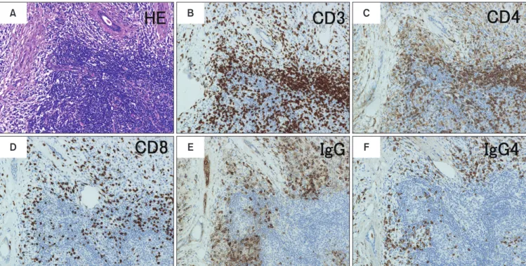
Histopathology is critical for diagnosis of IgG4-RD. Major central pathology features include lymphocytic infiltration, obliterate phlebitis, and storiform fibrosis in the affected le- sions.(Fig. 2. A) IgG4+ B cells and CD4+ and CD8+ T cells are commonly present (Fig. 2. B-D), and most IgG in the affected organs is IgG4.(Fig. 2. F, 2. G) The finding of abun- dant IgG4+ plasma cells is helpful to differentiate IgG4-RD from other mimic disorders with a similar presentation. How- ever, IgG4-RD cannot be diagnosed based only on IgG4+ cell infiltration because these plasma cells are present in many other inflammatory disorders
15.
IgG4 is generally considered a non-inflammatory immuno- globulin due to its limited ability to fix complement and bind activating Fc receptors
16. Notably, there is no evidence that IgG4 has a primary role in the pathophysiology of IgG4-RD.
Furthermore, the activity of IgG4-RD does not always corre- late well with serum IgG4 concentration
17.
5. Role of T-B interactions in IgG4-RD
It is important to determine which cells are crucial for
pathogenesis of IgG4-RD. The first reliable report on the pathophysiology of this disease was from preliminary stud- ies of rituximab (RTX) therapy (anti-CD20 B cell depletion therapy) showing that B cell depletion induced disease remis- sion and led to clinical improvement
18-20. Active and untreated IgG4-RD patients have an oligoclonally expanded population of circulating plasmablasts with a restricted oligoclonal B cell receptor repertoire
21, strongly indicating that IgG4-RD is an antigen-driven disease
22. Clonally expanded plasmablasts from IgG4-RD blood are a hallmark of active IgG4-RD. In flow cytometry studies after RTX therapy, clinical improve- ment correlated with selective depletion of this B cell sub- population. Oligoclonal proliferation of antibody-producing cells was correlated with disease activity, and marked clinical responsiveness to B cell depletion indicated the importance of B cells in the pathophysiology of IgG4-RD
21,23,24.
T cells are also implicated in the pathogenesis of IgG4- RD for several reasons, the most obvious is the histological observation that many CD4+ T cells are present in affected tissues
4.(Fig. 2. C) In addition, IgG4-RD was linked to HLA class II based on a GWAS of a Japanese population. T cell responses have long been considered central to the patho- physiology of IgG4-RD; however, interest regarding the T
A B C
D E F
Fig. 2. Histopathological features of a submandibular gland affected by fibro-inflammation in an immunoglobulin G4-related dacryoadenitis and sialoadeniti (IgG4-DS) patient. The inflammatory cell infiltrate mainly consists of lymphocytes and plasma cells, and fibrosis is evident throughout the tissue. A. Staining with H&E (×200) shows dense fibrosis with lymphocytes, plasma cells, and occasional eosinophils em- bedded within. B-D. Immunostaining for CD3 (×200), CD4 (×200), and CD8 (×200) shows that T cells are diffusely distributed. E, F. Immu- nostaining for IgG (×200) and IgG4 (×200) shows that most IgG-positive cells in affected tissues are also IgG4-positive.
Takashi Maehara et al: Review of a novel disease entity, immunoglobulin G4-related disease. J Korean Assoc Oral Maxillofac Surg 2020
cell population has recently shifted. IgG4-RD has long been considered a CD4+ type 2 helper T (Th2)/CD4+ regulatory helper T (Treg)-driven condition. The presence of dense fibrotic tissues and abundant IgG4+ plasma cells is consis- tent with an underlying “modified Th2 immune reaction,”
which is associated with production of both Th2 (interleukin [IL]-4 and IL-13) and Treg-related cytokines (transforming growth factor [TGF]-β1 and IL-10)
25. A novel population of effector memory CD4+ T cells with a cytotoxic function (CD4+ CTLs) was described in IgG4-RD patients using next- generation sequencing
20,26. CD4+ CTLs might be antigen- experienced T cells with features of both CD4+ and CD8+ T lymphocytes, which retain the ability to kill target cells in an major histocompatibility complex (MHC) class II-restricted manner
27. This cell population likely arises from chronic anti- genic stimulation. Notably, a significant reduction of circulat- ing CD4+ CTLs and circulating plasmablasts was observed following RTX therapy
20,28or glucocorticoid therapy in IgG4- RD
29. In contrast, circulating naïve CD4+ T cells remained stable after treatment interventions
20,28, indicating that this cell population has an important role in the pathophysiology of IgG4-RD.
Pillai et al.
30showed that B cell depletion therapy was ef- fective in autoimmune diseases with somatically hypermutat- ed B cells or plasmablasts at disease sites, which likely serve as critical antigen-presenting cells for a subset of disease- causing T cells. Taken together, these findings strongly indi- cate an antigen-driven process that requires a critical interac- tion between CD4+ CTLs and activated B cells
31. Regarding pathophysiology, dominantly expanded B cells possibly maintain or present antigens to a subset of expanded CD4+
CTLs in affected tissue sites of IgG4-RD patients
31.
A clonally expanded activated B cell population and multiple subsets of T cells are hallmarks of IgG4-RD. New molecular biological technologies have increased the un- derstanding of the pathogenesis of this disease related to activation-causing B cells and CD4+ CTLs. Currently, the focus of research is shifting to follicular helper T (Tfh) cells.
Generally, Tfh cells help B cells during T-dependent immune responses. Tfh cells are essential for germinal center forma- tion and affinity maturation, as well as development of most high-affinity antibodies and memory B cells, indicating that disease-specific Tfh cells promote specific class switching to IgG4
32. Recently, an increased number of blood memory type 2 Tfh (Tfh2) cells has been noted in patients with IgG4- RD
33-35. Especially, Akiyama et al.
34reported that circulating Tfh2 (cTfh2) cells, but not cTfh1 or cTfh17 cells, induced
differentiation of naïve B cells into CD19+ plasmablasts and enhanced production of IgG4 in patients with IgG4-RD. Tfh cells in germinal centers cooperate with B cells in production of antibodies. However, there is no evidence connecting sub- sets of Tfh cells in the blood with functional counterparts in secondary or tertiary lymphoid organs.
6. Autoantibodies in IgG4-RD
Four different autoantigens have been described as po- tential triggers for IgG4-RD: prohibitin, annexin A11, lam- inin 511, and galectin-3
36-38. Shiokawa et al.
38reported anti- laminin 511 in 51% of Japanese IgG4-RD patients. However, Liu et al.
39recently reported anti-laminin 511 in only 7% of Caucasian IgG4-RD patients. They reported that the higher prevalence of HLA class II molecules suggests that East Asians compared with Caucasians might be more efficient at presenting immune-dominant peptides from laminin 511 to activated CD4+ T cells, thereby permitting IgG or IgG4 antibody responses. This highlights the importance of cross- validation studies in patients of both East Asian and Western descent
39.
7. Innate immunity in IgG4-RD
Innate immunity was also recently shown to have a role in initiation of IgG4-RD. Macrophages, especially CD68+
CD163+ alternatively activated (M2) macrophages, were abundant in affected IgG4-RD tissues and expressed pro- fibrotic factors (CCL18 and TGF-β)
40-42. Furthermore, in several studies, B cell activating factor (BAFF) secreted by macrophages and basophils reportedly induced IgG4 produc- tion by B cells via activation of Toll-like receptors (TLR)
43,44. Thus, activated TLR signaling might promote IgG4 produc- tion in this disease. Although IgG4-RD model mice have not been established, we recently reported that human TLR7 transgenic (huTlr7) Tg mice developed fibrosis and lympho- cytic infiltration in the salivary mandibular glands (SMGs), pancreas, and lung
45. A more thorough understanding of how macrophages contribute to IgG4-RD might help elucidate the mechanism of fibrosis in this disease. However, additional research is required to establish a mouse model of IgG4-RD.
8. Targeted treatment results
Most clinical manifestations of IgG4-RD respond to glu-
cocorticoids, which are the first-line, standard care approach
for most patients
1,46. Masaki et al.
47reported a multicenter phase II prospective clinical trial of glucocorticoid therapy in Japanese patients with IgG4-RD. Hong et al.
48reported that glucocorticoid therapy was beneficial for induction and maintenance therapy in Chinese patients with IgG4-DS. In a randomized, controlled trial of long-term maintenance corticosteroid therapy, Masamune et al.
49reported that main- tenance glucocorticoid therapy was effective in reducing relapse in Japanese patients with IgG4-related pancreatitis.
One conventional treatment included an initial prednisolone dose of 0.6 to 1.0 mg/kg daily; after 2 to 4 weeks, the dose was tapered by 5 mg every 1 to 2 weeks based on clinical response
50. Clinical improvement after initiation of glucocor- ticoid therapy is rapid, and a follow-up serological evaluation should be performed approximately 2 weeks after therapy.
PET/CT may be useful for assessment of treatment responses.
A poor response to glucocorticoid therapy might be indicative of other diagnoses, particularly cancer. Furthermore, response to glucocorticoids varies with respect to affected organs and degree of fibrosis. After therapy, salivary secretion in patients with IgG4-DS is more likely to be improved, contrary to
glandular function in SS
51. These clinical conditions consis- tent with histologically ductal epithelial cell apoptosis are characteristic of SS but not IgG4-DS.
RTX therapy is typically used for patients who do not re- spond to glucocorticoids or who experience disease flares during or after glucocorticoid tapers. Important mechanistic insights correlating with pathogenesis of IgG4-RD have been reported after B cell depletion
18-21,31.
III. Conclusion
Fig. 3 shows immunological responses in IgG4-RD sub- jects who harbor plasmablasts or other activated B cells spe- cific for a subset of autoantigens, including galectin-3, which exhibit oligoclonal restriction resulting in clonal expansion of CD4+ CTLs in tissues. The B lineage cells might also present antigens to relevant Tfh cells
52. Furthermore, in IgG4-RD, IL- 4-secreting Tfh cells are increased in blood and tissues, which might enable a subset of B cells to undergo differentiation and somatic mutation. In recent studies, interactions among clon- ally expanded CD4+ CTLs, Tfh cells and B cells were criti-
Affected tissue
Antigen processing
Auto-antigens
CD4 CTL+ Tfh Naive T cell
Affected tissue
Lymph node CXCR5
CD4
PD-1 PD-L
CD40L CD40
IL-4, IL-10, IL-21
lgG4 lgG1
lgG4 Class switching to lgG4
B cell help
Tfh B
CD4 CTL+
Blood
Fibroblast activation
Macrophage activation Apoptosis
M2 macrophage
Clonal expansion
Perforin granzyme A/B Clonal expansion
CD4
lgG4 SLAMF7 CD19 SLAMF7
CD8 T+ CD4 CTL+
IL-1b, IFN-g, TGFb1
MHC II TCR
Fig. 3. Immunological responses in immunoglobulin G4-related disease (IgG4-RD). Chronic stimulation via activated antigen-presenting cells induces differentiation of naïve T cells into CD4+ CTLs and follicular helper T (Tfh) cells. In secondary lymphoid organs, Tfh cells col- laborate with B cells to drive IgG4 class switching, somatic hypermutation, and plasmablast differentiation of antigen-detecting B cells.
Clonally expanded CD4+ CTLs and activated B cells, including IgG4 secreting plasmablasts, might cause IgG4-RD. Reactivation of CD4+
CTLs may require presentation of antigens, including galectin-3, by plasmablasts or other activated B cells at affected tissue sites. Acti- vated CD4+ CTLs and CD8+ cytotoxic T cells may mediate fibrosis and inflammation associated with cytokine secretion or induction of cell death. Activated macrophages may contribute to fibrosis associated with pro-fibrotic cytokine expression.
Takashi Maehara et al: Review of a novel disease entity, immunoglobulin G4-related disease. J Korean Assoc Oral Maxillofac Surg 2020
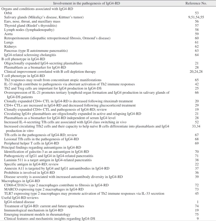
cal for pathogenesis of IgG4-RD. M2 macrophages are the dominant population in affected tissues and might contribute to fibrosis through production of pro-fibrotic cytokines. The important of several findings for the pathogenesis of IgG4- RD has been reported in Table 1
19-22,24,25,28,29,33,34,36-39,42,45,51-75.
ORCID
Takashi Maehara, https://orcid.org/0000-0002-7877-2161 Masafumi Moriyama, https://orcid.org/0000-0003-2911-2244 Seiji Nakamura, https://orcid.org/0000-0002-6373-8817
Table 1. Principal findings regarding the pathogenesis of IgG4-RD
Involvement in the pathogenesis of IgG4-RD Reference No.
Organs and conditions associated with IgG4-RD
Orbit 53
Salivary glands (Mikuliçz’s disease, Küttner’s tumor) 9,51,54,55
Ears, nose, throat, and maxillary mass 56
Thyroid gland (Riedel’s thyroiditis) 57
Lymph nodes (lymphadenopathy) 58
Aorta 59
Retroperitoneum (idiopathic retroperitoneal fibrosis, Ormond’s disease) 60
Lungs 61
Kidneys 62
Pancreas (type II autoimmune pancreatitis) 63
IgG4-related sclerosing cholangitis 64
B cell phenotype in IgG4-RD
Oligoclonally expanded IgG4-secreting plasmablasts 21
Plasmablasts as a biomarker for IgG4-RD 28
Clinical improvement correlated with B cell depletion therapy 20,24,28
T cell phenotype in IgG4-RD
Th2 responses may result from concomitant atopic manifestations 65
IL-33 might contribute to pathogenesis via aberrant activation of Th2 immune responses 42
Th2 and Treg cells are important for IgG4 production in IgG4-DS 25
Overexpression of IL-21 promotes tertiary lymphoid organ formation and IgG4 production in salivary glands of IgG4-DS patients
66 Clonally expanded CD4+ CTL in IgG4-RD is decreased following rituximab treatment 20 CD4+ CTLs are increased in IgG4-RD and decreased following glucocorticoid treatment 29
Clonally expanded CD4+ CTL and pathogenesis of IgG4-RD; review 19
Circulating IgG4+ plasmablasts are oligoclonally expanded in active and relapsing IgG4-RD 21
Plasmablasts as a biomarker for IgG4-RD independent of serum IgG4 level 28
Increased IL-4-secreting Tfh cells are associated with IgG4 class switching in vivo 52 Increased circulating Tfh2 cells and their capacity to help naïve B cells differentiate into plasmablasts and IgG4
production in vitro 33,34
Tfh cells in the pathogenesis of IgG4-RD; review 67
Lesional Tfh cells in the pathogenesis of IgG4-RD 68
Peripheral helper T cells in IgG4-RD 69
Principal findings regarding autoantigens in IgG4-RD
Identification of galectin-3 as an autoantigen in IgG4-RD 70
Pathogenicity of IgG1 and IgG4 in IgG4-related pancreatitis 71
Laminin 511 is a target antigen in IgG4-related pancreatitis 38
Specific antigen in IgG4-RD; review 22
Annexin A11 is targeted by IgG4 and IgG1 autoantibodies in IgG4-RD 37
Prohibitin is involved in IgG4-RD 36
Disease severity is associated with increased autoantibody diversity in IgG4-RD 39 Macrophages in IgG4-RD
CD68+CD163+ type 2 macrophages contribute to fibrosis in IgG4-RD 72
MARCO expressing type 2 macrophages in IgG4-RD 73
TLR7 expressing type 2 macrophages may promote activation of Th2 immune responses via IL-33 secretion 45 Useful IgG4-RD reviews
IgG4-related disease 1
Treatment of IgG4-RD: current and future approaches 74
Immunological mechanism in IgG4-RD 19
Emerging treatment models in rheumatology 75
Clinical features and mechanistic insights regarding IgG4-DS 9
(IgG4-RD: immunoglobulin G4-related disease, Th2: type 2 helper T, IL: interleukin, IgG4-DS: immunoglobulin G4-related dacryoadenitis and sialoadenitis, Tfh: follicular helper T, MARCO: macrophage receptor with collagenous structure, TLR: Toll-like receptor)
Takashi Maehara et al: Review of a novel disease entity, immunoglobulin G4-related disease. J Korean Assoc Oral Maxillofac Surg 2020
Authors’ Contributions
T.M. participated in conceptualization, investigation, and writing - original draft, review, and editing. M.M. and S.N.
participated in review and editing. All authors read and ap- proved the final manuscript.
Acknowledgements
This study was supported by JSPS KAKENHI Grant Num- bers 19H03854 and 18KK0260 and by the “Takeda Science Foundation” to T.M. We thank the Edanz Group (www.edan- zediting.com/ac) for editing a draft of this manuscript.
Ethics Approval and Consent to Participate
The study design and methods were approved by the Insti- tutional Review Board of the Center for Clinical and Transla- tional Research of Kyushu University Hospital (IRB Nos. 25- 287 and 26-86) and followed the tenets of the Declaration of Helsinki. Informed consent was obtained from all patients.
Conflict of Interest
No potential conflict of interest relevant to this article was reported.
References
1. Kamisawa T, Zen Y, Pillai S, Stone JH. IgG4-related disease. Lan- cet 2015;385:1460-71.
2. Mahajan VS, Mattoo H, Deshpande V, Pillai SS, Stone JH. IgG4- related disease. Annu Rev Pathol 2014;9:315-47.
3. Furukawa S, Moriyama M, Kawano S, Tanaka A, Maehara T, Hayashida JN, et al. Clinical relevance of Küttner tumour and IgG4-related dacryoadenitis and sialoadenitis. Oral Dis 2015;21:257-62.
4. Deshpande V, Zen Y, Chan JK, Yi EE, Sato Y, Yoshino T, et al.
Consensus statement on the pathology of IgG4-related disease.
Mod Pathol 2012;25:1181-92.
5. Khosroshahi A, Wallace ZS, Crowe JL, Akamizu T, Azumi A, Car- ruthers MN, et al. International consensus guidance statement on the management and treatment of IgG4-related disease. Arthritis Rheumatol 2015;67:1688-99.
6. Wallace ZS, Zhang Y, Perugino CA, Naden R, Choi HK, Stone JH.
Clinical phenotypes of IgG4-related disease: an analysis of two international cross-sectional cohorts. Ann Rheum Dis 2019;78:406- 7. Shimizu M, Okamura K, Kise Y, Takeshita Y, Furuhashi H, Weer-12.
awanich W, et al. Effectiveness of imaging modalities for screening IgG4-related dacryoadenitis and sialadenitis (Mikulicz's disease) and for differentiating it from Sjögren's syndrome (SS), with an emphasis on sonography. Arthritis Res Ther 2015;17:223.
8. Sakamoto M, Moriyama M, Shimizu M, Chinju A, Mochizuki
K, Munemura R, et al. The diagnostic utility of subman- dibular gland sonography and labial salivary gland biopsy in IgG4-related dacryoadenitis and sialadenitis: Its potential ap- plication to the diagnostic criteria. Mod Rheumatol 2019. doi:
10.1080/14397595.2019.1576271. [Epub ahead of print]
9. Maehara T, Pillai S, Stone JH, Nakamura S. Clinical features and mechanistic insights regarding IgG4-related dacryoadenitis and sialoadenitis: a review. Int J Oral Maxillofac Surg 2019;48:908-16.
10. Umehara H, Okazaki K, Nakamura T, Satoh-Nakamura T, Na- kajima A, Kawano M, et al. Current approach to the diagnosis of IgG4-related disease - combination of comprehensive diagnostic and organ-specific criteria. Mod Rheumatol 2017;27:381-91.
11. Li W, Chen Y, Sun ZP, Cai ZG, Li TT, Zhang L, et al. Clinicopath- ological characteristics of immunoglobulin G4-related sialadenitis.
Arthritis Res Ther 2015;17:186.
12. Moriyama M, Furukawa S, Kawano S, Goto Y, Kiyoshima T, Tanaka A, et al. The diagnostic utility of biopsies from the subman- dibular and labial salivary glands in IgG4-related dacryoadenitis and sialoadenitis, so-called Mikulicz's disease. Int J Oral Maxillo- fac Surg 2014;43:1276-81.
13. Kawa S, Ota M, Yoshizawa K, Horiuchi A, Hamano H, Ochi Y, et al. HLA DRB10405-DQB10401 haplotype is associated with auto- immune pancreatitis in the Japanese population. Gastroenterology 2002;122:1264-9.
14. Terao C, Ota M, Iwasaki T, Shiokawa M, Kawaguchi S, Kuri- yama K, et al. IgG4-related disease in the Japanese population: a genome-wide association study. Lancet Rheumatol 2019;1:PE14- 15. Strehl JD, Hartmann A, Agaimy A. Numerous IgG4-positive plas-22.
ma cells are ubiquitous in diverse localised non-specific chronic inflammatory conditions and need to be distinguished from IgG4- related systemic disorders. J Clin Pathol 2011;64:237-43.
16. Bruhns P, Iannascoli B, England P, Mancardi DA, Fernandez N, Jorieux S, et al. Specificity and affinity of human Fcgamma recep- tors and their polymorphic variants for human IgG subclasses.
Blood 2009;113:3716-25.
17. Carruthers MN, Khosroshahi A, Augustin T, Deshpande V, Stone JH. The diagnostic utility of serum IgG4 concentrations in IgG4- related disease. Ann Rheum Dis 2015;74:14-8.
18. Maehara T. [IgG4-related disease -mechanistic insights from both clinical and immunologic understanding of this condition]. Nihon Rinsho Meneki Gakkai Kaishi 2017;40:206-12. Japanese.
19. Mattoo H, Stone JH, Pillai S. Clonally expanded cytotoxic CD4+ T cells and the pathogenesis of IgG4-related disease. Autoimmunity 2017;50:19-24.
20. Mattoo H, Mahajan VS, Maehara T, Deshpande V, Della-Torre E, Wallace ZS, et al. Clonal expansion of CD4(+) cytotoxic T lym- phocytes in patients with IgG4-related disease. J Allergy Clin Im- munol 2016;138:825-38.
21. Mattoo H, Mahajan VS, Della-Torre E, Sekigami Y, Carruthers M, Wallace ZS, et al. De novo oligoclonal expansions of circulating plasmablasts in active and relapsing IgG4-related disease. J Allergy Clin Immunol 2014;134:679-87.
22. Haldar D, Hirschfield GM. Deciphering the biology of IgG4- related disease: specific antigens and disease? Gut 2018;67:602-5.
23. Carruthers MN, Topazian MD, Khosroshahi A, Witzig TE, Wallace ZS, Hart PA, et al. Rituximab for IgG4-related disease: a prospec- tive, open-label trial. Ann Rheum Dis 2015;74:1171-7.
24. Della-Torre E, Feeney E, Deshpande V, Mattoo H, Mahajan V, Ku- likova M, et al. B-cell depletion attenuates serological biomarkers of fibrosis and myofibroblast activation in IgG4-related disease.
Ann Rheum Dis 2015;74:2236-43.
25. Tanaka A, Moriyama M, Nakashima H, Miyake K, Hayashida JN, Maehara T, et al. Th2 and regulatory immune reactions contribute to IgG4 production and the initiation of Mikulicz disease. Arthritis Rheum 2012;64:254-63.
26. Maehara T, Mattoo H, Ohta M, Mahajan VS, Moriyama M, Yam-
auchi M, et al. Lesional CD4+ IFN-γ+ cytotoxic T lymphocytes in IgG4-related dacryoadenitis and sialoadenitis. Ann Rheum Dis 2017;76:377-85.
27. Tian Y, Sette A, Weiskopf D. Cytotoxic CD4 T cells: differentia- tion, function, and application to dengue virus infection. Front Im- munol 2016;7:531.
28. Wallace ZS, Mattoo H, Carruthers M, Mahajan VS, Della Torre E, Lee H, et al. Plasmablasts as a biomarker for IgG4-related dis- ease, independent of serum IgG4 concentrations. Ann Rheum Dis 2015;74:190-5.
29. Della-Torre E, Bozzalla-Cassione E, Sciorati C, Ruggiero E, Lan- zillotta M, Bonfiglio S, et al. A CD8α- subset of CD4+SLAMF7+
cytotoxic T cells is expanded in patients with IgG4-related disease and decreases following glucocorticoid treatment. Arthritis Rheu- matol 2018;70:1133-43.
30. Pillai S, Mattoo H, Cariappa A. B cells and autoimmunity. Curr Opin Immunol 2011;23:721-31.
31. Maehara T, Moriyama M, Nakamura S. Pathogenesis of IgG4- related disease: a critical review. Odontology 2019;107:127-32.
32. Crotty S. Follicular helper CD4 T cells (TFH). Annu Rev Immunol 2011;29:621-63.
33. Akiyama M, Suzuki K, Yamaoka K, Yasuoka H, Takeshita M, Kaneko Y, et al. Number of circulating follicular helper 2 T cells correlates with IgG4 and interleukin-4 levels and plasmablast num- bers in IgG4-related disease. Arthritis Rheumatol 2015;67:2476-81.
34. Akiyama M, Yasuoka H, Yamaoka K, Suzuki K, Kaneko Y, Kondo H, et al. Enhanced IgG4 production by follicular helper 2 T cells and the involvement of follicular helper 1 T cells in the pathogen- esis of IgG4-related disease. Arthritis Res Ther 2016;18:167.
35. Grados A, Ebbo M, Piperoglou C, Groh M, Regent A, Samson M, et al. T cell polarization toward TH2/TFH2 and TH17/TFH17 in patients with IgG4-related disease. Front Immunol 2017;8:235.
36. Du H, Shi L, Chen P, Yang W, Xun Y, Yang C, et al. Prohibi- tin is involved in patients with IgG4 related disease. PLoS One 2015;10:e0125331.
37. Hubers LM, Vos H, Schuurman AR, Erken R, Oude Elferink RP, Burgering B, et al. Annexin A11 is targeted by IgG4 and IgG1 au- toantibodies in IgG4-related disease. Gut 2018;67:728-35.
38. Shiokawa M, Kodama Y, Sekiguchi K, Kuwada T, Tomono T, Kuriyama K, et al. Laminin 511 is a target antigen in autoimmune pancreatitis. Sci Transl Med 2018;10:eaaq0997.
39. Liu H, Perugino CA, Ghebremichael M, Wallace ZS, Montesi SB, Stone JH, et al. Disease severity is linked to an increase in autoan- tibody diversity in IgG4-related disease. Arthritis Rheumatol 2019.
doi: 10.1002/art.41140. [Epub ahead of print]
40. Tsuboi H, Nakai Y, Iizuka M, Asashima H, Hagiya C, Tsuzuki S, et al. DNA microarray analysis of labial salivary glands in IgG4- related disease: comparison with Sjögren's syndrome. Arthritis Rheumatol 2014;66:2892-9.
41. Tsuboi H, Matsuo N, Iizuka M, Tsuzuki S, Kondo Y, Tanaka A, et al. Analysis of IgG4 class switch-related molecules in IgG4-related disease. Arthritis Res Ther 2012;14:R171.
42. Furukawa S, Moriyama M, Miyake K, Nakashima H, Tanaka A, Maehara T, et al. Interleukin-33 produced by M2 macrophages and other immune cells contributes to Th2 immune reaction of IgG4- related disease. Sci Rep 2017;7:42413.
43. Watanabe T, Yamashita K, Fujikawa S, Sakurai T, Kudo M, Shio- kawa M, et al. Involvement of activation of toll-like receptors and nucleotide-binding oligomerization domain-like receptors in enhanced IgG4 responses in autoimmune pancreatitis. Arthritis Rheum 2012;64:914-24.
44. Watanabe T, Yamashita K, Sakurai T, Kudo M, Shiokawa M, Uza N, et al. Toll-like receptor activation in basophils contributes to the development of IgG4-related disease. J Gastroenterol 2013;48:247- 45. Ishiguro N, Moriyama M, Furusho K, Furukawa S, Shibata T, 53.
Murakami Y, et al. Activated M2 macrophages contribute to the
pathogenesis of IgG4-related disease via toll-like receptor 7/inter- leukin-33 signaling. Arthritis Rheumatol 2020;72:166-78.
46. Kamisawa T, Shimosegawa T, Okazaki K, Nishino T, Watanabe H, Kanno A, et al. Standard steroid treatment for autoimmune pancre- atitis. Gut 2009;58:1504-7.
47. Masaki Y, Matsui S, Saeki T, Tsuboi H, Hirata S, Izumi Y, et al.
A multicenter phase II prospective clinical trial of glucocorticoid for patients with untreated IgG4-related disease. Mod Rheumatol 2017;27:849-54.
48. Hong X, Zhang YY, Li W, Liu YY, Wang Z, Chen Y, et al. Treat- ment of immunoglobulin G4-related sialadenitis: outcomes of glu- cocorticoid therapy combined with steroid-sparing agents. Arthritis Res Ther 2018;20:12.
49. Masamune A, Nishimori I, Kikuta K, Tsuji I, Mizuno N, Iiyama T, et al. Randomised controlled trial of long-term maintenance cor- ticosteroid therapy in patients with autoimmune pancreatitis. Gut 2017;66:487-94.
50. Shimosegawa T, Chari ST, Frulloni L, Kamisawa T, Kawa S, Mino- Kenudson M, et al. International consensus diagnostic criteria for autoimmune pancreatitis: guidelines of the International Associa- tion of Pancreatology. Pancreas 2011;40:352-8.
51. Moriyama M, Tanaka A, Maehara T, Ohyama Y, Shimizu M, Na- kashima H, et al. Clinical characteristics of Mikulicz's disease as an IgG4-related disease. Clin Oral Investig 2013;17:1995-2002.
52. Maehara T, Mattoo H, Mahajan VS, Murphy SJ, Yuen GJ, Ishiguro N, et al. The expansion in lymphoid organs of IL-4+ BATF+ T fol- licular helper cells is linked to IgG4 class switching in vivo. Life Sci Alliance 2018;1:e201800050.
53. Wallace ZS, Deshpande V, Stone JH. Ophthalmic manifestations of IgG4-related disease: single-center experience and literature re- view. Semin Arthritis Rheum 2014;43:806-17.
54. Baer AN, Gourin CG, Westra WH, Cox DP, Greenspan JS, Daniels TE, et al. Rare diagnosis of IgG4-related systemic disease by lip bi- opsy in an international Sjögren syndrome registry. Oral Surg Oral Med Oral Pathol Oral Radiol 2013;115:e34-9.
55. Yamamoto M, Takahashi H, Sugai S, Imai K. Clinical and patho- logical characteristics of Mikulicz's disease (IgG4-related plasma- cytic exocrinopathy). Autoimmun Rev 2005;4:195-200.
56. Hu EK, Parrish C, Wrobel B, Deshpande V, Stone JH. Immuno- globulin G4-related disease presenting as an ethmoid and maxillary mass. Ann Allergy Asthma Immunol 2013;111:75-7.
57. Dahlgren M, Khosroshahi A, Nielsen GP, Deshpande V, Stone JH.
Riedel's thyroiditis and multifocal fibrosclerosis are part of the IgG4-related systemic disease spectrum. Arthritis Care Res (Hobo- ken) 2010;62:1312-8.
58. Cheuk W, Chan JK. Lymphadenopathy of IgG4-related disease:
an underdiagnosed and overdiagnosed entity. Semin Diagn Pathol 2012;29:226-34.
59. Stone JH, Zen Y, Deshpande V. IgG4-related disease. N Engl J Med 2012;366:539-51.
60. Khosroshahi A, Carruthers MN, Stone JH, Shinagare S, Sainani N, Hasserjian RP, et al. Rethinking Ormond's disease: "idiopathic"
retroperitoneal fibrosis in the era of IgG4-related disease. Medicine (Baltimore) 2013;92:82-91.
61. Inoue D, Zen Y, Abo H, Gabata T, Demachi H, Kobayashi T, et al.
Immunoglobulin G4-related lung disease: CT findings with patho- logic correlations. Radiology 2009;251:260-70.
62. Saeki T, Nishi S, Imai N, Ito T, Yamazaki H, Kawano M, et al.
Clinicopathological characteristics of patients with IgG4-related tubulointerstitial nephritis. Kidney Int 2010;78:1016-23.
63. Stone JH, Khosroshahi A, Deshpande V, Chan JK, Heathcote JG, Aalberse R, et al. Recommendations for the nomenclature of IgG4- related disease and its individual organ system manifestations.
Arthritis Rheum 2012;64:3061-7.
64. Zen Y, Harada K, Sasaki M, Sato Y, Tsuneyama K, Haratake J, et al. IgG4-related sclerosing cholangitis with and without hepatic inflammatory pseudotumor, and sclerosing pancreatitis-associated
sclerosing cholangitis: do they belong to a spectrum of sclerosing pancreatitis? Am J Surg Pathol 2004;28:1193-203.
65. Mattoo H, Della-Torre E, Mahajan VS, Stone JH, Pillai S. Circulat- ing Th2 memory cells in IgG4-related disease are restricted to a defined subset of subjects with atopy. Allergy 2014;69:399-402.
66. Maehara T, Moriyama M, Nakashima H, Miyake K, Hayashida JN, Tanaka A, et al. Interleukin-21 contributes to germinal centre formation and immunoglobulin G4 production in IgG4-related dacryoadenitis and sialoadenitis, so-called Mikulicz's disease. Ann Rheum Dis 2012;71:2011-9.
67. Akiyama M, Suzuki K, Yasuoka H, Kaneko Y, Yamaoka K, Takeu- chi T. Follicular helper T cells in the pathogenesis of IgG4-related disease. Rheumatology (Oxford) 2018;57:236-45.
68. Kamekura R, Takano K, Yamamoto M, Kawata K, Shigehara K, Jitsukawa S, et al. Cutting edge: a critical role of lesional T fol- licular helper cells in the pathogenesis of IgG4-related disease. J Immunol 2017;199:2624-9.
69. Kamekura R, Yamamoto M, Takano K, Yabe H, Ito F, Ikegami I, et al. Circulating PD-1+CXCR5-CD4+ T cells underlying the immuno- logical mechanisms of IgG4-related disease. Rheumatol Adv Pract 2018;2:rky043.
70. Perugino CA, AlSalem SB, Mattoo H, Della-Torre E, Mahajan V, Ganesh G, et al. Identification of galectin-3 as an autoantigen in patients with IgG4-related disease. J Allergy Clin Immunol 2019;143:736-45.e6.
71. Shiokawa M, Kodama Y, Kuriyama K, Yoshimura K, Tomono T,
Morita T, et al. Pathogenicity of IgG in patients with IgG4-related disease. Gut 2016;65:1322-32.
72. Furukawa S, Moriyama M, Tanaka A, Maehara T, Tsuboi H, Iizuka M, et al. Preferential M2 macrophages contribute to fibrosis in IgG4-related dacryoadenitis and sialoadenitis, so-called Mikulicz's disease. Clin Immunol 2015;156:9-18.
73. Ohta M, Moriyama M, Maehara T, Gion Y, Furukawa S, Tanaka A, et al. DNA microarray analysis of submandibular glands in IgG4-related disease indicates a role for MARCO and other innate Immune-related proteins. Medicine (Baltimore) 2016;95:e2853.
74. Perugino CA, Stone JH. Treatment of IgG4-related disease: current and future approaches. Z Rheumatol 2016;75:681-6.
75. Perugino CA, Mattoo H, Mahajan VS, Maehara T, Wallace ZS, Pillai S, et al. Emerging treatment models in rheumatology: IgG4- related disease: insights into human immunology and targeted therapies. Arthritis Rheumatol 2017;69:1722-32.
How to cite this article: Maehara T, Moriyama M, Nakamura S.
Review of a novel disease entity, immunoglobulin G4-related dis- ease. J Korean Assoc Oral Maxillofac Surg 2020;46:3-11. https://
doi.org/10.5125/jkaoms.2020.46.1.3