This is an open-access article distributed under the terms of the Creative Commons Attribution Non-Commercial License (http://creativecommons.org/
licenses/by-nc/4.0/), which permits unrestricted non-commercial use, distribution, and reproduction in any medium, provided the original work is properly cited.
CC
Risk factors of medication-related osteonecrosis of the jaw:
a retrospective study in a Turkish subpopulation
Onur Şahin1, Onur Odabaşı2, Toghrul Aliyev1, Birkan Tatar1
1Department of Oral and Maxillofacial Surgery, Faculty of Dentistry, İzmir Katip Çelebi University, İzmir,
2Department of Oral and Maxillofacial Surgery, Ankara Yıldırım Beyazıt University, Ankara, Turkey
Abstract(J Korean Assoc Oral Maxillofac Surg 2019;45:108-115)
Objectives: Medication-related osteonecrosis of the jaw (MRONJ) is a well-known side effect of certain drugs that are used to influence bone metab- olism to treat osteometabolic disease or cancers. The purpose of our study was to investigate how high-concentration and low-concentration bisphos- phonate (BP) intake affects the disease severity.
Materials and Methods: Data collected from the medical records of 52 patients treated with BPs, antiresorptive, antiangiogenic drugs and diagnosed with MRONJ were included in this study. Age, sex, type of systemic disease, type of drug, duration of drug treatment, jaw area with MRONJ, drug administration protocol, and MRONJ clinical and radiological findings were obtained. Patients were divided into two groups: anti-neoplastic (Group I, n=23) and anti-osteoporotic (Group II, n=29). Statistical evaluations were performed using the IBM SPSS ver. 21.0 program.
Results: In both groups, more females had MRONJ. MRONJ was found in the mandibles of 30 patients (Group I, n=14; Group II, n=16). When we classified patients according to the American Association of Oral and Maxillofacial Surgeons staging system, significant differences were seen between groups (χ2=12.23, P<0.01). More patients with advanced stage (stage 2-3) MRONJ were found in Group I (60.9%).
Conclusion: According to our results, high-concentration BP intake, age and duration of drug intake increased disease severity.
Key words: Bisphosphonate-associated osteonecrosis of the jaw, Osteonecrosis, Risk factors, Zoledronic acid
[paper submitted 2018. 7. 2 / revised 2018. 7. 30 / accepted 2018. 7. 31]
Copyright © 2019 The Korean Association of Oral and Maxillofacial Surgeons. All rights reserved.
I. Introduction
Bisphosphonates (BPs) are the most commonly used anti- resorptive drugs for the prevention of skeletal complications in many diseases. The term bisphosphonate-related osteo- necrosis of the jaw (BRONJ) was first coined by Marx1 in 2003. An increase in localized osteonecrosis in jaw bones is related to BPs. The adverse effects of bisphosphonates on quality of life and increased morbidity, the researchers have led to the investigation of early diagnosis and effective treat-
ment strategies. In addition to BPs, other antiresorptive (e.g., denosumab) and antiangiogenic drugs (e.g., bevacizumab and sunitinib) can cause osteonecrosis, so the American Associa- tion of Oral and Maxillofacial Surgeons (AAOMS) proposed using medication-related osteonecrosis of the jaw (MRONJ) as the terminology to replace BRONJ2. Drugs that cause os- teonecrosis of the jaw are defined as antiresorptive and anti- angiogenic drugs. Drugs such as high-concentration and low- concentration BPs and denosumab are antiresorptive drugs.
Intravenous (IV) BPs are frequently used to treat hypercalce- mia associated with malignant tumors due to their antitumor- al effects in the treatment of cancers with bone metastases including breast, prostate and lung, and to prevent skeletal complications in multiple myeloma. Oral BPs are frequently associated with osteoporosis and osteopenia and rarely, with treatment of Paget’s disease and osteogenesis imperfecta3. Osteonecrosis of the jaw is a rare condition that develops as a result of impaired blood supply to the mandible and maxilla.
The incidence of osteonecrosis in individuals treated with BP therapy varies between 1% and 21%4. Bisphosphonates and other antiresorptive drugs increase apoptosis by inhibiting Onur Şahin
Department of Oral and Maxillofacial Surgery, Faculty of Dentistry, İzmir Katip Çelebi University, Aydınlıkevler Mahallesi, Cemil Meriç Bulvarı, 6780 Sokak, #48, İzmir 35640, Turkey
TEL: +90-(232)-325-25-35 FAX: +90-5054410192 E-mail: onursahin43@hotmail.com
ORCID: https://orcid.org/0000-0001-7816-1443
osteoclast differentiation and function, and these events cause bone resorption and reduced bone remodeling. Although osteoclast differentiation and function play a crucial part in the remodeling of all the bones in the skeletal system, osteo- necrosis is most commonly observed in jaw bones owing to high bone metabolism in jaw bones5.
In 2014, three criteria were defined by the AAOMS for dif- ferential diagnosis of MRONJ from other diseases that can cause clinical osteonecrosis. These criteria are: the presence of antiresorptive and antiangiogenic drug use in the patient’s medical history, clinical exposure of the bone site for more
than 8 weeks, and no radiotherapy or metastasis in the jaw bone2. In 2006, a staging system and treatment protocol was developed by Ruggiero et al.6. This staging system was up- dated by the AAOMS in 2007, 2009, and 2014. Our study was based on the staging system updated in 20142.(Table 1)
The purpose of our study was to investigate how high- concentration and low-concentration BP intake affected the severity of MRONJ. Patient records were evaluated retro- spectively for demographic data, medical information, clini- cal and radiological examination findings, drug type, usage information, and related disease.
Table 1. American Association of Oral and Maxillofacial Surgeons staging system At-risk category Asymptomatic stage without necrotic bone
Stage 0 No exposure bone, but clinical and radiological nonspecific findings and symptoms Stage 1 In asymptomatic patients with no signs of infection, presence of exposed and necrotic bone
Stage 2 Symptomatic stage, radiological findings, exposed, necrotic bone, fistula and infection are localized in the alveolar bone region Stage 3 The exposure may range from necrotic bone, fistula and infection, symptomatic stage, necrotic bone from the alveolar bone to
the lower border of the mandible, to the ramus or to the maxillary sinus, to the zygomatic bone. There may be pathological fractures, extra-oral fistula, oral-antral or oral-nazal relationship and osteolytic area extending to the lower border of the mandible or sinus base.
Data from the article of Ruggiero et al. (J Oral Maxillofac Surg 2014;72:1938-56)2.
Onur Şahin et al: Risk factors of medication-related osteonecrosis of the jaw: a retrospective study in a Turkish subpopulation. J Korean Assoc Oral Maxillofac Surg 2019
A B
C D
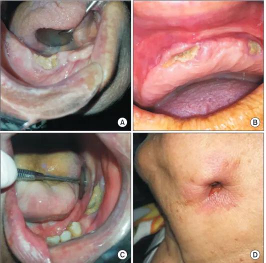
Fig. 1. Clinical findings of some med- ication-related osteonecrosis of the jaw (MRONJ) patients in this study. A.
MRONJ lesion developed that follow- ing tooth extraction at right mandibular premolar area. B. Clinical image of maxillary exposed, necrotic bone in a MRONJ patient due to prosthesis trauma. C. Stage II MRONJ in a female patient treated with alendronate for os- teoporosis. D. Extra-oral draining fistula at the submandibular region of the jaw in a patient with stage 3 MRONJ.
Onur Şahin et al: Risk factors of medication-related osteonecrosis of the jaw: a retrospective study in a Turkish subpopulation. J Korean Assoc Oral Maxillofac Surg 2019
II. Materials and Methods
This retrospective study included 52 patients over a 5-year period (January 2013 to December 2017) using database re- cords of the Department of Oral and Maxillofacial Surgery, Faculty of Dentistry, İzmir Katip Çelebi University (İzmir, Turkey). Patients who were diagnosed with MRONJ were in- cluded. The study was approved by the Ethical Committee of İzmir Katip Çelebi University (approval No. 207), and it was conducted in accordance with the Declaration of Helsinki.
Data collected from the medical records of patients in- cluded age, sex, type of systemic disease, type of drug, du- ration of drug treatment, jaw area seen with MRONJ, drug administration protocol, MRONJ staging and radiological findings. According to information collected on patients, we determined staging and treatment systems as defined by the AAOMS position paper, on MRONJ for each patient2.(Table 1) Patients with head and neck radiotherapy or necrotic bone areas less than 8 weeks were not included. Several stud- ies have been published using the AAOMS staging system updated in 2014. However, as far as we know, few studied
the use of the staging system to classify patients into anti- neoplastic (Group I, n=23) and anti-osteoporotic (Group II, n=29) groups. We analyzed sex, age, related disease, type of drug, location of MRONJ, local etiological factors and clinical and radiological findings and their relationships to in- vestigate how high-concentration and low-concentration BP intake affected disease severity.(Fig. 1, 2, Table 2)
Statistical evaluations were performed with the IBM SPSS Statistics program (ver. 21.0; IBM Corp., Armonk, NY, USA). The Kolmogorov–Smirnov test was applied to all con- tinuous variables to determine if distributions met the nor- mality assumption. Parametric tests were used for variables that met normality assumptions. Comparisons of categorical variables were made with chi-square or Fisher’s exact tests.
The Student’s t-test was used to compare quantitative vari- ables between Group I and Group II. All data were evaluated at a significance level of P<0.05.
III. Results
Of the 52 patients included in the study, 7 were male and
A B
C D
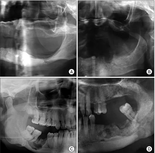
Fig. 2. Radiological findings of some medication-related osteonecrosis of the jaw (MRONJ) patients in this study. A.
Panoramic radiograph demonstrating a pathologic fracture of the left molar region of the mandible in a patient with metastatic breast and stage 3 MRONJ.
B. Panoramic radiography demon- strated osteolytic process at the left mandible. C. Panoramic radiography showing persisting alveolar socket in the mandible. D. Panoramic radiogra- phy demonstrated diffuse osteolysis of the jaw with areas of bone sclerosis.
Onur Şahin et al: Risk factors of medication-related osteonecrosis of the jaw: a retrospective study in a Turkish subpopulation. J Korean Assoc Oral Maxillofac Surg 2019
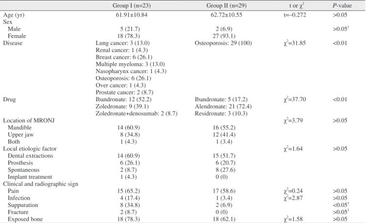
45 were female. Of these, 29 were given BP or antiresorp- tive drugs orally (2 males and 27 females) and 23 were given drugs by IV (5 males and 18 females). The mean age of pa- tients was 61.91±10.84 years (range, 44-80 years) in Group I and 62.72±10.55 years (range, 40-76 years) in Group II.
In Group I, osteonecrosis was observed in the mandible in 14 patients, in the maxilla in 8 patients and in both jaws in one patient. In Group II, 16 patients had mandibular and 12 patients had maxillary MRONJ, and one patient had MRONJ findings in both jaws. MRONJ was found in the mandibles in 30 patients. No significant difference was seen between the groups by location (P>0.05).
All patients in Group II were taking medications to prevent osteoporosis. Patients in Group I were taking high-concen- tration BPs for oncological reasons. Of the patients, 3 were treated for lung cancer, 1 for renal cancer, 6 for breast cancer, 3 for multiple myeloma, 1 for nasopharynx cancer, 6 for os- teoporosis, 1 for ovarian cancer, and 2 for prostate cancer.
All patients in Group II were treated for osteoporosis. In Group I, 6 patients were taking parenteral medication for
osteoporosis, 3 using ibandronate and 3 zoledronate. Patients in Group I were treated 2 to 10 years (mean, 29 months) and patients in Group II were treated 1 to 8 years (mean, 44 months).
Of patients in Group I, 12 were using ibandronate (52.2%), 9 zoledronate (39.1%), and 2 a combination of zoledronate and denosumab (8.7%). Of the patients in Group II, 5 were using ibandronate (17.2%), 21 alendronate (72.4%), and 3 residronate (10.3%).
When local etiologic factors causing MRONJ were exam- ined, 14 patients in Group I showed MRONJ after tooth ex- traction, 6 after receiving dental prosthesis, 2 spontaneously and 1 after dental implant treatment. Of patients in Group II, 15 had MRONJ post tooth extraction, 6 after receiving prosthetics, and 8 had spontaneous osteonecrosis. In both groups, the etiologic factor that caused MRONJ was mainly tooth extraction. The difference between the groups was not significant.
When clinical and radiological findings were examined for pain, infection, suppuration, fracture and exposed bone, pain
Table 2. Patient characteristics and outcomes
Group I (n=23) Group II (n=29) t or χ2 P-value
Age (yr) 61.91±10.84 62.72±10.55 t=–0.272 >0.05
Sex Male 5 (21.7) 2 (6.9) >0.051
Female 18 (78.3) 27 (93.1)
Disease Lung cancer: 3 (13.0) Osteoporosis: 29 (100) χ2=31.85 <0.01
Renal cancer: 1 (4.3) Breast cancer: 6 (26.1) Multiple myeloma: 3 (13.0) Nasopharynx cancer: 1 (4.3) Osteoporosis: 6 (26.1) Over cancer: 1 (4.3) Prostate cancer: 2 (8.7)
Drug Ibandronate: 12 (52.2) Ibandronate: 5 (17.2) χ2=37.70 <0.01
Zoledronate: 9 (39.1) Alendronate: 21 (72.4) Zoledronate+denosumab: 2 (8.7) Residronate: 3 (10.3)
Location of MRONJ χ2=3.79 >0.05
Mandible 14 (60.9) 16 (55.2)
Upper jaw 8 (34.8) 12 (41.4)
Both 1 (4.3) 1 (3.4)
Local etiologic factor χ2=1.64 >0.05
Dental extractions 14 (60.9) 15 (51.7)
Prosthesis 6 (26.1) 6 (20.7)
Spontaneous 2 (8.7) 8 (27.6)
Implant treatment 1 (4.3) 0 (0)
Clinical and radiographic sign
Pain 15 (65.2) 17 (58.6) χ2=0.24 >0.05
Infection 4 (17.4) 1 (3.4) χ2=2.87 >0.05
Suppuration 8 (34.8) 2 (6.9) <0.051
Fracture 2 (8.7) 0 (0) >0.051
Exposed bone 18 (78.3) 18 (62.1) χ2=1.58 >0.05
(Group I: anti-neoplastic, Group II: anti-osteoporotic, MRONJ: medication-related osteonecrosis of the jaw)
1Fisher’s exact test.
Values are presented as mean±standard deviation or number (%).
Onur Şahin et al: Risk factors of medication-related osteonecrosis of the jaw: a retrospective study in a Turkish subpopulation. J Korean Assoc Oral Maxillofac Surg 2019
and exposed bone were observed in both groups. No signifi- cant difference was found between the groups except for sup- puration (P>0.05).(Table 2)
When we classified patients according to the AAOMS staging system, significant differences were seen between the groups (χ2=12.23, P<0.01). Advanced stage (stage 2-3) disease was found at higher rates in Group I (60.9%). Early stage cases were significantly higher in Group II (82.7%).
(Table 3)
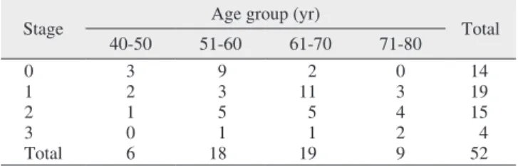
A significant difference was found in the relationship between age and stage. The incidence of stage 2 and 3 dis- ease increased with age in patients with MRONJ (χ2=17.73, P<0.05).(Table 4) We found that 3 patients smoked a package of 20 cigarettes per day for more than 10 years: 2 had stage 2 and 1 patient had stage 3 disease.
IV. Discussion
Although the first case of MRONJ was reported in 2003, MRONJ physiopathology is not fully understood, and poten- tial mechanisms are still being discussed1. The physiopathol- ogy of MRONJ is explained as modified bone remodeling or suppression of bone resorption, inflammation or infection, inhibition of angiogenesis, soft tissue toxicity, congenital or acquired immunodeficiency, microtrauma, and vitamin D in- sufficiency7.
According to the AAOMS classification, risk factors for osteonecrosis formation in jaw bones due to use of BPs are medication-related, local, and demographic/systemic8. Risk factors associated with drug use include type of BP used, method of drug administration, and duration of treatment. In particularly, intravenous use of BPs increases risk9,10. Local risk factors include periodontal diseases, inflammatory dental diseases (such as dental abscesses), torus mandibularis and palatinus, dental caries, dental alveolar surgery (tooth extrac- tion, dental implant application, periodontal surgery including bone tissue, periapical surgery), and traumatic factors such as
bone exostoses, mylohyoid protrusion, or unadapted dental prostheses11.
Demographic and systemic risk factors are defined as dia- betes mellitus, immunosuppression, anemia and thalassemia, malnutrition, osteoporosis/osteopenia diagnosed with cancer, sex, chronic corticosteroid use, radiotherapy, estrogen thera- py, smoking, cancer diagnosis, coagulation disorders, blood anomalies, vascular and connective tissue diseases, and hypo- thyroidism12. This study also investigated how these risk fac- tors affect patient low-concentration and high-concentration BP intake.
Osteoporosis can occur in individuals over 50 years of age and antiresorptive agents are often preferred for treatment.
In patients with osteoporosis using low-concentration BPs, the incidence of MRONJ is between 0.00038% and 0.1%.
For use longer than 4 years, the incidence is reported to be 0.21%13.
In patients with osteoporosis using high-concentration BPs, MRONJ incidence is reported to be 0.017% in a 3-year follow-up period. This ratio remained unchanged in a 6-year follow-up14. In our study, 23 patients with MRONJ were using high-concentration and 29 patients were using low- concentration BPs. In Group I, the duration of use of BPs or antiresorptive drugs was 29 months. In Group II, duration of use was 44 months, similar to Zervas et al.12 (24 months).
Our results are not consistent with those of Berenson et al.15 (18 months of zoledronate) and Dimopoulos et al.16 (53.4 months of pamidronate and zoledronate therapy). The high MRONJ incidence for patients taking low-concentration BPs could be linked to a longer duration of drug use and greater influence of local etiological factors. Also, we examined pa- tients who were diagnosed with MRONJ. When we classified patients according to the AAOMS staging system, Group I had a higher rate of advanced disease stage than Group II.
We found 14 patients (60.9%) with advanced-stage (2 or 3) disease in Group I and 5 patients (17.2%) in Group II.
Table 3. Staging of patients with high-concentration (Group I) and low-concentration (Group II) bisphosphonate use
Stage Group I (n=23) Group II (n=29) Total (n=52)
0 3 (13.0) 11 (37.9) 14 (26.9)
1 6 (26.1) 13 (44.8) 19 (36.5)
2 10 (43.5) 5 (17.2) 15 (28.8)
3 4 (17.4) 0 (0) 4 (7.7)
χ2=12.23, P<0.01.
Onur Şahin et al: Risk factors of medication-related osteonecrosis of the jaw: a retrospective study in a Turkish subpopulation. J Korean Assoc Oral Maxillofac Surg 2019
Table 4. Relationship between disease stage and age groups
Stage Age group (yr)
Total
40-50 51-60 61-70 71-80
0 3 9 2 0 14
1 2 3 11 3 19
2 1 5 5 4 15
3 0 1 1 2 4
Total 6 18 19 9 52
χ2=17.73, P<0.05.
Onur Şahin et al: Risk factors of medication-related osteonecrosis of the jaw: a retrospective study in a Turkish subpopulation. J Korean Assoc Oral Maxillofac Surg 2019
In Group I, rates of use for different drugs that cause MRONJ were similar. Half of patients were using ibandro- nate (52.2%) and the other half denosumab (47.8%) together with zoledronate. In Group II, alendronate (72.4%) was the most common drug causing MRONJ.
The reason that BPs cause osteonecrosis in the jaw bones rather than other bones is that BPs accumulate at high levels in alveolar bones with high regenerative capacity and in soft tissue adjacent to the bones. The inability to create an aseptic environment due to the large microflora of the oral cavity, the constant association with this microflora through the peri- odontal gap, and a high trauma incidence increase the risk of osteonecrosis in the jaw bones17. Marx et al.18 reported that 68.1% of MRONJ cases were mandibular and 27.7% were maxillary in their study. Similar results were reported by Badros et al.19. According to their study on multiple myeloma patients with BRONJ, 15 patients had mandibular, 2 had only maxillary and 5 had both mandibular and maxillary lesions.
In our study, 14 cases were mandibular, 8 cases were maxil- lary and 1 case was in both jaws in Group I. In Group II, 16 cases were mandibular, 12 cases were maxillary and 1 case of MRONJ was in both jaws. MRONJ was found in the man- dibles of 30 patients.
In the cohort study conducted by Vahtsevanos et al.20, they calculated the relative risk for osteonecrosis of the jaw (ONJ) development to be 18 times higher in patients who experienced dental extractions and patients who use dentures had an at least two-fold increased risk for ONJ development.
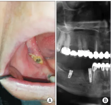
Dental extraction alone is a predisposing factor for 52% to 61% of examples of MRONJ formation21. The incidence of osteonecrosis after tooth extraction in patients using low- concentration BPs is reported to be 0.5%; it is reported to be 1.6% to 14.8% in patients receiving high-concentration BPs22. In our study, the factor that caused the formation of MRONJ was usually tooth extraction, in accordance with previous study20-22. In Group I, 14 of patients had MRONJ af- ter tooth extraction, 6 after receiving dental prosthetics and 2 had spontaneous MRONJ. In addition, we observed osteone- crosis of the right mandible after dental implant therapy due to use of zoledronate and denosumab.(Fig. 3) Of patients in Group II, 15 had MRONJ post tooth extraction, 6 because of prosthetics, and 8 had spontaneous MRONJ. In both groups, the etiologic factor that caused MRONJ was mostly tooth ex- traction.
Lesions were staged by AAOMS classification. The param- eters examined were pain, suppuration, presence of exposed bone, infection, and jaw fractures. Bagan et al.23 and Fedele et al.24 reported the most frequently encountered symptoms as pain and necrotic bone. Bagan et al.23 found pain in 77% and necrotic bone in 69% of patients in their study. These two signs of MRONJ were the most common clinical symptoms in our study in accordance with previous studies. However, 70% of patients in our study had bone exposure and 63%
had pain. In our study, exposed bone was more common than pain in both groups, but no significant difference was found between groups except for suppuration (P=0.024).
Demographic and systemic factors and other drug use also affect MRONJ formation. Age and sex are risk factors for MRONJ25. The types of drugs that cause osteonecrosis are given more frequently to women due to their indicational sta- tus. In our study, the majority of patients with MRONJ were female: 78.3% in Group I and 93.1% in Group II. The mean age of patients in this present study is in accordance with the age of those in the literature23,25. We also observed correla- tions between patient age and lesion stage. The older the age, the higher the incidence of stage 2 and 3 disease. A signifi- cant difference was found in the relationship between age and stage. The incidence of stage 2 and 3 increased with age in patients with MRONJ (χ2=17.73, P<0.05).
Whether smoking is a risk factor for MRONJ is still be-
A B
Fig. 3. Clinical and radiologic findings after dental implant treat- ment in medication-related osteonecrosis of the jaw (MRONJ) patients using zoledronate and denosumab. A. Clinical view after dental implant treatment in MRONJ patient using denosumab. B.
Panoramic radiography showed sequestrum line around of the dental implant at the right mandible molar area.
Onur Şahin et al: Risk factors of medication-related osteonecrosis of the jaw: a retrospective study in a Turkish subpopulation. J Korean Assoc Oral Maxillofac Surg 2019
ing discussed in the literature. Some studies find it to have an effect while others report that it is not a risk factor for MRONJ17. In our study, 3 patients smoked 20 cigarettes per day for more than 10 years: 2 had stage 1 disease and one had stage 3 MRONJ lesions.
V. Conclusion
According to results of our study, high-concentration BP intake, age, and duration of drug intake increased disease se- verity. According to the AAOMS staging system, updated in 2014, clinical and radiological findings have great importance for diagnosing MRONJ. In studies to date, the pathogenesis of MRONJ was explained theoretically but the pathophysiol- ogy of this disease is not exactly understood, despite the long time since the first reported case of MRONJ in the litera- ture. Future studies should aim to explain the pathogenesis of MRONJ and find effective treatment strategies in larger groups of patients. Before intake of any BP or antiresorptive drugs, patients with cancer or osteoporosis should have an extensive dental examination, especially by an oral and max- illofacial surgeon who is experienced in the prevention and treatment of MRONJ. Patients with a challenging dental situ- ation should participate in dental care before starting these medications.
ORCID
Onur Şahin, https://orcid.org/0000-0001-7816-1443 Onur Odabaşı, https://orcid.org/0000-0001-7771-048X Toghrul Aliyev, https://orcid.org/0000-0002-6312-476X Birkan Tatar, https://orcid.org/0000-0002-8917-575X
Authors’ Contributions
O.Ş., T.A., and B.T. participated in data collection and wrote the manuscript. O.Ş. and O.O. participated in the study design and coordination. O.O. and B.T. performed the statisti- cal analysis. O.O. helped to draft the manuscript. All authors read and approved the final manuscript.
Ethics Approval and Consent to Participate
The study was approved by the Ethical Committee of İzmir Katip Çelebi University (approval No. 207).
Consent for Publishing Photographs
Written informed consent was obtained from the patients for publication of this article and accompanying images.
Conflict of Interest
No potential conflict of interest relevant to this article was reported.
References
1. Marx RE. Pamidronate (Aredia) and zoledronate (Zometa) induced avascular necrosis of the jaws: a growing epidemic. J Oral Maxil- lofac Surg 2003;61:1115-7.
2. Ruggiero SL, Dodson TB, Fantasia J, Goodday R, Aghaloo T, Mehrotra B, et al.; American Association of Oral and Maxillofacial Surgeons. American Association of Oral and Maxillofacial Sur- geons position paper on medication-related osteonecrosis of the jaw--2014 update. J Oral Maxillofac Surg 2014;72:1938-56.
3. Rosen LS, Gordon D, Tchekmedyian NS, Yanagihara R, Hirsh V, Krzakowski M, et al. Long-term efficacy and safety of zole- dronic acid in the treatment of skeletal metastases in patients with nonsmall cell lung carcinoma and other solid tumors: a random- ized, Phase III, double-blind, placebo-controlled trial. Cancer 2004;100:2613-21.
4. Benhamou CL. Effects of osteoporosis medications on bone qual- ity. Joint Bone Spine 2007;74:39-47.
5. Woo SB, Hellstein JW, Kalmar JR. Narrative [corrected] review:
bisphosphonates and osteonecrosis of the jaws. Ann Intern Med 2006;144:753-61.
6. Ruggiero S, Gralow J, Marx RE, Hoff AO, Schubert MM, Huryn JM, et al. Practical guidelines for the prevention, diagnosis, and treatment of osteonecrosis of the jaw in patients with cancer. J On- col Pract 2006;2:7-14.
7. Hokugo A, Christensen R, Chung EM, Sung EC, Felsenfeld AL, Sayre JW, et al. Increased prevalence of bisphosphonate-related osteonecrosis of the jaw with vitamin D deficiency in rats. J Bone Miner Res 2010;25:1337-49.
8. Ruggiero SL, Dodson TB, Assael LA, Landesberg R, Marx RE, Mehrotra B; Task Force on Bisphosphonate-Related Osteonecrosis of the Jaws, American Association of Oral and Maxillofacial Sur- geons. American Association of Oral and Maxillofacial Surgeons position paper on bisphosphonate-related osteonecrosis of the jaw - 2009 update. Aust Endod J 2009;35:119-30.
9. Campisi G, Di Fede O, Musciotto A, Lo Casto A, Lo Muzio L, Fulfaro F, et al. Bisphosphonate-related osteonecrosis of the jaw (BRONJ): run dental management designs and issues in diagnosis.
Ann Oncol 2007;18 Suppl 6:vi168-72.
10. Kim TH, Seo WG, Koo CH, Lee JH. Evaluation of the predispos- ing factors and involved outcome of surgical treatment in bisphos- phonate-related osteonecrosis of the jaw cases including bone biopsies. J Korean Assoc Oral Maxillofac Surg 2016;42:193-204.
11. Ruggiero SL, Fantasia J, Carlson E. Bisphosphonate-related osteo- necrosis of the jaw: background and guidelines for diagnosis, stag- ing and management. Oral Surg Oral Med Oral Pathol Oral Radiol Endod 2006;102:433-41.
12. Zervas K, Verrou E, Teleioudis Z, Vahtsevanos K, Banti A, Mihou D, et al. Incidence, risk factors and management of osteonecrosis of the jaw in patients with multiple myeloma: a single-centre expe- rience in 303 patients. Br J Haematol 2006;134:620-3.
13. Pazianas M, Miller P, Blumentals WA, Bernal M, Kothawala P. A
review of the literature on osteonecrosis of the jaw in patients with osteoporosis treated with oral bisphosphonates: prevalence, risk factors, and clinical characteristics. Clin Ther 2007;29:1548-58.
14. Lo JC, O'Ryan FS, Gordon NP, Yang J, Hui RL, Martin D, et al. Prevalence of osteonecrosis of the jaw in patients with oral bisphosphonate exposure. J Oral Maxillofac Surg 2010;68:243-53.
15. Berenson JR, Yellin O, Crowley J, Makary A, Gravenor DS, Yang HH, et al. Prognostic factors and jaw and renal complications among multiple myeloma patients treated with zoledronic acid. Am J Hematol 2011;86:25-30.
16. Dimopoulos MA, Kastritis E, Anagnostopoulos A, Melakopoulos I, Gika D, Moulopoulos LA, et al. Osteonecrosis of the jaw in patients with multiple myeloma treated with bisphosphonates: evi- dence of increased risk after treatment with zoledronic acid. Hae- matologica 2006;91:968-71.
17. Ruggiero SL, Mehrotra B, Rosenberg TJ, Engroff SL. Osteonecro- sis of the jaws associated with the use of bisphosphonates: a review of 63 cases. J Oral Maxillofac Surg 2004;62:527-34.
18. Marx RE, Sawatari Y, Fortin M, Broumand V. Bisphosphonate- induced exposed bone (osteonecrosis/osteopetrosis) of the jaws:
risk factors, recognition, prevention, and treatment. J Oral Maxil- lofac Surg 2005;63:1567-75.
19. Badros A, Weikel D, Salama A, Goloubeva O, Schneider A, Rapo- port A, et al. Osteonecrosis of the jaw in multiple myeloma pa- tients: clinical features and risk factors. J Clin Oncol 2006;24:945-
20. Vahtsevanos K, Kyrgidis A, Verrou E, Katodritou E, Triaridis S, 52.
Andreadis CG, et al. Longitudinal cohort study of risk factors in cancer patients of bisphosphonate-related osteonecrosis of the jaw.
J Clin Oncol 2009;27:5356-62.
21. Henry DH, Costa L, Goldwasser F, Hirsh V, Hungria V, Prausova J, et al. Randomized, double-blind study of denosumab versus zoledronic acid in the treatment of bone metastases in patients with advanced cancer (excluding breast and prostate cancer) or multiple myeloma. J Clin Oncol 2011;29:1125-32.
22. Yamazaki T, Yamori M, Ishizaki T, Asai K, Goto K, Takahashi K, et al. Increased incidence of osteonecrosis of the jaw after tooth extraction in patients treated with bisphosphonates: a cohort study.
Int J Oral Maxillofac Surg 2012;41:1397-403.
23. Bagan JV, Jimenez Y, Murillo J, Hernandez S, Poveda R, Sanchis JM, et al. Jaw osteonecrosis associated with bisphosphonates: mul- tiple exposed areas and its relationship to teeth extractions. Study of 20 cases. Oral Oncol 2006;42:327-9.
24. Fedele S, Porter SR, D'Aiuto F, Aljohani S, Vescovi P, Manfredi M, et al. Nonexposed variant of bisphosphonate-associated osteone- crosis of the jaw: a case series. Am J Med 2010;123:1060-4.
25. Hoff AO, Toth BB, Altundag K, Johnson MM, Warneke CL, Hu M, et al. Frequency and risk factors associated with osteonecrosis of the jaw in cancer patients treated with intravenous bisphospho- nates. J Bone Miner Res 2008;23:826-36.