CASE REPORT
위에서 발생한 선암과 미만성 거대 B세포 림프종의 동시성 중복암 1예
박수역, 은창수, 변영상, 윤지영, 전용철, 한동수, 손주현, 오영하
1한양대학교 의과대학 내과학교실, 병리학교실1
A Case of Synchronous Double Primary Cancer of Gastric Adenocarcinoma and Diffuse Large B Cell Lymphoma
Soo Yuck Park, Chang Soo Eun, Young Sang Byun, Ji Young Yoon, Yong Cheol Jeon, Dong Soo Han, Joo Hyun Sohn and Young-Ha Oh1
Departments of Internal Medicine and Pathology1, Hanyang University College of Medicine, Guri, Korea
The simultaneous occurrence of primary gastric lymphoma and adenocarcinoma is rarely reported. We here report a case of synchronous double primary tumor of advanced gastric cancer and diffuse large B cell lymphoma. A 65-year-old woman underwent an esophagogastroduodenoscopy for the evaluation of abdominal discomfort of two months’ duration. The endoscopic examination showed an ulcerating tumor in the gastric antrum and thickened folds in the fundus and the microscopic examination revealed an adenocarcinoma in the antrum and a diffuse large B-cell lymphoma in the fundus. She has had total gastrectomy and CHOP chemotherapy with rituximab. Since the cases of synchronous double primary gastric tumors have been increased on the recent medical advances, when a gastric tumor is detected for the endoscopic examination, an endoscopist has to make every endeavor not to miss another tumor in the stomach. (Korean J Gastroenterol 2011;57:115-119)
Key Words: Synchronous neoplasms; Adenocarcinoma; Lymphoma, Large B-Cell, Diffuse
Received July 23, 2010. Revised July 23, 2010. Accepted August 5, 2010.
CC This is an open access article distributed under the terms of the Creative Commons Attribution Non-Commercial License (http://creativecommons.org/licenses/
by-nc/3.0) which permits unrestricted non-commercial use, distribution, and reproduction in any medium, provided the original work is properly cited.
교신저자: 은창수, 471-701, 경기도 구리시 교문동 249-1, 한양대학교 구리병원 소화기내과
Correspondence to: Chang Soo Eun, Division of Gastroenterology, Department of Internal Medicine, Hanyang University Guri Hospital, 249-1, Gyomun-dong, Guri 471-701, Korea. Tel: +82-31-560-2219, Fax: +82-31-555-2998, E-mail: [email protected]
Financial support: None. Conflict of interest: None.
서 론
위선암은 위에서 발생하는 악성 종양 중 가장 흔한 형태이 며, 위 림프종은 비교적 드문 암으로 약 3%의 빈도를 보인다.1 위 림프종 중에서 미만성 거대 B세포 림프종(DLBCL, diffuse large B cell lymphoma)은 조직학적으로 비호지킨 림프종의 일종으로 원발성 위 림프종 중에서 40-70%를 차지하는 가장 흔한 유형이다.2 위의 림프종과 선암이 원발성 중복암으로 공 존하는 경우는 매우 드물며, 대부분 이시성으로 선행암의 진 단 후에 이에 대한 치료나 추적 검사를 통해 발견된다.3 동시 성으로 진단된 경우는 점막연관 림프조직형(MALT, muco-
sa-associated lymphoid tissue) 위 림프종과 위선암의 중복 암이 종종 보고되었으나, 악성 림프종에 해당하는 DLBCL이 위선암과 동시성으로 보고된 사례는 매우 드물다.4 또한 최근 진단 기술의 발달로 위의 원발성 중복암이 점차 증가하는 추 세에 있으나, 각기 다른 두 종류의 중복암에 대한 상관성이나 치료 방향은 아직까지 명확히 언급된 바 없다. 이에 저자들은 복부 불편감으로 내원한 환자에서 상부위장관 내시경을 통해 위의 각각 다른 부위에서 진행성 위선암과 DLBCL을 동시에 진단하고 치료한 증례를 경험하여 문헌고찰과 함께 보고한다.
116
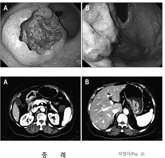
박수역 등. 위에서 발생한 선암과 미만성 거대 B세포 림프종의 동시성 중복암 1예Fig. 1. Gastroscopic findings. (A) Initial gastroscopic finding showed huge ulcerative mass on the anterior wall of antrum. (B) Gastric mucosal folds were enlarged on the posterior wall of upper body and fundus, and several irregular ero- sions were seen on that mucosal surface.
Fig. 2. Abdominal CT findings. (A) Abdominal CT showed gastric antral wall thickening with 5 cm sized excavating ulcer. (B) It also showed another lobular thickening of gas- tric wall and thickened gastric folds in the cardia and fundus.
증 례
64세 여자가 2개월 전부터 시작된 식 후 복부 불편감으로 내원하였다. 복부 불편감은 식 후 약 30분부터 시작되었으며, 상복부가 단단히 뭉치고 간헐적인 통증이 느껴지기도 하였다.
이러한 증상은 약간의 구역 증상과 동반되었으나 구토는 하지 않았고, 공복 시에는 증상이 소실되었다. 과거력 및 가족력에 서는 당뇨병 이외에 특이 소견은 없었다.
신체검사에서 활력 징후는 정상이었고, 두경부, 흉부 및 복 부에 이상소견은 관찰되지 않았다. 말초혈액검사에서 백혈구 5,700/mm3, 혈색소 12.2 g/dL, 혈소판 242,000/mm3이었고, 일반화학검사에서 총 단백 6.8 g/dL, 알부민 4.0 g/dL, 총 빌 리루빈 0.6 mg/dL, ALP 77 IU/L, AST 30 IU/L, ALT 29 IU/L, LDH 402 IU/L이었다. 종양표지자 검사 및 단순흉부촬 영 검사는 정상이었다.
상부위장관 내시경에서는 전정부 전벽에 경계가 불규칙한 5 cm 크기의 궤양성 종괴가 관찰되었고, 분문부와 기저부의 점막 주름은 두꺼워져 있었으며, 표면에 다양한 형태의 미란 들을 동반하고 있었다(Fig. 1).
복부전산화단층촬영에서 전정부의 전벽은 두꺼워져 있고 5 cm 크기의 궤양성 병변이 관찰되었으며, 주변으로 1.5 cm 크기의 림프절병증이 보였다. 기저부와 분문부의 위벽도 두꺼 워져 있었으며, 주변 림프절병증은 2 cm 크기로 여러 개 관찰
되었다(Fig. 2).
상부위장관 내시경 생검 결과, 전정부 병변은 cytokeratin (CK) 양성, leukocyte common antigen (LCA) 음성인 선암 으로 진단되었고, 분문부와 기저부 병변은 림프구의 미만성 침윤을 보였으며, LCA (CD45) 양성, B세포 표지자인 CD79a 양성, CK 음성인 B세포 림프종으로 확인되었다.
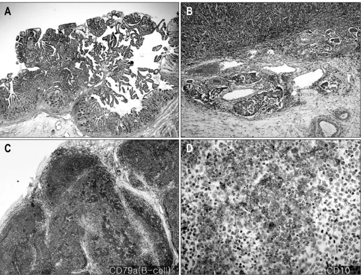
환자는 골수검사와 흉부 컴퓨터단층촬영에서 정상 소견을 보여 위전절제술과 식도공장 문합술 및 비장절제술을 시행 받 았다. 수술 후 병리 소견에서 전정부 종괴는 5×4 cm으로 중 앙이 깊게 함몰되고 변연은 융기되었으며 위벽의 장막하측까 지 암세포가 관찰되었다. 주변 연부 조직 침윤은 없었으며 조 직학적으로는 중등도의 분화를 보이거나 분화가 좋지 않은 선 암으로 확인되었다(Fig. 3A, 3B). 분문부와 기저부에는 경계 가 비교적 좋은 8×5 cm의 종괴가 점막과 점막하층에 분포하 였으며, 면역조직화학염색에서 림프구들은 CD79a와 CD20 에 양성을 보이는 B 림프구로 확인되었고, CD10, Bcl-2, Bcl-6에 대해서 양성으로 관찰되어 DLBCL으로 진단되었다 (Fig. 3C, 3D). 그 외에 절제 후 확인된 총 50개의 림프절 중 에 전정부 주변 소만측에서 3개, 대만측에서 2개의 선암 전이 가 관찰되었고, 분문부와 기저부 주변에서는 6개의 림프종 전 이가 관찰되었다.
환자는 전정부의 선암에 대해서 American Joint Commi- ttee on Cancer 분류에 따라 진행성 위암 3기(T3N2M0)와
Fig. 3. Microscopic findings and immunohistochemical stain. Histologic finding of gastric antral mass reveals moderately (A) (H&E,
×2) to poorly (B) (H&E, ×100) differentiated adenocarcinoma. The lymphoid cells infiltrated in cardia and fundus have diffusely positive cytoplasmic reaction for (C) CD79a (×40) and (D) CD10 (×400).
분문부와 기저부의 림프종에 대해서는 Modified Ann Arbor 분류 2기의 미만성 거대 B세포 림프종으로 최종 확진되었다.
환자는 수술 치료 후에 R-CHOP (rituximab, cyclophos- phamide, doxorubicin, vincristine, and prednisone) 항암 치료를 6회 받았고, 1년 후 시행한 복부컴퓨터단층촬영과 상 부위장관 내시경에서 문합부에 위선암이 재발하였으나 심한 전신 쇠약으로 수술 치료 및 복합항암화학요법 치료를 거절하 여 현재는 보존 치료를 하면서 경과 관찰 중이다.
고 찰
원발성 위 림프종이 위선암과 함께 동시성으로 발견되는 경우는 흔하지 않으며, MALT림프종에 한하여 간간이 보고되 어 왔다.5,6 더욱이 DLBCL이 위선암과 병발되는 경우는 매우 드물며, 그 중에서도 동시성보다는 이시성으로 발견된 경우가 대부분이었다.7-10 국내 보고에서는 위의 DLBCL과 진행성 위
선암이 동시에 진단된 경우가 1예 있었으나, 이번 증례와 달 리 중복암에 대한 치료와 경과 관찰을 시행하지 못한 단순 진단 증례였다.4
위에서 병발된 선암과 림프종에 대한 병인론적 연관성은 아직까지 확립되지 않았으나 위선암과 위 림프종이 병발된 12명을 대상으로 한 연구에서, 위선암은 조기 위암이 진행성 위암보다 많았고, 림프절 전이는 림프종에 의한 것이 위선암 종에 의한 것보다 많았으며, 림프종의 병변 크기가 위선암보 다 더 크고 침윤 깊이도 더 깊었다. 따라서 림프종이 위선암보 다 선행한 것으로 생각하였고, 선암 발생의 유발인자로 추측 하였다.11 이러한 부분은 아직 명확히 밝혀지진 않았지만 선행 하는 MALT림프종 내의 선상피가 지속적인 증식을 유발하는 자극이나 암 발생 유발 인자에 의해 암으로 변형되기 쉬운 경향을 보이기 때문이라는 가설이 있다.12 그러나 저자들이 경 험한 증례는 진행성 선암과 DLBCL이 위에서 같은 시점에 진 단된 매우 드문 경우였다. 두 암종의 침윤 깊이는 위선암이
118
박수역 등. 위에서 발생한 선암과 미만성 거대 B세포 림프종의 동시성 중복암 1예장막하층, 림프종이 점막하층으로 위선암의 침윤 깊이가 더 깊었으며 림프절 전이도 위선암이 5개, 림프종이 6개로 별다 른 차이를 보이지 않아 기존의 문헌 고찰에서 제시했던 가설 과는 다른 양상이라 할 수 있다.
동시 다발성 위암의 국내 보고에 의하면, 전체 8,101명의 위암 환자 중 동시 다발성 위암의 발생 빈도는 2.33%였으며, 최근 의학 기술의 발달과 위암 검진의 확대 추세로 미루어 볼 때 향후 위선암과 동시 병발하는 위암의 빈도는 증가할 것으로 예상된다.13 또한 외국 문헌에 따르면, 동시 다발성 위 암의 빈도가 육안적으로 관찰하였을 경우 5.8%였으나, 연속 적으로 위를 절제하여 생검하였을 경우 13.2%까지 증가하였 다. 따라서 위암을 진단할 당시 주의깊은 관찰과 조직검사를 통해 또 다른 종양의 존재 가능성을 간과해서는 안된다.14
일반적으로 위 림프종의 치료는 MALT림프종과 악성도가 높은 림프종에 따라 치료 방법의 차이가 있는데, MLAT림프 종에서는 대부분 Helicobacter pylori 제균 치료만으로 완전 관해에 도달하는 경우가 많지만, 악성도가 높은 림프종에서는 복합 항암화학요법이 적절하다. 국소성 림프종의 경우에도 장 기 생존율이나 삶의 질 모두에서 수술 치료보다는 복합 항암 화학요법이나 방사선 치료의 병용이 적절하다.15,16 한편, 진행 성 위암의 경우 수술의 적응증이 되면, 위 아전절제술이나 전 절제술이 가장 좋은 치료이며, 수술 후 보조 항암화학요법은 생존율 향상에 매우 적은 영향만을 미친다.17 따라서 이 환자 에서는 진행성 위선암에 대해 수술 처치를 시행한 후 복합 항암화학요법은 위선암보다는 DLBCL에 대해서만 시행하였 으며, 림프종 주변의 림프절 전이가 있었기 때문에 방사선 치 료는 하지 않았다.17,18
위의 원발성 중복암에 대한 치료는 아직까지 정립되어 있 지는 않다. 하나의 종양이 발견되어 치료하고 추적 검사 중에 새로운 종양을 진단하는 경우는 각각의 종양에 대해 별도로 생각하여 근치적 치료를 계획하는 것이 좋을 것이다. 이번 증 례와 같이 단일 시점에 두 개의 암을 진단한 경우는 암의 종류 와 치료에 대한 반응도 및 환자의 전신 상태 등을 고려하여 결정해야 할 것이고 그 중에서도 환자의 생존을 위협하는 암 부터 우선적으로 치료의 중점에 두어야 할 것이다. 위의 선암 과 림프종이 동시에 발견된 경우는 매우 드물고, 같은 위에서 발견되었다 할지라도 위선암의 경우 가능하다면 수술 치료가 선호되는 반면에 위 림프종은 복합 항암화학요법이 더 좋은 예후를 보인다. 치료가 상이한 중복암이 동시성으로 발견되었 을 때의 치료 원칙이나 방향에 대해 아직까지 정립된 것이 없으나 최근 그 빈도가 증가하고 있는 점을 고려하여 이에 대한 적극적인 논의가 필요할 것으로 보인다.
REFERENCES
1. Amer MH, el-Akkad S. Gastrointestinal lymphoma in adults:
clinical features and management of 300 cases. Gastroen- terology 1994;106:846-858.
2. Ferreri AJ, Montalbán C. Primary diffuse large B-cell lym- phoma of the stomach. Crit Rev Oncol Hematol 2007;63:
65–71.
3. Noda T, Akashi H, Matsueda S, Katsuki N, Shirahashi K, Kojiro M. Collision of malignant lymphoma and multiple early adenocarcinomas of the stomach. Arch Pathol Lab Med 1989;113:419–422.
4. Lee YJ, Kim YJ, Choi US, et al. A case of advanced gastric adenocarcinoma with synchronous gastric diffuse large B cell lymphoma. Korean J Gastrointest Endosc 2010;40:
181-185.
5. Wotherspoon AC, Isaacson PG. Synchronous adenocarcino- ma and low grade B-cell lymphoma of mucosa associated lymphoid tissue (MALT) of the stomach. Histopathology 1995;27:325-331.
6. Goteri G, Ranaldi R, Rezai B, Baccarini MG, Bearzi I. Syn- chronous mucosa-associated lymphoid tissue lymphoma and adenocarcinoma of the stomach. Am J Surg Pathol 1997;21:505-509.
7. Hamaloglu E, Topaloglu S, Ozdemir A, Oznec A. Synchro- nous and metachronous occurrence of gastric adenocar- cinoma and gastric lymphoma: a review of the literature.
World J Gastroenterol 2006;12:3564-3574.
8. Prabhash K, Biswas G, Nair R, et al. Metachronous diffuse large B-cell lymphoma and adenocarcinoma. Indian J Gas- troenterol 2006;25:261-262.
9. Travoto C, Sonzogni A, Ravizza D, et al. Confocal laser en- domicroscopy diagnosis of gastric adenocarcinoma in a patient treated for gastric diffuse large-B-cell lymphoma.
Dig Liver Dis 2009;41:447–449.
10. Seo DB, Kwon KS, Park HS, et al. Metachronous gastric MALT lymphoma and early gastric cancer: a case report.
Korean J Gastroenterol 2007;49:245-250.
11. Nakamura S, Aoyagi K, Iwanaga S, Yao T, Tsuneyoshi M, Fujishima M. Synchronous and metachronous primary gas- tric lymphoma and adenocarcinoma: a clinicopathological study of 12 patients. Cancer 1997;79:1077-1085.
12. Copie-Bergman C, Locher C, Levy M, et al. Metachronous gastric MALT lymphoma and early gastric cancer: is re- sidual lymphoma a risk factor for the development of gas- tric carcinoma? Ann Oncol 2005;16:1232-1236.
13. Kim JB, Choi MK, Lee JH, et al. Clinicopathologic features of multiple synchronous gastric cancer. J Korean Cancer Assoc 1998;30:652-659.
14. Kosaka T, Miwa K, Yonemura Y, et al. A clinicopathologic study on multiple gastric cancers with special reference to distal gastrectomy. Cancer 1990;65:2602-2605.
15. Hwang CY, Ryu MH, Kang YK, et al. Complete remission of high grade gastric MALT lymphoma after Helicobacter pylo-
ri eradication. Korean J Med 2004;66:95-99.
16. Maor MH, Velasquez WS, Fuller LM, Silvermintz KB. Sto- mach conservation in stages IE and IIE gastric non- Hodgkin's lymphoma. J Clin Oncol 1990;8:266-271.
17. GASTRIC (Global Advanced/Adjuvant Stomach Tumor Re- search International Collaboration) Group, Paoletti X, Oba K, et al. Benefit of adjuvant chemotherapy for resectable
gastric cancer: a meta-analysis. JAMA 2010;303:1729- 1737.
18. Psyrri A, Papageorgiou S, Economopoulos T. Primary extra- nodal lymphomas of stomach: clinical presentation, diag- nostic pitfalls and management. Ann Oncol 2008;19:
1992-1999.