ORIGINAL ARTICLE
대장직장 종양에 대한 내시경 점막하 절제술의 연대순 임상 결과와 학습 곡선
이상민, 김은수, 박경식, 조광범, 김동춘, 강유진, 이유진, 이정민, 최은성, 최재혁, 이호영
계명대학교 의과대학 내과학교실 소화기내과
Chronological Outcomes and Learning Curve of Endoscopic Submucosal Dissection for Colorectal Tumors
Sang Min Lee, Eun Soo Kim, Kyung Sik Park, Kwang Bum Cho, Dong Choon Kim, Yu Jin Kang, Yoo Jin Lee, Jung Min Lee, Eun Sung Choi, Jae Hyuk Choi and Ho Young Lee
Division of Gastroenterology and Hepatology, Department of Internal Medicine, Keimyung University School of Medicine, Daegu, Korea
Background/Aims: Endoscopic submucosal dissection (ESD) is an effective procedure for en-bloc curative resection of the colorectal tumor. As it requires high technical skills and experience in therapeutic endoscopy, it is important to understand learning curve of ESD technique. The aim of this study was to retrospectively describe the clinical results of ESD and to evaluate learning curve for the ESD of colorectal tumors.
Methods: A total of 90 patients with 90 colorectal neoplasms, who had undergone ESD at a tertiary referral hospital from July 2009 to December 2012, were enrolled. The ESD was performed by a single endoscopist. All ESD cases were divided into three periods: first, cases 1-30; second, cases 31-60; and third, cases 61-90.
Results: The en-bloc resection rates in third period (100%) was significantly higher than that of the first (93.3%) and second period (80%) (p=0.025). The perforation rate in third period (0%) also significantly decreased compared with that of the first (13.3%) and second period (20%) (p=0.032). To calibrate the difference of tumor size among periods, proficiency was calculated, as the procedure time per specimen area (min/cm2). The proficiency in third period (4.3) was significantly shorter than that of the first (16.8) and second period (10.2) (p=0.004).
Conclusions: The learning curve of colorectal ESD in our study shows that at least 60 cases of ESD have to be conducted to acquire sufficient skill of degree without perforation. (Korean J Gastroenterol 2014;64:198-205)
Key Words: Learning curve; Colon; Endoscopy; Dissection
Received May 1, 2014. Revised August 1, 2014. Accepted August 5, 2014.
CC This is an open access article distributed under the terms of the Creative Commons Attribution Non-Commercial License (http://creativecommons.org/licenses/
by-nc/3.0) which permits unrestricted non-commercial use, distribution, and reproduction in any medium, provided the original work is properly cited.
교신저자: 김은수, 700-712, 대구시 중구 달성로 56, 계명대학교 의과대학 내과학교실 소화기내과
Correspondence to: Eun Soo Kim, Division of Gastroenterology and Hepatology, Department of Internal Medicine, Keimyung University School of Medicine, 56 Dalseong-ro, Jung-gu, Daegu 700-712, Korea. Tel: +82-53-250-8096, Fax: +82-53-250-7442, E-mail: [email protected]
Financial support: None. Conflict of interest: None.
INTRODUCTION
Colorectal cancer is a global health problem and the fourth leading cause of death from cancer worldwide.1 As most colorectal cancers are developed from adenomatous lesions through adenoma-carcinoma sequence, they can be prevented by colonoscopy with endoscopic resection at the
stage of adenoma.2 Sessile polyps and flat adenoma can be treated effectively by endoscopic mucosal resection (EMR) using a snare.3 Although EMR using a snare is a simple and time-saving procedure, this method tends to remove color- ectal tumors larger than 20 mm in a piecemeal manner.4,5 Because it is difficult to determine the depth of invasion, lym- phovascular invasion, and marginal involvement of tumors
resected by endoscopic piecemeal resection, a precise pathologic evaluation is not possible to obtain.6 Hence, it may result in relatively high incidence of local recurrence.
In an effort to resolve the problems associated with EMR, a new method, endoscopic submucosal dissection (ESD), has been developed.7-9 ESD is an effective procedure for en-bloc resection and curative resection of large colorectal tumors that are difficult to resect by EMR.10-13 However, it re- quires high-level technical skills and experience in ther- apeutic endoscopy. It also has a relatively higher rate of perfo- ration and longer procedural time than EMR.14,15 Therefore, it is important to understand the learning curve of the color- ectal ESD technique in order to achieve good results with a tolerable complication rate. The aim of this study was to retro- spectively describe the chronological clinical results of ESD and to evaluate the learning curve for ESD of colorectal tumors.
MATERIALS AND METHODS
1. Patients
A total of 90 patients with 90 colorectal neoplasms, who had undergone ESD at Keimyung University Dongsan Hospital, Daegu, Korea from July 2009 to December 2012, were consecutively enrolled in our study. ESD was performed by a single endoscopist (E.S.K.), who had experienced ap- proximately 2,000 colonoscopies, 200 colonic EMR, and 15 gastric ESD before starting colorectal ESD. In addition, he performed gastric ESD in 170 cases during the study period.
Chronologically all ESD cases were divided according to three periods: first, cases 1-30; second, cases 31-60; and third, cases 61-90. In the first five cases, ESD was performed under the supervision of an expert endoscopist (K.B.C.) who was a regular staff member at our center. The institutional review board of our hospital approved this retrospective study (DSMC 2014-04-046-001).
2. Indication for ESD
ESD procedure can completely remove superficial color- ectal neoplasms larger than 20 mm in diameter that might have a malignant potential without lymph node metastases.
Rectal carcinoid tumors less than 20 mm can also be effec- tively removed by ESD.16 Therefore, in this study, the in- dications for ESD included: (i) laterally spreading tumor (LST)
of any type >20 mm; (ii) the protruded type of tumor >20 mm; and (iii) rectal carcinoid tumor, lesion <20 mm. LST or protruded type lesions with an irregular or non-structural shaped pit pattern accompanying a non-lifting sign were con- sidered not suitable for ESD.17
3. ESD procedure
All patients took three liters of polyethylene glycol for bowel preparation one day before colorectal ESD. ESD was per- formed under unconscious sedation using intravenous mid- azolam and propofol.
All ESD were performed with single channel colonoscopies (CF-H260AI or CF-Q260AI; Olympus Optical Co. Ltd., Tokyo, Japan) and an air insufflation system. An attachment cap was used in order to facilitate dissection in all periods. The proce- dure sequence was submucosal injection, mucosal incision, and submucosal dissection with simultaneous hemostasis.
Sodium hyaluronate or saline solution containing epi- nephrine (0.01 mg/mL) mixed with 0.8% indigo carmine was injected into the submucosal layer, just outside of the margin of the tumor in order to lift the lesions. Then, mucosal incision was made using a clear cut knife (I-type-1820; Finemedix Co.
Ltd., Daegu, Korea). The clear cut knife or hook knife (KD- 620LR; Olympus Optical Co. Ltd.) was used primarily for the submucosal dissection. If there was bleeding during the ESD procedure or the need to prevent delayed bleeding after the procedure, hemostasis was performed using hemostatic for- ceps (Coagrasper, FD-410LR; Olympus Optical Co. Ltd.). The high-frequency generator for electrical cutting and coagu- lation was VIO300D (ERBE Elektromedizin, Tübingen, Germany). The endocut mode was used for mucosal incision while swift coagulation mode was used for submucosal dissection.
4. Assessment of learning curve
We retrospectively reviewed the prospectively registered data of patients in order to assess the learning curve of color- ectal ESD. We analyzed en-bloc resection rate, R0 resection rate, and perforation rate. R0 resection was defined as an en-bloc resection without tumor involvement to the lateral and vertical margins of the resected specimen in a pathologic diagnosis.18 We used the proficiency that was calculated as the procedure time per specimen area (min/cm2).19 Compli- cations related to ESD procedures such as perforation and
Table 1. Patient Demographics and Endoscopic Characteristics of the Lesions by Treatment Period
Characteristic 1st period 2nd period 3rd period p-value
Age (yr) 59.1 (36-79) 58.9 (41-76) 62.2 (35-81) 0.538
Gender (n, male:female) 14:16 18:12 21:9 0.183
Comobidity
Hypertension 12 (40.0) 12 (40.0) 9 (30.0) 0.650
Type 2 DM 8 (26.7) 3 (10.0) 5 (16.7) 0.236
CLD 2 (6.7) 0 (0.0) 1 (3.3) 0.770
CVD 0 (0.0) 0 (0.0) 2 (6.7) 0.326
CRD 2 (6.7) 1 (3.3) 0 (0.0) 0.770
Othersa 2 (6.7) 3 (10.0) 2 (6.7) 1.000
Location of tumorsb 0.426
Right colon 7 (23.3) 7 (23.3) 10 (33.3)
Left colon 3 (10.0) 6 (20.0) 7 (23.3)
Rectum 20 (66.7) 17 (56.7) 13 (43.3)
Endoscopic diagnosis 0.314
LST-G 11 (36.7) 15 (50.0) 14 (46.7)
LST-NG 3 (10.0) 4 (13.3) 8 (26.7)
Polypoid 6 (20.0) 3 (10.0) 4 (13.3)
Carcinoid 10 (33.3) 8 (26.7) 4 (13.3)
Tumor size, long axis (mm) 20.9 (9-40) 24.9 (10-55) 23.8 (9-45) 0.359
Duration per ESD (day) 16.5 13.1 12.1
Values are presented as mean (range), n (%), or mean only.
Chronologically all cases were divided according to three periods: first period, cases 1-30; second period, cases 31-60; third period, cases 61-90.
Type 2 DM, type 2 diabetes mellitus; CLD, chronic liver disease; CVD, cardiovascular disease; CRD, chronic renal disease; LST-G, laterally spreading tumor-granular type; LST-NG, laterally spreading tumor-nongranular type; ESD, endoscopic submucosal dissection.
aFirst period: uterine cervical cancer, stomach cancer; second period: stomach cancer, hypothyroidism; third period: stomach cancer.
bRight colon: cecum, ascending colon, transverse colon; left colon: descending colon, sigmoid colon.
bleeding were also estimated. In addition, we sub-analyzed clinical outcomes of ESD for colorectal epithelial tumor only, excluding carcinoid tumor cases. As performing ESD in the right colon is challenging, it is important to know the learning curve of ESD in the right colon.20 Therefore, we also analyzed clinical outcomes of ESD for right colon tumors.
5. Statistical analysis
All statistical analyses of the data were performed using PASW Statistics version 18 for Windows (IBM Co., Armonk, NY, USA). Categorical data were analyzed using the chi- square test or Fisher’s exact test. Numerical data were ana- lyzed by one way analysis of variance (ANOVA). p-value of
<0.05 was considered statistically significant.
RESULTS
The patient demographics and endoscopic characteristics of the lesion are described in Table 1. The mean age of the patients in each period was 59.1, 58.9, and 62.2 years, respectively. The majority of lesions in each period were lo-
cated in the rectum (first: 66.7%, second: 56.7%, third:
43.3%), followed by right colon (first: 23.3%, second: 23.3%, third: 33.3%), and left colon (first: 10.0%, second: 20.0%, third: 23.3%). Based on macroscopic examination, LST was the most common type in all periods. Mean tumor size in each period was 20.9 mm, 24.9 mm, and 23.8 mm, respectively.
No statistically significant difference in the demographics, lo- cation of tumors, macroscopic finding, and tumor size was observed among the three periods.
The clinicopathological outcomes according to treatment period are summarized in Table 2. The histological diagnosis was adenoma in 38 cases (42.2%), adenocarcinoma in 30 cases (33.3%), and carcinoid tumor in 19 cases (21.1%). The majority of adenocarcinoma cases were intramucosal can- cer (23, 76.7%). On the basis of 1,000 m, submucosal can- cer was divided according to shallow and deep invasion.
There were three patients with adenocarcinoma of deep in- vasion and two of them were managed by subsequent surgi- cal operation. The other patient refused to undergo surgery.
No difference with respect to pathology was observed among the three periods.
Table 2. Clinicopathological Outcomes by Treatment Period
1st period 2nd period 3rd period p-value
Pathology 0.217
Adenoma 11 (36.7) 13 (43.3) 14 (46.7)
Adenocarcinoma 8 (26.7) 9 (30.0) 13 (43.3)
Intramucosal cancer 6 (20.0) 6 (20.0) 11 (36.7)
Submucosal cancer
Shallow (<1,000 m) 1 (3.3) 2 (6.7) 1 (3.3)
Deep (≥1,000 m) 1 (3.3) 1 (3.3) 1 (3.3)
Carcinoid tumor 9 (30.0) 8 (26.7) 2 (6.7)
Othersa 2 (6.7) 0 (0.0) 1 (3.3)
Procedure time, min 39 (10-100) 31.5 (8-133) 15 (5-100) 0.034
En-bloc resection 28 (93.3) 24 (80.0) 30 (100.0) 0.025
R0 resectionb 27 (90.0) 21 (70.0) 27 (90.0) 0.056
Perforation 4 (13.3) 6 (20.0) 0 (0.0) 0.032
Values are presented as n (%) or median (range).
Chronologically all cases were divided according to three periods: first period, cases 1-30; second period, cases 31-60; third period, cases 61-90.
aFirst period: chronic colitis, granular cell tumor; third period: atypical cells.
bEn-bloc resection without tumor involvement to the margins of the resected specimen.
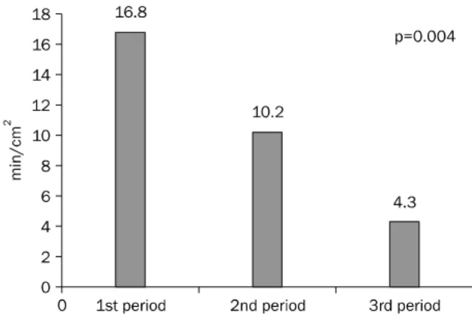
Fig. 1. Learning curve of procedure time per specimen area (min/cm2). The values represent mean proficiency in each period.
The en-bloc resection rate was significantly higher in the third period (100%) compared with that of the first (93.3%) and second period (80%) (p=0.025). The R0 resection rates in each period were 90.0%, 70.0%, and 90.0%, respectively (p=0.056). The perforation rate also showed significant im- provement in the third period (0%) compared with that of the first (13.3%) and second period (20%) (p=0.032). All perfo- ration cases were treated by endoscopic clipping and con- servative management, including intravenous antibiotics and nothing per mouth without the surgical intervention.
Significant difference in the median procedure time was observed among the three periods (39 min, 32 min, and 15 min, p=0.034). To calibrate the difference of tumor size among periods, proficiency was calculated, as the procedure time divided by specimen area (min/cm2). The learning curve of proficiency is shown in Fig. 1. Proficiency decreased over time and the proficiency was significantly shorter in the third period (4.3) compared with that of the first (16.8) period and second (10.2) period (p=0.004).
Excluding carcinoid tumors, we sub-analyzed clinical out- comes for colorectal epithelial tumors only. Mean tumor size in each period was 25.6 mm, 30.2 mm, and 26.9 mm. No sig- nificant difference in R0 resection rate (first, 85%; second, 63.6%; third, 88.5%) was observed among the three periods.
However, en-bloc resection rate was significantly higher in the third period (100%) compared with that of the first (90.0%) and second period (72.7%) (p=0.008). The profi-
ciency also showed significant improvement in the third peri- od (4.2) compared with that of the first (12.7) and second pe- riod (7.4) (p=0.02). The perforation rate was significantly bet- ter in the third period (0%) compared with that of the first (20.0%) and second period (27.3%) (p=0.008) (Table 3).
We also sub-analyzed the results of the right colon ESD. No significant difference in en-bloc resection rates (first, 71.4%
[5/7]; second, 71.4% [5/7]; third, 100% [10/10]), R0 re- section rates (first, 71.4% [5/7]; second, 57.1% [4/7]; third, 80.0% [8/10]) and perforation rates (first, 42.9% [3/7]; sec- ond, 28.6% [2/7]; third, 0% [0/10]) was observed among the three periods. However, the proficiency of the right colon ESD was significantly shorter in the third period (3.6) compared
Table 3. Clinicopathological Outcomes by Treatment Period in Colorectal Epithelial Tumors
1st period 2nd period 3rd period p-value
Tumor size, long axis (mm), mean (range) 25.6 (20-40) 30.2 (21-55) 26.9 (20-47) 0.199 Procedure time (min), median (range) 51.6 (15-100) 47.4 (8-133) 27.7 (5-100) 0.023
En-bloc resection, n (%) 18 (90.0) 16 (72.7) 26 (100.0) 0.008
R0 resectiona, n (%) 17 (85.0) 14 (63.6) 23 (88.5) 0.124
Perforation, n (%) 4 (20.0) 6 (27.3) 0 (0.0) 0.008
Proficiency (min/cm2), mean only 12.7 7.4 4.2 0.02
Chronologically all cases were divided according to three periods: first period, 20 cases; second period, 22 cases; third period, 26 cases.
aEn-bloc resection without tumor involvement to the margins of the resected specimen.
with that of the first (15.1) and second (6.4) period (p=0.034).
DISCUSSION
The results of our study demonstrated that the proficiency of the novice in performing colorectal ESD improved signifi- cantly without serious complications. Competent and expert level of technical skill was achieved after 60 cases of color- ectal ESD represented by en-bloc resection and perforation rate.
The ESD procedure was initially developed for early gastric neoplasm, which was difficult to remove by EMR.21 ESD facili- tates complete en-bloc resection and leads to lower re- currence rate than EMR.14 However, colorectal ESD proce- dure requires high-level technical skills and significant expe- rience in therapeutic endoscopy compared with gastric ESD because it has a relatively higher risk of complication due to anatomical characteristics of the colon.16 Therefore, it is im- portant to understand the learning curve of colorectal ESD.
Learning curves of colorectal ESD have been evaluated in several studies. Sakamoto et al.22 demonstrated that color- ectal ESD can be performed without serious complications by trainee endoscopists after a certain degree of preparatory training and experience with 30 cases. Hotta et al.19 used a measurement: the procedure time divided by square centi- meter of specimen area (min/cm2) for evaluating the learn- ing curve. After 40 procedures, the proficiency improved sig- nificantly with the measurement. The current study was also conducted with this measurement, and the proficiency im- proved significantly in the third period compared with that of the first and second period. In other words, the endoscopist had to experience at least 60 cases of colorectal ESD proce- dure before performing ESD with a stable level of technical
competency. However, the number of cases of threshold in the current study was higher than that of previous Japanese studies. There are several plausible explanations for this vari- ous number of cases required for good colorectal ESD from different studies. First, the duration of guidance by the experi- enced endoscopist differed between studies. The study by Hotta et al.19 included 20 ESD cases guided by an expert while only five cases were included in the current study. In an- other Japanese study, the trainee experienced all colorectal ESD with the assistance of an expert. If the cases were as- sisted by experts during the trainee’s performance of the pro- cedure, it might affect clinical outcomes favorably.18 Second, different endoscopic equipment for ESD was used in each study. A former Japanese study used a pediatric-type colono- scope (PCF-Q260JI; Olympus Optical Co.) with a built-in water jet system and a carbon dioxide insufflation system while we used a conventional colonoscope with an air insufflation system. A small diameter pediatric-type colonoscope with good maneuverability might be useful for the situation requir- ing retroversion of the scope and water-jet function is con- venient for bleeding control, particularly in a large tumor that is accompanied by many nutrient vessels.23 In addition, car- bon dioxide insufflation has been suggested to be effective during colorectal ESD.24 Despite no definite evidence of the impact of these equipments on the ESD procedure, the di- verse clinical outcomes of each study might be attributed to the various devices used in different settings.
The trainee endoscopist must have a certain degree of eli- gibility to achieve a sufficient self-completion for submucosal dissection. This includes skillful insertion of the colonoscope without looping, EMR, and bleeding or perforation control with clips. Before performing the colonic ESD, experience in gastric ESD is also required, since gastric ESD is relatively easy to perform and removal of lesions is less time-
consuming. Trainees with gastric ESD experience showed better self-completion rate and satisfactory clinical out- comes in the introductory period of colorectal ESD compared to those without gastric ESD experience.18 In the current study, the novice had competent skills in therapeutic proce- dures and management of complications (2,000 colonos- copies, 200 colonic EMR, and 15 gastric ESD) before starting colorectal ESD.
Piecemeal resection is associated with high incomplete re- section rate and local recurrence rate.4,7 ESD enables per- formance of en-bloc resection and allows a low recurrence rate. Therefore, en-bloc resection rate is also an important factor in evaluating colorectal ESD skills. The en-bloc re- section rate in several clinical studies concerning the effi- ciency and safety of ESD in the colon and rectum was 80-98.6%.11,12,25-29 The current study showed an en-bloc re- section rate of 91.1%, similar to previous studies, and especially 100% in the third period after performing 60 cases of ESD.
In the meantime, ESD has a relatively high perforation rate compared with EMR. In particular, because of the tenuity of the colorectal wall, colorectal ESD has a higher risk of perfo- ration than gastric ESD.30 Several studies reported perfo- ration rates of 1.4-10.4% in colorectal ESD.31 The majority of cases were treated successfully by conservative manage- ment without emergency surgery and small perforations were closed by endoscopic clipping.15,32 Compared with pre- vious studies, our study shows a relatively high perforation rate in the first and second periods. However, the endo- scopist was able to treat all perforation cases by endoscopic clipping and conservative management without surgical intervention. Furthermore, in the third period, the perforation rate was down to 0%. Delayed perforation as a serious com- plication after ESD was demonstrated in several studies, with rates ranging from 0.3-0.7%.25,26,33 No case of delayed perfo- ration was verified in our study.
Of particular interest, clinical outcomes including both perforation and en-bloc resection rates in the second period were worse than those of the first period. Although the reason for the poor outcomes in the second period was unclear, we postulated that the accumulation of experiences of color- ectal ESD during the first period might cause the operator to perform relatively drastic submucosal incision and dis- section in the following second period.
In the current study, ESD was performed not only for color- ectal epithelial tumors but also carcinoid tumors. All 22 cas- es of carcinoid tumor were located in the rectum and mean tumor size in the three periods was 11.4 mm, 10.3 mm, and 14.5 mm, respectively. All carcinoid tumors were removed by complete en-bloc resection without complications of perfo- ration or hemorrhage. In general, EMR was the treatment of choice for small rectal carcinoid tumors. However, histologi- cally completeness of resection is difficult for EMR, because the majority of carcinoid tumors were not confined to the mu- cosa, but extended to the submucosa.34 Therefore, ESD that can dissect tumors directly along the submucosal layer can be an effective treatment for carcinoid tumors.25 Several studies reported that ESD could be a feasible and safe proce- dure for treatment of carcinoid tumors showing good clinical outcomes.34-38
Our study has several limitations. First, the study was a ret- rospective analysis. Although there could be a possibility of selection bias, there was no significant difference in varia- bles among the three groups. Second, we included cases of carcinoid tumor in the study. Due to the small tumor size, car- cinoid tumors could be relatively easily removed by ESD, com- pared to colorectal epithelial tumors. Therefore, the clinical outcomes such as proficiency and procedure time in our study might be greater than those of previous reports.19,22 As colorectal epithelial tumors in our study were relatively small- er than those of previous reports, they would also affect the clinical outcomes.19 The third limitation is that the study was designed for just one endoscopist in a single center. However, it is not easy to conduct a multicenter study with multiple en- doscopists because there might be heterogeneity in basic skill and ability to acquire the procedure among endo- scopists from different institutes. Therefore, our study can provide a good example for endoscopists just beginning the colorectal ESD. Nevertheless, competence in therapeutic procedures and ability to manage complications should be a prerequisite in training for colorectal ESD.
In conclusion, the ESD procedure can be performed safely, although it requires high-level technical skills. The learning curve of colorectal ESD in our study shows that be performed in at least 60 cases in order to acquire sufficient skill of de- gree without perforation.
REFERENCES
1. Edwards BK, Ward E, Kohler BA, et al. Annual report to the nation on the status of cancer, 1975-2006, featuring colorectal cancer trends and impact of interventions (risk factors, screening, and treatment) to reduce future rates. Cancer 2010;116:544-573.
2. Byeon JS, Yang DH, Kim KJ, et al. Endoscopic submucosal dis- section with or without snaring for colorectal neoplasms.
Gastrointest Endosc 2011;74:1075-1083.
3. Puli SR, Kakugawa Y, Gotoda T, Antillon D, Saito Y, Antillon MR.
Meta-analysis and systematic review of colorectal endoscopic mucosal resection. World J Gastroenterol 2009;15:4273-4277.
4. Hotta K, Fujii T, Saito Y, Matsuda T. Local recurrence after endo- scopic resection of colorectal tumors. Int J Colorectal Dis 2009;24:225-230.
5. Tanaka S, Haruma K, Oka S, et al. Clinicopathologic features and endoscopic treatment of superficially spreading colorectal neo- plasms larger than 20 mm. Gastrointest Endosc 2001;54:62-66.
6. Thorlacius H, Uedo N, Toth E. Implementation of endoscopic submucosal dissection for early colorectal neoplasms in Sweden. Gastroenterol Res Pract 2013;2013:758202.
7. Hotta K, Saito Y, Matsuda T, Shinohara T, Oyama T. Local re- currence and surveillance after endoscopic resection of large colorectal tumors. Dig Endosc 2010;22(Suppl 1):S63-S68.
8. Yoshida N, Naito Y, Sakai K, et al. Outcome of endoscopic sub- mucosal dissection for colorectal tumors in elderly people. Int J Colorectal Dis 2010;25:455-461.
9. Zhou PH, Yao LQ, Qin XY. Endoscopic submucosal dissection for colorectal epithelial neoplasm. Surg Endosc 2009;23:1546- 1551.
10. Saito Y, Sakamoto T, Fukunaga S, Nakajima T, Kiriyama S, Matsuda T. Endoscopic submucosal dissection (ESD) for color- ectal tumors. Dig Endosc 2009;21(Suppl 1):S7-S12.
11. Saito Y, Uraoka T, Matsuda T, et al. Endoscopic treatment of large superficial colorectal tumors: a case series of 200 endoscopic submucosal dissections (with video). Gastrointest Endosc 2007;66:966-973.
12. Tanaka S, Oka S, Kaneko I, et al. Endoscopic submucosal dis- section for colorectal neoplasia: possibility of standardization.
Gastrointest Endosc 2007;66:100-107.
13. Jamg MY, Cho JW, Oh WG, et al. A case of pneumorrhachis and pneumoscrotum following colon endoscopic submucosal dissection. Intest Res 2013;11:208-212.
14. Saito Y, Fukuzawa M, Matsuda T, et al. Clinical outcome of endo- scopic submucosal dissection versus endoscopic mucosal re- section of large colorectal tumors as determined by curative resection. Surg Endosc 2010;24:343-352.
15. Uraoka T, Kawahara Y, Kato J, Saito Y, Yamamoto K. Endoscopic submucosal dissection in the colorectum: present status and fu- ture prospects. Dig Endosc 2009;21(Suppl 1):S13-S16.
16. Saito Y, Otake Y, Sakamoto T, et al. Indications for and technical aspects of colorectal endoscopic submucosal dissection. Gut Liver 2013;7:263-269.
17. Matsuda T, Fujii T, Saito Y, et al. Efficacy of the invasive/non-in- vasive pattern by magnifying chromoendoscopy to estimate the
depth of invasion of early colorectal neoplasms. Am J Gastroen- terol 2008;103:2700-2706.
18. Ohata K, Ito T, Chiba H, Tsuji Y, Matsuhashi N. Effective training system in colorectal endoscopic submucosal dissection. Dig Endosc 2012;24(Suppl 1):84-89.
19. Hotta K, Oyama T, Shinohara T, et al. Learning curve for endo- scopic submucosal dissection of large colorectal tumors. Dig Endosc 2010;22:302-306.
20. Nishiyama H, Isomoto H, Yamaguchi N, et al. Endoscopic sub- mucosal dissection for colorectal epithelial neoplasms. Dis Colon Rectum 2010;53:161-168.
21. Ohkuwa M, Hosokawa K, Boku N, Ohtu A, Tajiri H, Yoshida S. New endoscopic treatment for intramucosal gastric tumors using an insulated-tip diathermic knife. Endoscopy 2001;33:221-226.
22. Sakamoto T, Saito Y, Fukunaga S, Nakajima T, Matsuda T.
Learning curve associated with colorectal endoscopic sub- mucosal dissection for endoscopists experienced in gastric en- doscopic submucosal dissection. Dis Colon Rectum 2011;
54:1307-1312.
23. Tanaka S, Oka S, Chayama K. Colorectal endoscopic sub- mucosal dissection: present status and future perspective, in- cluding its differentiation from endoscopic mucosal resection.
J Gastroenterol 2008;43:641-651.
24. Saito Y, Uraoka T, Matsuda T, et al. A pilot study to assess the safe- ty and efficacy of carbon dioxide insufflation during colorectal en- doscopic submucosal dissection with the patient under con- scious sedation. Gastrointest Endosc 2007;65:537-542.
25. Fujishiro M, Yahagi N, Kakushima N, et al. Outcomes of endo- scopic submucosal dissection for colorectal epithelial neo- plasms in 200 consecutive cases. Clin Gastroenterol Hepatol 2007;5:678-683; quiz 645.
26. Isomoto H, Nishiyama H, Yamaguchi N, et al. Clinicopatho- logical factors associated with clinical outcomes of endoscopic submucosal dissection for colorectal epithelial neoplasms.
Endoscopy 2009;41:679-683.
27. Niimi K, Fujishiro M, Kodashima S, et al. Long-term outcomes of endoscopic submucosal dissection for colorectal epithelial neoplasms. Endoscopy 2010;42:723-729.
28. Tamegai Y, Saito Y, Masaki N, et al. Endoscopic submucosal dis- section: a safe technique for colorectal tumors. Endoscopy 2007;39:418-422.
29. Yoshida N, Wakabayashi N, Kanemasa K, et al. Endoscopic sub- mucosal dissection for colorectal tumors: technical difficulties and rate of perforation. Endoscopy 2009;41:758-761.
30. Huang C, Huang RX, Xiang P, Qiu ZJ. Current research status of endoscopic submucosal dissection for colorectal neoplasms.
Clin Invest Med 2012;35:E158-E164.
31. Yoshida N, Yagi N, Naito Y, Yoshikawa T. Safe procedure in endo- scopic submucosal dissection for colorectal tumors focused on preventing complications. World J Gastroenterol 2010;16:
1688-1695.
32. Kawabe T, Ichinose M, Omata M. Successful nonsurgical man- agement of perforation complicating endoscopic submucosal dissection of gastrointestinal epithelial neoplasms. Endoscopy 2006;38:1001-1006.
33. Toyanaga T, Man-I M, Ivanov D, et al. The results and limitations
of endoscopic submucosal dissection for colorectal tumors.
Acta Chir Iugosl 2008;55:17-23.
34. Park HW, Byeon JS, Park YS, et al. Endoscopic submucosal dis- section for treatment of rectal carcinoid tumors. Gastrointest Endosc 2010;72:143-149.
35. Lee DS, Jeon SW, Park SY, et al. The feasibility of endoscopic sub- mucosal dissection for rectal carcinoid tumors: comparison with endoscopic mucosal resection. Endoscopy 2010;42: 647-651.
36. Moon SH, Hwang JH, Sohn DK, et al. Endoscopic submucosal
dissection for rectal neuroendocrine (carcinoid) tumors. J Laparoendosc Adv Surg Tech A 2011;21:695-699.
37. Yamaguchi N, Isomoto H, Nishiyama H, et al. Endoscopic sub- mucosal dissection for rectal carcinoid tumors. Surg Endosc 2010;24:504-508.
38. Zhou PH, Yao LQ, Xu MD, et al. Endoscopic submucosal dis- section for rectal carcinoid tumors. Zhonghua Wei Chang Wai Ke Za Zhi 2007;10:319-322.